Comparative Study
. 2006 Feb;80(3):1513-23.
doi: 10.1128/JVI.80.3.1513-1523.2006.

High-resolution molecular and antigen structure of the VP8* core of a sialic acid-independent human rotavirus strain

Affiliations
Comparative Study

High-resolution molecular and antigen structure of the VP8* core of a sialic acid-independent human rotavirus strain

Nilah Monnier et al. J Virol. 2006 Feb.

Abstract

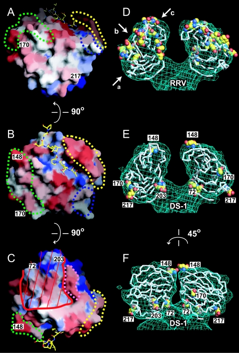
The most intensively studied rotavirus strains initially attach to cells when the "heads" of their protruding spikes bind cell surface sialic acid. Rotavirus strains that cause disease in humans do not bind this ligand. The structure of the sialic acid binding head (the VP8* core) from the simian rotavirus strain RRV has been reported, and neutralization epitopes have been mapped onto its surface. We report here a 1.6-A resolution crystal structure of the equivalent domain from the sialic acid-independent rotavirus strain DS-1, which causes gastroenteritis in humans. Although the RRV and DS-1 VP8* cores differ functionally, they share the same galectin-like fold. Differences between the RRV and DS-1 VP8* cores in the region that corresponds to the RRV sialic acid binding site make it unlikely that DS-1 VP8* binds an alternative carbohydrate ligand in this location. In the crystals, a surface cleft on each DS-1 VP8* core binds N-terminal residues from a neighboring molecule. This cleft may function as a ligand binding site during rotavirus replication. We also report an escape mutant analysis, which allows the mapping of heterotypic neutralizing epitopes recognized by human monoclonal antibodies onto the surface of the VP8* core. The distribution of escape mutations on the DS-1 VP8* core indicates that neutralizing antibodies that recognize VP8* of human rotavirus strains may bind a conformation of the spike that differs from those observed to date.


Figures

FIG. 1.
FIG. 1.
The rotavirus VP4 spike. The Cα traces of the VP8* core (white) and a globular domain of VP5* (yellow) from RRV are fitted to the molecular envelope of the VP4 spike in an approximately 12-Å-resolution electron cryomicroscopy image reconstruction of a primed SA11-4F rotavirus virion. The arrow indicates the perspective of the depictions in Fig. 3 and 4B.
FIG. 2.
FIG. 2.
Size exclusion chromatography and SDS-PAGE of purified VP8* cores. (A) Chromatograms of RRV (red) and DS-1 (blue) VP8* cores, which were separated on a Superdex 200 10/30 column at 4°C in TNE after storage for 2 years at 4°C in TNE with 0.02% sodium azide and 0.1 mM benzamidine. The inset image of a Coomassie blue-stained SDS-PAGE gel shows stored samples prior to chromatography. (B) Chromatogram of a freshly prepared sample of the KU VP8* core, which was separated on a Hi-Load Superdex 200 16/60 column at 4°C in TNE. The inset image of a Coomassie-blue stained SDS-PAGE gel shows protein from the pooled peak fractions. Apparent molecular masses that correspond to peak elution volumes are listed in Table 1.
FIG. 3.
FIG. 3.
Comparison of DS-1 and RRV VP8* cores. (A) Ribbon diagram of the DS-1 VP8* core. Labeling of secondary-structure elements is as previously described for the RRV VP8* core (14), except that strand βH, which splits into strands βH and βH′ in RRV, is continuous in DS-1. The EF β-hairpin is red, the six-stranded β-sheet is green, and the five-stranded β-sheet is blue. (B) Superimposed Cα traces of the DS-1 VP8* core (blue) and the RRV VP8* core (red). Residue Q135 of RRV, which lacks a structural equivalent in DS-1, is indicated. The blue and red arrows indicate the widths of surface clefts, measured in Å between Cα atoms of the labeled residues, for the DS-1 and RRV VP8* cores, respectively. (C) Surface representation of the DS-1 VP8* core colored by electrostatic potential. Blue is positive; red is negative. The bound leader of an adjacent molecule in the crystal is depicted with a ball-and-stick model. Residues in the space-filling model are labeled in white text boxes. Residues in the ball-and-stick model are labeled in yellow text boxes. (D) Surface representation of the RRV VP8* core colored by electrostatic potential. The bound sialoside is depicted with a ball-and-stick model. (E) Molecular details of the site in DS-1 VP8* that corresponds to the RRV SA binding pocket. The depicted area is indicated by the dashed outline in panel C. (F) Molecular details of the SA binding pocket of RRV VP8*. The depicted area is indicated by a dashed outline in panel D. Selected hydrogen bonds are indicated by dashed gray lines. In panels E and F, residues on strand βK are depicted with backbone and side chain atoms; only Cα and side chain atoms of other residues are shown. The perspective of all the panels is indicated by the arrow in Fig. 1.
FIG. 4.
FIG. 4.
Leader binding in the DS-1 VP8* core crystal. (A) Cα traces of representative molecules in the crystal. The cleft between the β-hairpin and the six-stranded β-sheet of each molecule binds the leader of an adjacent molecule. (B) Molecular details of the cleft-to-leader interaction. The leader is depicted in yellow. Hydrogen bonds are depicted as dotted black lines. Residues on strand βF are depicted without side chains. The perspective and coloring match Fig. 3A.
FIG. 5.
FIG. 5.
Neutralization surfaces of the RRV and DS-1 VP8* cores. Panels A to C show three views of a surface representation of the DS-1 VP8* core, colored by conservation among a set of VP8* sequences that contains one representative from each of 14 SA-independent P genotypes (Table 3). Blue is conserved; red is variable. Residue numbers mark neutralization escape mutations selected in SA-independent human rotavirus strains. Dashed outlines mark the previously described (14) neutralization epitopes of SA-dependent strains: green, 8-1; blue, 8-2; yellow, 8-3; pink, 8-4. The white asterisk in panel B indicates a hydrophobic pocket at the base of the peptide binding cleft. Red lines in panel C mark a surface that is inaccessible to antibody binding when the DS-1 VP8* core is fitted to the head of the SA11-4F VP4 spike. The bound leader is depicted with a ball-and-stick model. The perspectives of panels A, B, and C are indicated by arrows a, b, and c, respectively, in panel D. An observer looking down arrow c would be “head down” to view the perspective in panel C. In panels D to F, Cα traces of VP8* cores are fitted to the molecular envelope of the head from an electron cryomicroscopy image reconstruction of the SA11-4F spike on primed virions. Residues selected in neutralization escape mutants are indicated with space-filling models. (D) The RRV VP8* core with escape mutations selected in SA-dependent animal rotavirus strains. (E) The DS-1 VP8* core with escape mutations selected in SA-independent human rotavirus strains. Exposed escape mutations are labeled by residue number. (F) The model from panel E rotated 45° to show more clearly the inaccessible escape mutations in the cleft between the heads.

Similar articles

Cited by

References

    1. Bartels, C., T.-H. Xia, M. Billeter, P. Guntert, and K. Wuthrich. 1995. The program XEASY for computer-supported NMR spectral analysis of biological macromolecules. J. Biomol. NMR 5:1-10. - PubMed
    1. Bastardo, J. W., and I. H. Holmes. 1980. Attachment of SA-11 rotavirus to erythrocyte receptors. Infect. Immun. 29:1134-1140. - PMC - PubMed
    1. Brunger, A. T., P. D. Adams, G. M. Clore, W. L. DeLano, P. Gros, R. W. Grosse-Kunstleve, J. S. Jiang, J. Kuszewski, M. Nilges, N. S. Pannu, R. J. Read, L. M. Rice, T. Simonson, and G. L. Warren. 1998. Crystallography and NMR system: a new software suite for macromolecular structure determination. Acta Crystallogr. D 54:905-921. - PubMed
    1. Ciarlet, M., S. E. Crawford, and M. K. Estes. 2001. Differential infection of polarized epithelial cell lines by sialic acid-dependent and sialic acid-independent rotavirus strains. J. Virol. 75:11834-11850. - PMC - PubMed
    1. Ciarlet, M., and M. K. Estes. 1999. Human and most animal rotavirus strains do not require the presence of sialic acid on the cell surface for efficient infectivity. J. Gen. Virol. 80:943-948. - PubMed

Publication types

MeSH terms

Associated data

LinkOut - more resources