. 2006 Feb;80(3):1537-48.
doi: 10.1128/JVI.80.3.1537-1548.2006.

Mutational analysis of the herpes simplex virus triplex protein VP19C

Affiliations

Mutational analysis of the herpes simplex virus triplex protein VP19C

Walt E Adamson et al. J Virol. 2006 Feb.

Abstract

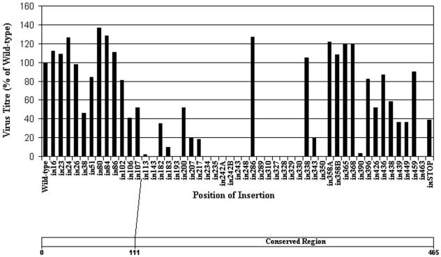
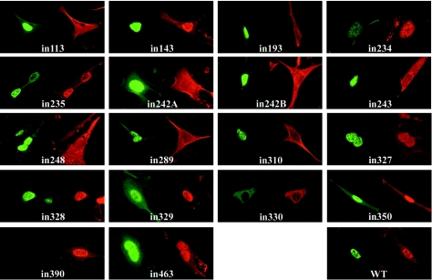
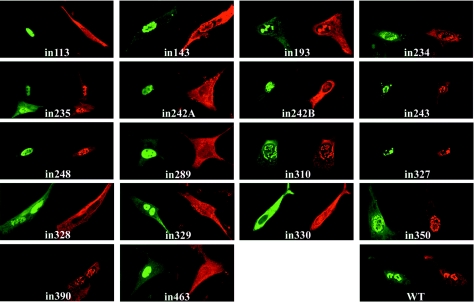
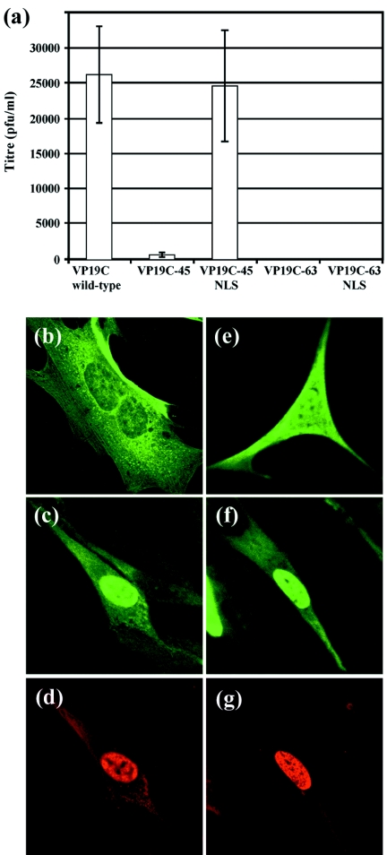
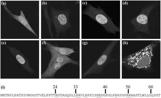
Herpes simplex virus type 1 (HSV-1) capsids have an icosahedral structure with capsomers formed by the major capsid protein, VP5, linked in groups of three by distinctive structures called triplexes. Triplexes are heterotrimers formed by two proteins in a 1:2 stoichiometry. The single-copy protein is called VP19C, and the dimeric protein is VP23. We have carried out insertional and deletional mutagenesis on VP19C and have examined the effects of the mutations on virus growth and capsid assembly. Insertional mutagenesis showed that the N-terminal approximately 100 amino acids of the protein, which correspond to a region that is poorly conserved among herpesviruses, are insensitive to disruption and that insertions into the rest of the protein had various effects on virus growth. Some, but not all, severely disabled mutants were compromised in the ability to bind VP23 or VP5. Analysis of deletion mutants revealed the presence of a nuclear localization signal (NLS) near the N terminus of VP19C, and this was mapped to a 33-amino-acid region by fusion of specific sequences to a green fluorescent protein marker. By replacing the endogenous NLS with that from the simian virus 40 large T antigen, we were able to show that the first 45 amino acids of VP19C were not essential for assembly of functional capsids and infectious virus particles. However, removing the first 63 amino acids resulted in formation of aberrant capsids and prevented virus growth, suggesting that the poorly conserved N-terminal sequences have some as-yet-unidentified function.


Figures

FIG. 1.
FIG. 1.
Functional analysis of VP19C insertional mutants. (Top) Complementation of growth of the VP19C-minus mutant vΔ38YFP by transfected plasmids expressing the VP19C insertional mutants was carried out in BHK cells. The progeny virus was titrated on UL38RSC cells. The results are shown as percentages of the titer obtained with the wild-type VP19C control plasmid. (Bottom) Scale drawing of the UL38 ORF showing the extent of the poorly conserved N-terminal sequences (Fig. 7a).
FIG. 2.
FIG. 2.
Influence of VP19C insertional mutants on the distribution of VP23. BHK cells were cotransfected with pE18 (expressing VP23) and either pUL38FBpCI (wild-type [WT] VP19C) or one of the VP19C insertional mutants as indicated. VP19C was detected with the monoclonal antibody mAb02040 and visualized using FITC GAM (green). VP23 was detected with the antiserum rAb186 and visualized using TRITC-conjugated GAR (red).
FIG. 3.
FIG. 3.
Influence of VP19C insertional mutants on the distribution of VP5. BHK cells were cotransfected with pE19 (expressing VP5) and either pUL38FBpCI (wild-type [WT] VP19C) or one of the VP19C insertional mutants as indicated. VP19C was detected with the monoclonal antibody mAb02040 and visualized using FITC GAM (green). VP5 was detected with the antiserum rAb184 and visualized using TRITC-conjugated GAR (red).
FIG. 4.
FIG. 4.
Roles of the VP19C N-terminal sequences. (a) Complementation of growth of the VP19C-minus mutant vΔ38YFP by transfected plasmid pUL38-45FBpCI (expressing VP19C-45), pUL38-63FBpCI (VP19C-63), pUL38-45NLSFBpCI (VP19C-45NLS), or pUL38-63NLSFBpCI (VP19C-63NLS) was carried out in BHK cells. The progeny virus was titrated on UL38RSC cells. The error bars indicate the standard errors of the means. (b to g) Intracellular localization of N-terminal mutants of VP19C. BHK cells were transfected singly with plasmids (b) pUL38-45FBpCI (expressing VP19C-45) and (e) pUL38-63FBpCI (VP19C-63) or were cotransfected with (c and d) pUL38-45NLSFBpCI (VP19C-45NLS) and pE18 (VP23) or with (f and g) pUL38-63NLSFBpCI (VP19C-63NLS) and pE18. VP19C (b, c, e, and f) was detected with the monoclonal antibody mAb02040 and visualized using FITC-conjugated GAM (green). VP23 (d and g) was detected with the antiserum rAb186 and visualized using TRITC-conjugated GAR (red).
FIG. 5.
FIG. 5.
Mapping the VP19C NLS. BHK cells were transfected with plasmids expressing the GFP protein fused in frame to the N-terminal 83 (b), 76 (c), 66 (d), 56 (e), or 46 (f) amino acids of VP19C. (g and h) GFP is fused to amino acids 24 to 66 and 33 to 66 of VP19C. (a) GFP control. (i) N-terminal 70 amino acids of VP19C with the positions of various truncations indicated. Arginine residues are underlined.
FIG. 6.
FIG. 6.
Effects of VP19C N-terminal mutations on capsid assembly. (a and b) Thirty-five-millimeter plates of U2OS cells were transduced with 50 PFU/cell of baculovirus UL38FBacpCI (a) or UL38-45NLSFBacpCI (b) and incubated at 20°C for 1 h before being infected with 5 PFU/cell of vΔ38YFP. Infection was continued at 37°C for 16 h. (c to e) Thirty-five-millimeter plates of Sf21 cells were infected with 5 PFU/cell each of baculoviruses AcUL19, AcUL26.5, and AcAB3.12 and with 5 PFU/cell of AcUL38 (c), UL38-45FBac (d), or UL38-63FBac (e) and incubated at 28°C for 48 h. At the appropriate times after infection, the cells were harvested and prepared for electron microscopy. The majority of capsids in panels a to d are B capsids and are unlabeled. In panels a and b, the C (DNA-containing) capsids are indicated by triangles. In panel d, concentrations of incomplete capsids are indicated with arrowheads. Only incomplete capsids are present in panel e, and they are not labeled. Baculovirus capsids (c to e) are indicated by arrows. Scale bar = 500 nm. (f) Expression of VP19C in the infected cells was confirmed by Western blotting. Protein samples were separated on a 10% sodium dodecyl sulfate-polyacrylamide gel and detected by enhanced chemiluminescence using mAb02040 and protein A-peroxidase. Lanes 1 and 2 show U2OS cell samples corresponding to panels a and b. Lane 3 shows a sample from U2OS cells transduced with UL38-63NLSFBacpCI and infected with vΔ38YFP. Lanes 4 to 6 show Sf21 cell samples corresponding to panels c to e.
FIG. 7.
FIG. 7.
Sequence comparison of VP19C homologues. We used the CLUSTAL W (http://www.ebi.ac.uk/clustalw/) multiple sequence alignment program (34) to align the triplex α-subunit sequences of 15 alphaherpesviruses, 9 betaherpesviruses, and 12 gammaherpesviruses identified by a BLAST search from the VIDA database (http://www.biochem.ucl.ac.uk/bsm/virus_database/VIDA.html) using the HSV-1, HCMV, and Kaposi’s sarcoma-associated herpesvirus triplex α-subunit sequences as input. The CLUSTAL W output was used as the basis for subsequent analyses. (a) Levels of sequence conservation among the triplex α-subunits in alphaherpesviruses were analyzed and plotted using the JalView (http://www.jalview.org/) multiple sequence editor program (4). (b) Schematic representation of the consensus alignment between alpha-, beta- and gammaherpesvirus triplex α-subunit sequences, showing the locations of additional sequences (insert 1 and insert 2 comprising residues 140 to 176 and 357 to 405, respectively, in HSV-1) in the alphaherpesvirus proteins. The consensus sequence was calculated using the program Consensus (http://www.bork.embl-heidelberg.de/Alignment/consensus.html). (c) Secondary-structure predictions for triplex α-subunits of alpha- (top), beta- (middle), and gammaherpesviruses (bottom). Secondary-structure predictions were carried out on each individual sequence using PredictProtein (28) (http://cubic.bioc.columbia.edu/predictprotein/). Structural elements present in >50% of examples were plotted relative to the alignments shown in Fig. 7b for each subfamily. The positions of α-helices are shown in red, β-sheet in blue, and coil in green.

Similar articles

Cited by

References

    1. Bishop, D. H. L. 1992. Baculovirus expression vectors. Semin. Virol. 3:253-264.
    1. Bowman, B. R., M. L. Baker, F. J. Rixon, W. Chiu, and F. A. Quiocho. 2003. Structure of the herpesvirus major capsid protein. EMBO J. 22:757-765. - PMC - PubMed
    1. Chen, D. H., H. Jiang, M. Lee, F. Y. Liu, and Z. H. Zhou. 1999. Three-dimensional visualization of tegument/capsid interactions in the intact human cytomegalovirus. Virology 260:10-16. - PubMed
    1. Clamp, M., J. Cuff, S. M. Searle, and G. J. Barton. 2004. The Jalview Java alignment editor. Bioinformatics 20:426-427. - PubMed
    1. Desai, P., N. A. Deluca, J. C. Glorioso, and S. Person. 1993. Mutations in herpes simplex virus type 1 genes encoding VP5 and VP23 abrogate capsid formation and cleavage of replicated DNA. J. Virol. 67:1357-1364. - PMC - PubMed

Publication types

MeSH terms

LinkOut - more resources