. 2006 Feb;80(3):1549-62.
doi: 10.1128/JVI.80.3.1549-1562.2006.

A genetic system for rhesus monkey rhadinovirus: use of recombinant virus to quantitate antibody-mediated neutralization

Affiliations

A genetic system for rhesus monkey rhadinovirus: use of recombinant virus to quantitate antibody-mediated neutralization

John P Bilello et al. J Virol. 2006 Feb.

Abstract

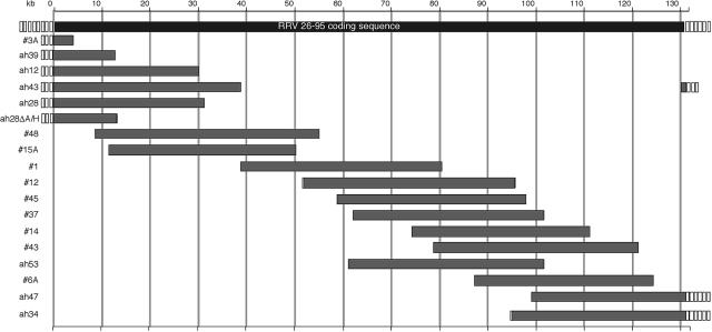
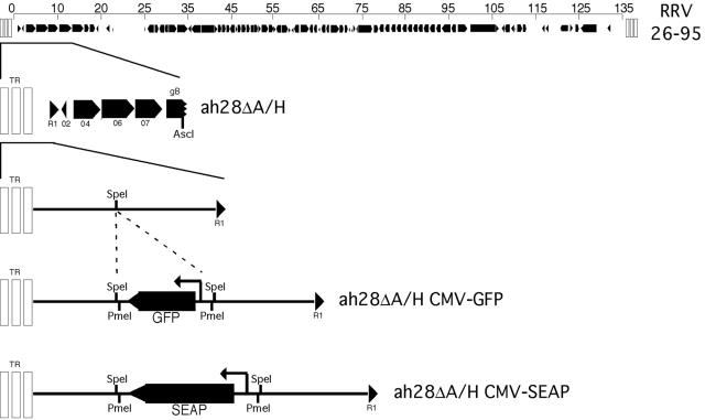
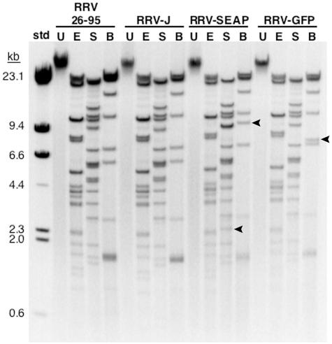
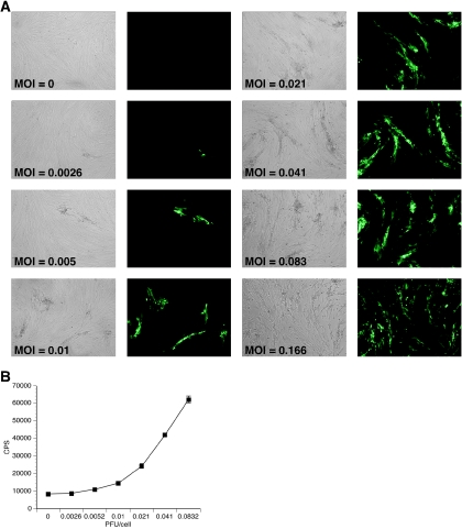
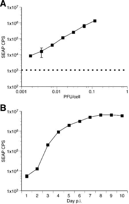
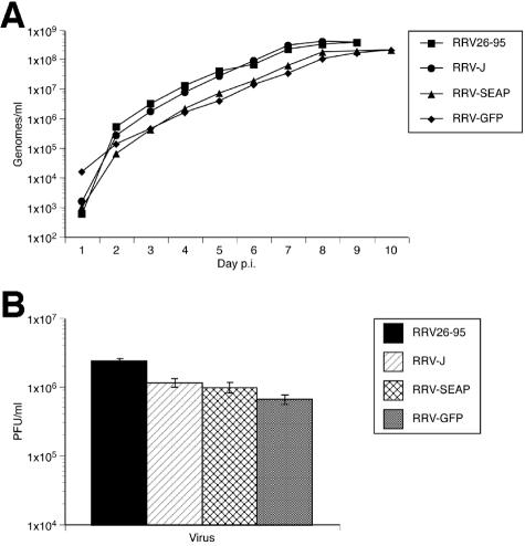
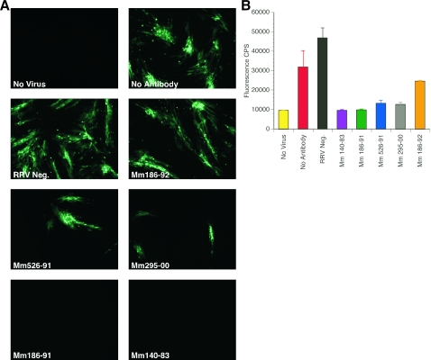
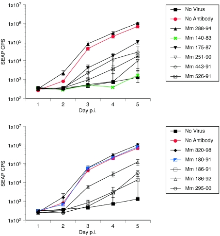
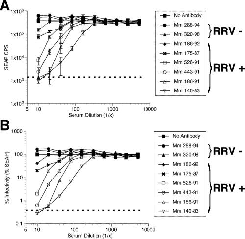
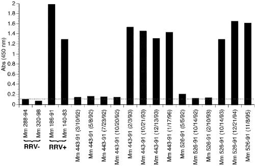
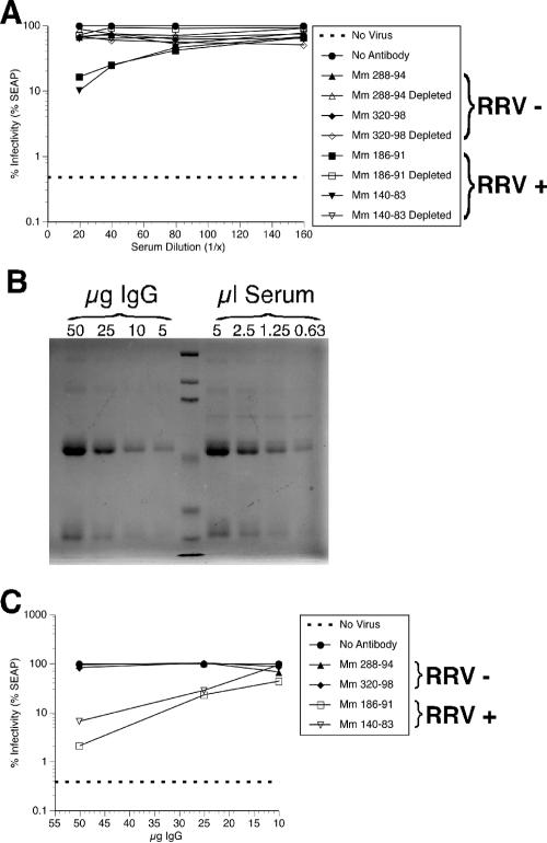
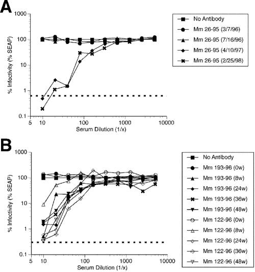
Rhesus monkey rhadinovirus (RRV), a simian gamma-2 herpesvirus closely related to the Kaposi sarcoma-associated herpesvirus, replicates lytically in cultured rhesus monkey fibroblasts and establishes persistence in B cells. Overlapping cosmid clones were generated that encompass the entire 130-kilobase-pair genome of RRV strain 26-95, including the terminal repeat regions required for its replication. Cloned RRV that was produced by cotransfection of overlapping cosmids spanning the entire RRV26-95 genome replicated with growth kinetics and to titers similar to those of the parental, uncloned, wild-type RRV26-95. Expression cassettes for secreted-engineered alkaline phosphatase (SEAP) and green fluorescent protein (GFP) were inserted upstream of the R1 gene, and the cosmid-based system for RRV genome reconstitution was used to generate replication-competent, recombinant RRV that expressed either the SEAP or GFP reporter gene. Using the SEAP and GFP recombinant RRVs, assays were developed to monitor RRV infection, neutralization, and replication. Heat-inactivated sera from rhesus monkeys that were naturally or experimentally infected with RRV were assayed for their ability to neutralize RRV-SEAP and RRV-GFP infectivity using rhesus monkey fibroblasts. Sera from RRV-positive monkeys, but not RRV-negative monkeys, were consistently able to neutralize RRV infectivity when assayed by the production of SEAP activity or by the ability to express GFP. The neutralizing activity was present in the immunoglobulin fraction. Of the 17 rhesus monkeys tested, sera from rhesus monkey 26-95, i.e., the monkey that yielded the RRV 26-95 isolate, had the highest titer of neutralizing activity against RRV26-95. This cosmid-based genetic system and the reporter virus neutralization assay will facilitate study of the contribution of individual RRV glycoproteins to entry into different cell types, particularly fibroblasts and B cells.


Figures

FIG. 1.
FIG. 1.
Schematic representation of the cosmids derived from RRV26-95. Open boxes represent the terminal repeat regions of various lengths ranging from <1 kb to approximately 28 kb. The gray bars indicate the extent and location of each cosmid insert within the parental RRV26-95 genome (top black bar). Each cosmid insert was cloned into the pSuperCos 1 vector following the addition of an ICeuI homing endonuclease linker. The cosmid insert designated ah28ΔA/H was derived from the removal of the AscI-HindIII fragment from the ah28 cosmid within the pSuperCos 1 vector.
FIG. 2.
FIG. 2.
Insertion of GFP and SEAP transgene cassettes into the RRV ah28ΔA/H cosmid. An oligonucleotide linker containing the PmeI restriction endonuclease recognition site was inserted into the SpeI site of the ah28ΔA/H cosmid. A cassette containing either CMV-GFP or CMV-SEAP and flanked by PmeI restriction endonuclease sites was cloned into the PmeI site of ah28ΔA/H to generate ah28ΔA/H CMV-GFP or ah28ΔA/H CMV-SEAP. The size of the CMV-GFP and CMV-SEAP transgene cassettes are 1,636 and 3,882 bp, respectively.
FIG. 3.
FIG. 3.
Restriction digests of RRV genomic DNA. DNA from purified RRV26-95 or cosmid-derived recombinant RRV-J, RRV-SEAP, and RRV-GFP was digested with either EcoRV (E), SphI (S), or the nicking endonucleases BbvCIA and BbvCIB (B). Restriction endonuclease-digested and undigested (U) virion DNA were separated on a 0.5% agarose electrophoretic gel and stained with ethidium bromide. Bands marked with an arrow represent altered digestion products resulting from insertion of the CMV-SEAP or CMV-GFP reporter gene cassettes. The molecular size standards (std) are HindIII-digested λ DNA.
FIG. 4.
FIG. 4.
GFP expression in rhesus fibroblasts after infection with RRV-GFP is MOI dependent. At 24 h postseeding, rhesus fibroblasts were infected with increasing MOIs of RRV-GFP. At day 4 p.i., cultures were examined for GFP expression. (A) Fluorescence microscopy of rhesus fibroblasts after infection with increasing MOIs of RRV-GFP. (B) GFP expression by RRV-GFP as measured by emission intensity. Rhesus fibroblasts were seeded into 96-well plates and infected with increasing MOIs of RRV-GFP at 24 h postseeding. At day 4 p.i., cultures were examined for GFP expression using a multiwell plate reader (n = 8). The results show the GFP counts/s (CPS) with standard deviation at the indicated PFU/cell. Where no error bar is shown, the error falls within the size of the symbol.
FIG. 5.
FIG. 5.
SEAP expression in rhesus fibroblasts after infection with RRV-SEAP. (A) SEAP expression in rhesus fibroblasts after infection with RRV-SEAP is MOI dependent. At 24 h postseeding, triplicate cultures of rhesus fibroblasts were infected with increasing MOIs of RRV-SEAP. At day 4 p.i., cultures were examined for SEAP expression (r2 = 0.9973). The results show the SEAP counts/s (CPS) with standard deviation for each PFU/cell indicated. The background SEAP CPS is represented by the dashed line. (B) Time course of SEAP expression after infection of rhesus fibroblasts with RRV-SEAP. At 24 h postseeding, triplicate cultures of rhesus fibroblasts were infected with 0.002 PFU/cell of RRV-SEAP. At day 1 p.i., cultures were rinsed five times with HBSS and refed with DH20 medium. One milliliter of cell-free supernatant was removed each day and replaced with 1 ml of DH20. The SEAP counts/s (CPS), with standard deviation, are shown for each day p.i. Where no error bar is shown, the error falls within the size of the symbol.
FIG. 6.
FIG. 6.
Growth kinetics of parental and recombinant RRV in rhesus fibroblasts. (A) Recombinant RRVs replicate with similar kinetics to the parental RRV26-95 as measured by real-time PCR. At 24 h postseeding, triplicate cultures of rhesus fibroblasts were infected at 0.002 PFU/cell with RRV26-95, RRV-J, RRV-SEAP, or RRV-GFP. At day 1 p.i., cultures were rinsed five times with HBSS and refed with DH20. One milliliter of cell-free supernatant was removed each day and replaced with 1 ml of DH20 medium. Triplicate samples of viral DNA isolated at each day were examined by real-time PCR. Results show the average genomes/ml, with standard deviation, at the indicated day p.i. (B) End-point titers of recombinant RRVs are similar to RRV26-95. Cell-free supernatant isolated at either day 9 (RRV26-95 and RRV-J) or 10 (RRV-SEAP and RRV-GFP) p.i. was diluted in DH20 medium and used to determine the end-point titer for each virus at the completion of lytic replication in rhesus fibroblasts. Each bar represents the average PFU/ml, with standard deviation, for the indicated virus.
FIG. 7.
FIG. 7.
Neutralization of RRV-GFP by sera from rhesus monkeys naturally infected with RRV. (A) Fluorescence microscopy of RRV-GFP neutralization. RRV-GFP (MOI of 0.04 PFU/cell) was incubated with either DH20 medium (No Antibody), sera from a RRV-negative rhesus monkey (RRV Neg.) or sera from rhesus monkeys naturally infected with RRV (Mm 186-92, 526-91, 295-00, 186-91, and 140-83). After a 3-h incubation at 37°C, rhesus fibroblasts were inoculated with either DH20 medium (No Virus) or the virus-sera mixture. At 24 h p.i., cultures were rinsed five times with HBSS and refed with DH20. At day 4 p.i., cultures were examined by fluorescence microscopy for GFP expression. (B) Detection of RRV-GFP neutralization by GFP emission intensity. Each bar represents the average GFP counts/s (CPS), with standard deviation, for each rhesus monkey sera tested.
FIG. 8.
FIG. 8.
SEAP expression by RRV-SEAP following neutralization by sera from rhesus monkeys naturally infected with RRV. RRV-SEAP (MOI of 0.006 PFU/cell) was incubated with either DH20 medium (No Antibody), sera from RRV-negative rhesus monkeys (Mm 288-94 and 320-98) or sera from rhesus monkeys naturally infected with RRV (Mm 140-83, 175-87, 180-91, 186-91, 186-92, 251-90, 295-00, 443-91, and 526-91). After a 3-h incubation at 37°C, rhesus fibroblasts were inoculated with either DH20 medium (No Virus) or the virus-sera mixture. At 24 h p.i., cultures were rinsed five times with HBSS and refed with DH20. At days 1 to 5 p.i., an aliquot of medium was removed to determine the level of SEAP expression and replaced with DH20 medium. The results show the average SEAP counts/s (CPS), with standard deviation, for each day p.i.
FIG. 9.
FIG. 9.
Neutralization of RRV-SEAP by sera from rhesus monkeys naturally infected with RRV. RRV-SEAP (MOI of 0.006 PFU/cell) was incubated with either DH20 medium (No Antibody), serial dilutions of sera from RRV-negative rhesus monkeys (Mm 288-94 and 320-98) or serial dilutions of sera from rhesus monkeys naturally infected with RRV (Mm 186-92, 175-87, 526-91, 443-91, 186-91, and 140-83). After a 3-h incubation at 37°C, rhesus fibroblasts were inoculated with either DH20 medium (dotted line) or the virus-sera mixture. At 24 h p.i., cultures were rinsed five times with HBSS and refed with DH20. At day 4 p.i., an aliquot of medium was removed to determine the level of SEAP expression. The results show the average SEAP counts/s (CPS) (A), with standard deviation, and the percent infectivity (B), for each serum dilution.
FIG. 10.
FIG. 10.
ELISA for RRV antibodies in sera from naturally infected rhesus monkeys. Maxi-sorb plates coated with RRV were incubated with sera from Mm 443-91 and 526-91 isolated at different dates after birth. After exposure, RRV positivity was determined for these sera by comparison to sera from RRV-negative (Mm 288-94 and 320-98) and RRV-positive (Mm 186-91 and 140-83) monkeys.
FIG. 11.
FIG. 11.
Neutralization of RRV-SEAP is antibody mediated. (A) Depletion of IgG from rhesus monkey serum abrogated neutralization of RRV. To remove IgG from the serum, serum from either RRV-negative rhesus monkeys (Mm 288-94 and 320-98) or rhesus monkeys naturally infected with RRV (Mm 186-91 and 140-83) was incubated with immobilized protein A/G overnight at 4°C. The resulting IgG-depleted serum and nondepleted serum were diluted as indicated and analyzed for their ability to neutralize RRV-SEAP as described previously. At each indicated serum dilution, the percent infectivity or RRV-SEAP is shown. (B) Coomassie staining to reveal relative levels of IgG in serum (results from Mm 186-91 is shown). Known amounts of previously isolated IgG and decreasing volumes of sera from Mm 186-91 were immunoprecipitated with immobilized protein A/G. After polyacrylamide gel electrophoresis, gels were stained with Coomassie blue to reveal the resulting proteins. (C) Purified IgG from the sera of rhesus monkeys naturally infected with RRV neutralizes RRV-SEAP. RRV-SEAP (MOI of 0.006 PFU/cell) was incubated with either DH20 medium (No Antibody) or known amounts of IgG isolated from either RRV-negative rhesus monkeys (Mm 288-94 and 320-98) or rhesus monkeys naturally infected with RRV (Mm 186-91 and 140-83) diluted in DH20 medium. After a 3-h incubation at 37°C, rhesus fibroblasts were inoculated with either DH20 medium (No Virus) or the virus-IgG mixture. At 24 h p.i., cultures were rinsed five times with HBSS and refed with DH20. At day 4 p.i., an aliquot of medium was removed to determine the level of SEAP expression. The results show the percent infectivity for each IgG concentration.
FIG. 12.
FIG. 12.
Neutralization of RRV-SEAP by sera from Mm 26-95 and rhesus monkeys experimentally infected with RRV26-95. (A) Neutralization of RRV-SEAP by Mm 26-95 sera. RRV-SEAP (MOI of 0.006 PFU/cell) was incubated with either DH20 medium (No Antibody) or various dilutions of serum from Mm 26-95 isolated at different dates. After a 3-h incubation at 37°C, rhesus fibroblasts were inoculated with either DH20 medium (dotted line) or the virus-sera mixture. At 24 h p.i., cultures were rinsed five times with HBSS and refed with DH20. At day 4 p.i., an aliquot of medium was removed to determine the level of SEAP expression. The results show the percent infectivity for each serum dilution at the serum isolation dates indicated. (B) Neutralization of RRV-SEAP by sera from rhesus monkeys experimentally infected with RRV26-95. At 8, 24, 36, and 48 weeks after inoculation of RRV-naïve rhesus monkeys with RRV26-95, sera was collected and used to determine its efficacy in neutralizing RRV-SEAP as previously described. The results show the percent infectivity for each serum dilution at the indicated week postinoculation.

Similar articles

Cited by

References

    1. Alexander, L., L. Denekamp, A. Knapp, M. R. Auerbach, B. Damania, and R. C. Desrosiers. 2000. The primary sequence of rhesus monkey rhadinovirus isolate 26-95: sequence similarities to Kaposi's sarcoma-associated herpesvirus and rhesus monkey rhadinovirus isolate 17577. J. Virol. 74:3388-3398. - PMC - PubMed
    1. Auerbach, M. R., S. C. Czajak, W. E. Johnson, R. C. Desrosiers, and L. Alexander. 2000. Species specificity of macaque rhadinovirus glycoprotein B sequences. J. Virol. 74:584-590. - PMC - PubMed
    1. Bergquam, E. P., N. Avery, S. M. Shiigi, M. K. Axthelm, and S. W. Wong. 1999. Rhesus rhadinovirus establishes a latent infection in B lymphocytes in vivo. J. Virol. 73:7874-7876. - PMC - PubMed
    1. Bestman-Smith, J., and G. Boivin. 2003. Drug resistance patterns of recombinant herpes simplex virus DNA polymerase mutants generated with a set of overlapping cosmids and plasmids. J. Virol. 77:7820-7829. - PMC - PubMed
    1. Chang, Y., E. Cesarman, M. S. Pessin, F. Lee, J. Culpepper, D. M. Knowles, and P. S. Moore. 1994. Identification of herpesvirus-like DNA sequences in AIDS-associated Kaposi's sarcoma. Science 266:1865-1869. - PubMed

Publication types

MeSH terms

LinkOut - more resources