

Deterministic model of dermal wound invasion incorporating receptor-mediated signal transduction and spatial gradient sensing
- PMID: 16415056
- PMCID: PMC1403196
- DOI: 10.1529/biophysj.105.077610
Deterministic model of dermal wound invasion incorporating receptor-mediated signal transduction and spatial gradient sensing
Erratum in
- Biophys J. 2007 Jan 15;92(2):696
Abstract
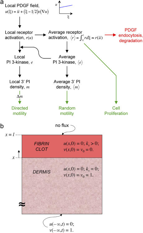
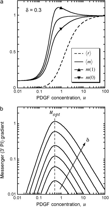
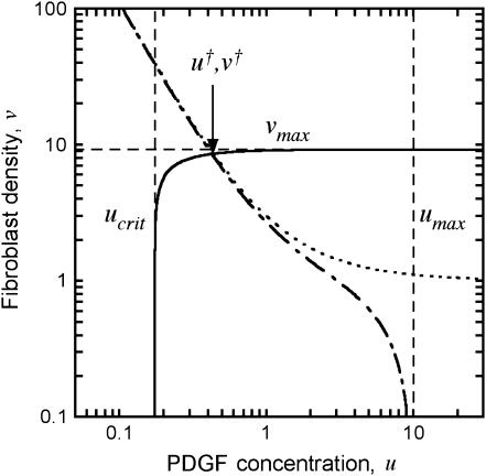
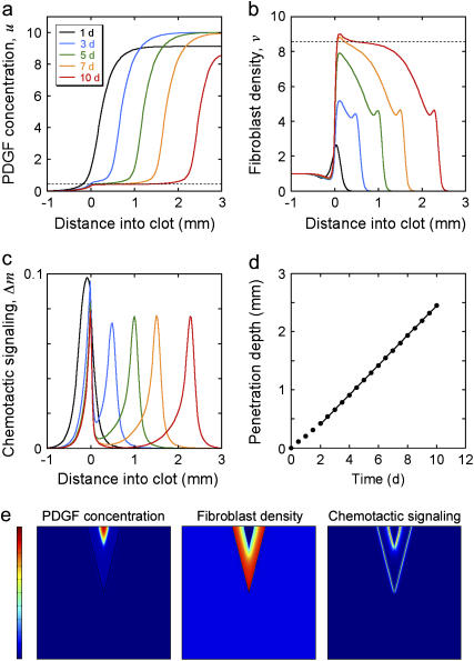
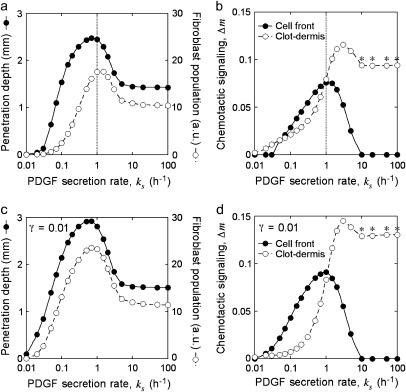
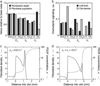
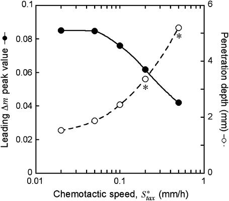
During dermal wound healing, platelet-derived growth factor (PDGF) serves as both a chemoattractant and mitogen for fibroblasts, potently stimulating their invasion of the fibrin clot over a period of several days. A mathematical model of this process is presented, which accurately accounts for the sensitivity of PDGF gradient sensing through PDGF receptor/phosphoinositide 3-kinase-mediated signal transduction. Analysis of the model suggests that PDGF receptor-mediated endocytosis and degradation of PDGF allows a constant PDGF concentration profile to be maintained at the leading front of the fibroblast density profile as it propagates, at a constant rate, into the clot. Thus, the constant PDGF gradient can span the optimal concentration range for asymmetric phosphoinositide 3-kinase signaling and fibroblast chemotaxis, with near-maximal invasion rates elicited over a relatively broad range of PDGF secretion rates. A somewhat surprising finding was that extremely sharp PDGF gradients do not necessarily stimulate faster progression through the clot, because maintaining such a gradient through PDGF consumption is a potentially rate-limiting process.
Figures
Similar articles
-
Quantitative elucidation of a distinct spatial gradient-sensing mechanism in fibroblasts.J Cell Biol. 2005 Dec 5;171(5):883-92. doi: 10.1083/jcb.200509028. Epub 2005 Nov 28. J Cell Biol. 2005. PMID: 16314431 Free PMC article.
-
Constitutive diffuse activation of phosphoinositide 3-kinase at the plasma membrane by v-Src suppresses the chemotactic response to PDGF by abrogating the polarity of PDGF receptor signalling.Exp Cell Res. 2007 Apr 1;313(6):1090-105. doi: 10.1016/j.yexcr.2007.01.020. Epub 2007 Feb 6. Exp Cell Res. 2007. PMID: 17335807
-
Cell population-based model of dermal wound invasion with heterogeneous intracellular signaling properties.Cell Adh Migr. 2008 Apr-May;2(2):137-46. doi: 10.4161/cam.2.2.6511. Epub 2008 Apr 26. Cell Adh Migr. 2008. PMID: 19262100 Free PMC article.
-
Platelet derived growth factor/tyrosine kinase receptor mediated proliferation.Growth Regul. 1993 Sep;3(3):172-9. Growth Regul. 1993. PMID: 8220110 Review.
-
Mechanisms of gradient sensing and chemotaxis: conserved pathways, diverse regulation.Cell Cycle. 2006 Jun;5(11):1130-4. doi: 10.4161/cc.5.11.2770. Epub 2006 Jun 1. Cell Cycle. 2006. PMID: 16760661 Review.
Cited by
-
Systems-based approaches toward wound healing.Pediatr Res. 2013 Apr;73(4 Pt 2):553-63. doi: 10.1038/pr.2013.3. Epub 2013 Jan 11. Pediatr Res. 2013. PMID: 23314298 Free PMC article. Review.
-
Stability of a two-dimensional biomorphoelastic model for post-burn contraction.J Math Biol. 2023 Mar 24;86(4):59. doi: 10.1007/s00285-023-01893-w. J Math Biol. 2023. PMID: 36964257 Free PMC article.
-
A Reaction-Diffusion Model Explains Amplification of the PLC/PKC Pathway in Fibroblast Chemotaxis.Biophys J. 2017 Jul 11;113(1):185-194. doi: 10.1016/j.bpj.2017.05.035. Biophys J. 2017. PMID: 28700916 Free PMC article.
-
Self-Generated Chemoattractant Gradients: Attractant Depletion Extends the Range and Robustness of Chemotaxis.PLoS Biol. 2016 Mar 16;14(3):e1002404. doi: 10.1371/journal.pbio.1002404. eCollection 2016 Mar. PLoS Biol. 2016. PMID: 26981861 Free PMC article.
-
Epithelial cell guidance by self-generated EGF gradients.Integr Biol (Camb). 2012 Mar;4(3):259-69. doi: 10.1039/c2ib00106c. Epub 2012 Feb 8. Integr Biol (Camb). 2012. PMID: 22314635 Free PMC article.
References
-
- Martin, P. 1997. Wound healing- aiming for perfect skin regeneration. Science. 276:75–81. - PubMed
-
- Singer, A. J., and R. A. F. Clark. 1999. Mechanisms of disease: cutaneous wound healing. N. Engl. J. Med. 341:738–746. - PubMed
-
- Gharaee-Kermani, M., and S. H. Phan. 2001. Role of cytokines and cytokine therapy in wound healing and fibrotic diseases. Curr. Pharm. Des. 7:1083–1103. - PubMed
-
- Gabbiani, G. 2003. The myofibroblast in wound healing and fibrocontractive diseases. J. Pathol. 200:500–503. - PubMed
Publication types
MeSH terms
Substances
LinkOut - more resources
Full Text Sources
