. 2006 Jan 24;103(4):1018-23.
doi: 10.1073/pnas.0510347103. Epub 2006 Jan 13.

The tumor suppressor menin regulates hematopoiesis and myeloid transformation by influencing Hox gene expression

Affiliations

The tumor suppressor menin regulates hematopoiesis and myeloid transformation by influencing Hox gene expression

Ya-Xiong Chen et al. Proc Natl Acad Sci U S A. .

Abstract

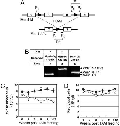
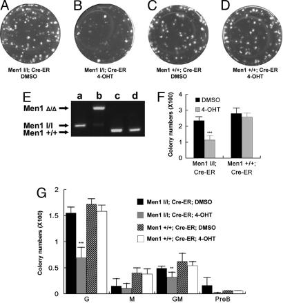
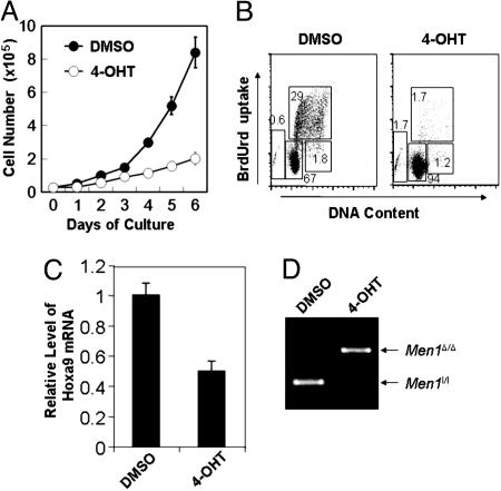
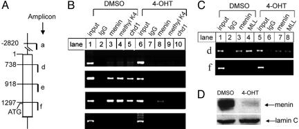
Menin is the product of the tumor suppressor gene Men1 that is mutated in the inherited tumor syndrome multiple endocrine neoplasia type 1 (MEN1). Menin has been shown to interact with SET-1 domain-containing histone 3 lysine 4 (H3K4) methyltransferases including mixed lineage leukemia proteins to regulate homeobox (Hox) gene expression in vitro. Using conditional Men1 knockout mice, we have investigated the requirement for menin in hematopoiesis and myeloid transformation. Men1 excision causes reduction of Hoxa9 expression, colony formation by hematopoietic progenitors, and the peripheral white blood cell count. Menin directly activates Hoxa9 expression, at least in part, by binding to the Hoxa9 locus, facilitating methylation of H3K4, and recruiting the methylated H3K4 binding protein chd1 to the locus. Consistent with signaling downstream of menin, ectopic expression of both Hoxa9 and Meis1 rescues colony formation defects in Men1-excised bone marrow. Moreover, Men1 excision also suppresses proliferation of leukemogenic mixed lineage leukemia-AF9 fusion-protein-transformed myeloid cells and Hoxa9 expression. These studies uncover an important role for menin in both normal hematopoiesis and myeloid transformation and provide a mechanistic understanding of menin's function in these processes that may be used for therapy.


Figures

Fig. 1.
Fig. 1.
Excision of floxed Men1 in mice reduces the peripheral WBC counts. (A) A diagram for the floxed Men1 and primers for detecting excision of the floxed Men1 in genome. P1, P2, and P3 denote primers 1, 2, and 3, respectively. F1 and F2 are DNA fragments representing the unexcised and excised floxed Men1 loci, respectively. The numbered boxes denote exon numbers. (B) Tight control of TAM induced the floxed Men1 excision. Men1+/+;Cre-ER mice (lane 1) and Men1 l/l;Cre-ER mice (lane 2) were treated with TAM, whereas Men1l/l;Cre-ER mice (lane 3) were treated with corn oil. BM genomic DNA from each of the mice was used for genotyping. The results are representative of three mice for each condition. Men1Δ/Δ (F2), excised Men1 locus; Men1l/l (F1), unexcised floxed Men1.(C) Total peripheral WBC of control Men1+/+;Cre-ER mice (•) and Men1l/l;Cre-ER mice (○) are plotted, after treatment with TAM for various periods of time (weeks). (D) Time course of RBC counts in mice of both distinct genotypes. Five to 20 mice for each group were used for each time point over 12 weeks.
Fig. 2.
Fig. 2.
Men1 excision in vitro reduces colony formation of BM. (AD) Representative plates with visible colonies from colony formation assays. BM pooled from Men1+/+;Cre-ER mice or Men1l/l;Cre-ER mice were plated (2.5 × 10⁴ cells per plate) in complete medium supplemented with either DMSO or 4-OHT. The genotype of BM cells and treatment applied to the plate are indicated. (E) PCR genotyping with genomic DNA from the above plates as templates. (F) Summary of colony numbers from three independent experiments with duplicate plates. Asterisks indicate statistically significant differences between the adjacent groups (P < 3 × 10⁻⁴). (G) Average numbers of CFU-granulocytes (G), CFU-macrophages (M), CFU-granulocytes/macrophages (GM), and CFU-subset of B-lymphoid progenitors (PreB) were classified and recorded for each group from three independent experiments with duplicate plates as described in F. Identities of representative colonies were confirmed with Wright-Giemsa staining.
Fig. 3.
Fig. 3.
Men1 excision inhibits proliferation of MLL-AF9-transformed myeloid cells and suppresses Hoxa9 expression. (A) BM from multiple Men1l/l;Cre-ER mice was transformed by transduction with retroviruses expressing MLL-AF9. The transformed BM cells from a representative mouse, AT-1, were plated in triplicate at 2.5 × 10⁴ cells per well, treated with either DMSO or 4-OHT, and counted daily for 6 days. (B) The myeloid cells treated with either DMSO or 4-OHT for 4 days were pulsed with BrdUrd for 90 min, and then stained with an anti-BrdUrd antibody and 7-amino-actinomycin D, followed by flow cytometric analysis. (C) The transformed Men1l/l;Cre-ER cells were treated with either DMSO or TAM for 4 days, and then harvested for RNA isolation and real-time PCR to quantitate Hoxa9 mRNA. (D) Genomic DNA from the cells treated with either DMSO or 4-OHT was used for PCR to determine Men1 excision. Two independent experiments were performed for each of the above experiments.
Fig. 4.
Fig. 4.
Menin is important for methylation of H3K4 in the Hoxa9 locus and for binding of MLL and cdh1 to the locus. (A) A diagram of the murine Hoxa9 locus. (B and C) Myeloid cells harboring Men1l/l;Cre-ER were treated with DMSO or 4-OHT, and then processed for ChIP assays by using the indicated antibodies. Results are representative of two independent experiments. (D) Men1 excision induced by 4-OHT was confirmed by Western blotting using an antimenin antibody. Equal loading of the samples was revealed by blotting for Lamin C.

References

    1. Lemmens, I., Van de Ven, W. J., Kas, K., Zhang, C. X., Giraud, S., Wautot, V., Buisson, N., De Witte, K., Salandre, J., Lenoir, G., et al. (1997) Hum. Mol. Genet. 6, 1177–1183. - PubMed
    1. Chandrasekharappa, S. C., Guru, S. C., Manickam, P., Olufemi, S. E., Collins, F. S., Emmert-Buck, M. R., Debelenko, L. V., Zhuang, Z., Lubensky, I. A., Liotta, L. A., et al. (1997) Science 276, 404–407. - PubMed
    1. Marx, S. J. & Stratakis, C. A. (2005) J. Intern. Med. 257, 2–5. - PubMed
    1. Pannett, A. A. & Thakker, R. V. (1999) Endocr. Relat. Cancer 6, 449–473. - PubMed
    1. Poisson, A., Zablewska, B. & Gaudray, P. (2003) Cancer Lett. 189, 1–10. - PubMed

Publication types

MeSH terms