Review
. 2006 May;34(5):527-36.
doi: 10.1165/rcmb.2005-0436SF. Epub 2006 Jan 13.

Airway mucus: From production to secretion

Affiliations
Review

Airway mucus: From production to secretion

Olatunji W Williams et al. Am J Respir Cell Mol Biol. 2006 May.

Abstract

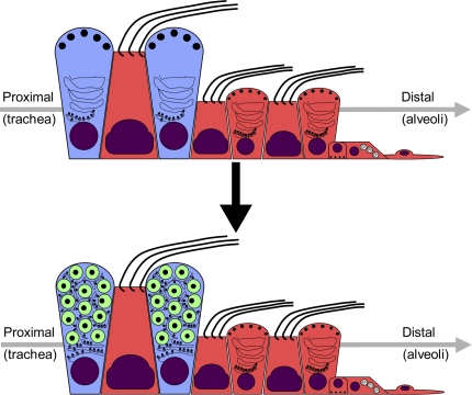
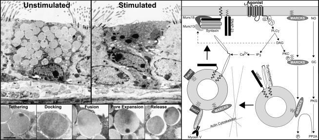
Mucus hypersecretion is a phenotype associated with multiple obstructive lung diseases. However, in spite of its nefarious reputation under pathologic conditions, there are significant benefits to having low levels of mucus present in the airways at baseline, such as the ability to trap and eliminate inhaled particles and to prevent desiccation of airway surfaces. Mucins are high-molecular-weight glycoproteins that are the chief components that render viscoelastic and gel-forming properties to mucus. Recent advances in animal models and in vitro systems have provided a wealth of information regarding the identification of the mucin genes that are expressed in the lungs, the signal transduction pathways that regulate the expression of these mucins, and the secretory pathways that mediate their release into the airways. In addition, the clinical and pathologic literature has corroborated many of the basic laboratory findings. As a result, mucin overproduction and hypersecretion are moving away from being markers of disease and toward being testable as functional components of lung disease processes.


Figures

<b>Figure 1.</b>
Figure 1.
Temporal and spatial regulation of mucin expression in the airways. At baseline (top), mouse airways are lined by a tall columnar epithelium consisting of ciliated and nonciliated cells. The nonciliated cells are secretory cells that express the Clara cell marker CCSP and contain abundant smooth endoplasmic reticulum (ER). After allergic stimulation (bottom), there is a dramatic alteration in the morphology of nonciliated cells in the proximal airway epithelium (blue) characterized by the upregulation of their secretory product contents and rough ER abundance, with the addition of mucin expression occurring as the central event during this differentiation process. Clilated cells, bronchiolar Clara cells, neuroendocrine cells, and type I and II pneumocytes (red) are restricted from initiating mucin glycoprotein synthesis.
<b>Figure 2.</b>
Figure 2.
Secretion of mucin from epithelial cells by regulated exocytosis. Left. (Top) Transmission electron microscopy (TEM) shows massive release of mucin-containing SGs from metaplastic goblet cells in response to stimulation by ATP aerosol. The remaining SG in the stimulated cell localizes to the apical plasma membrane (PM), suggesting that the default intracellular trafficking pathway for mucin SGs in airway goblet cells positions these in a readily releasable pool for rapid secretion in response to further stimulation. (Bottom) Morphologic demonstration of the of late steps of vesicular traffic resulting in close apposition of SGs with the inner leaflet of the apical PM (Tethering and Docking), mixing of the lipid bilayer surrounding mucin SGs with the apical PM (Fusion), and ultimately in mucin content release from the fused SG's (Expansion and Release). Right: Schematic demonstration of the molecular machinery involved in mucin SG trafficking and fusion. Agonists (e.g., ATP) activate cell surface receptors (e.g., the purinergic seven transmembrane spanning G protein–coupled P2Y2 receptor) and activate the regulated exocytic machinery thus mediating the docking and fusion of SGs with the apical PM. Magnification bar = 1 μm and 500 nm in the upper and lower TEM images, respectively.

References

    1. Sheehan JK, Thornton DJ, Somerville M, Carlstedt I. Mucin structure: the structure and heterogeneity of respiratory mucus glycoproteins. Am Rev Respir Dis 1991;144:S4–S9. - PubMed
    1. Li Y, Bharti A, Chen D, Gong J, Kufe D. Interaction of glycogen synthase kinase 3beta with the DF3/MUC1 carcinoma-associated antigen and beta-catenin. Mol Cell Biol 1998;18:7216–7224. - PMC - PubMed
    1. Carraway KL, Perez A, Idris N, Jepson S, Arango M, Komatsu M, Haq B, Price-Schiavi SA, Zhang J, Carraway CA. Muc4/sialomucin complex, the intramembrane ErbB2 ligand, in cancer and epithelia: to protect and to survive. Prog Nucleic Acid Res Mol Biol 2002;71:149–185. - PubMed
    1. Moniaux N, Escande F, Batra SK, Porchet N, Laine A, Aubert JP. Alternative splicing generates a family of putative secreted and membrane-associated MUC4 mucins. Eur J Biochem 2000;267:4536–4544. - PubMed
    1. Evans CM, Williams OW, Tuvim MJ, Nigam R, Mixides GP, Blackburn MR, DeMayo FJ, Burns AR, Smith C, Reynolds SD, et al. Mucin is produced by clara cells in the proximal airways of antigen-challenged mice. Am J Respir Cell Mol Biol 2004;31:382–394. - PMC - PubMed

Publication types