. 2006 Mar;3(3):e46.
doi: 10.1371/journal.pmed.0030046.

Excess circulating angiopoietin-2 may contribute to pulmonary vascular leak in sepsis in humans

Affiliations

Excess circulating angiopoietin-2 may contribute to pulmonary vascular leak in sepsis in humans

Samir M Parikh et al. PLoS Med. 2006 Mar.

Abstract

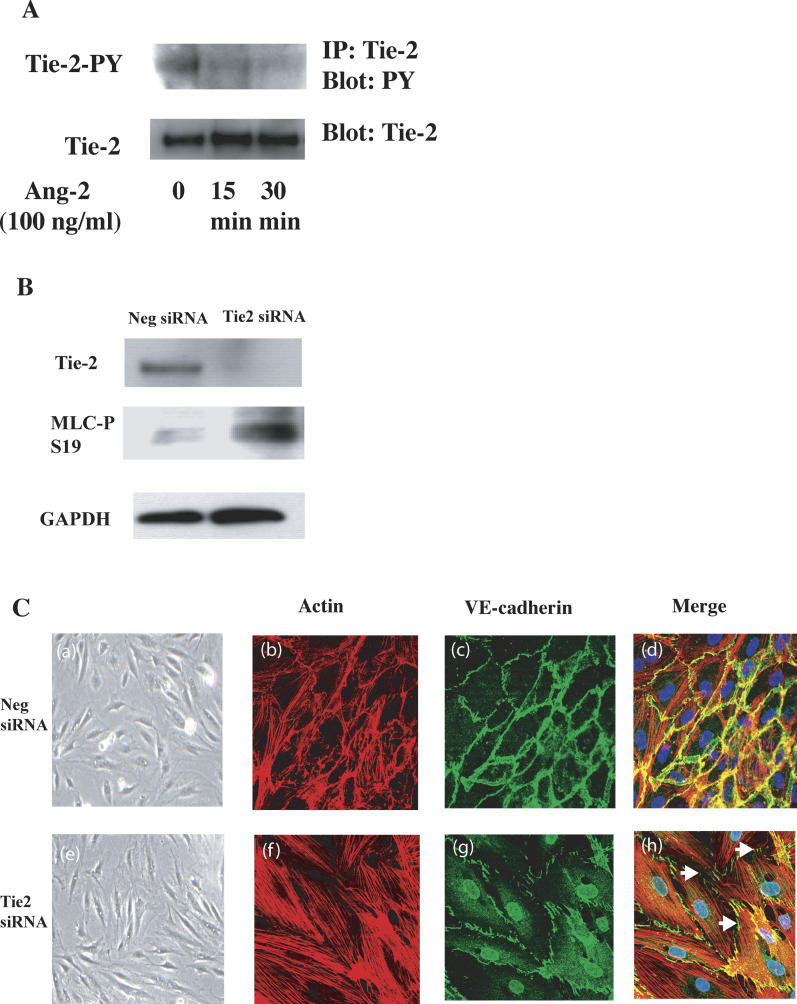
Background: Acute respiratory distress syndrome (ARDS) is a devastating complication of numerous underlying conditions, most notably sepsis. Although pathologic vascular leak has been implicated in the pathogenesis of ARDS and sepsis-associated lung injury, the mechanisms promoting leak are incompletely understood. Angiopoietin-2 (Ang-2), a known antagonist of the endothelial Tie-2 receptor, was originally described as a naturally occurring disruptor of normal embryonic vascular development otherwise mediated by the Tie-2 agonist angiopoietin-1 (Ang-1). We hypothesized that Ang-2 contributes to endothelial barrier disruption in sepsis-associated lung injury, a condition involving the mature vasculature.

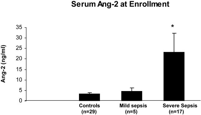
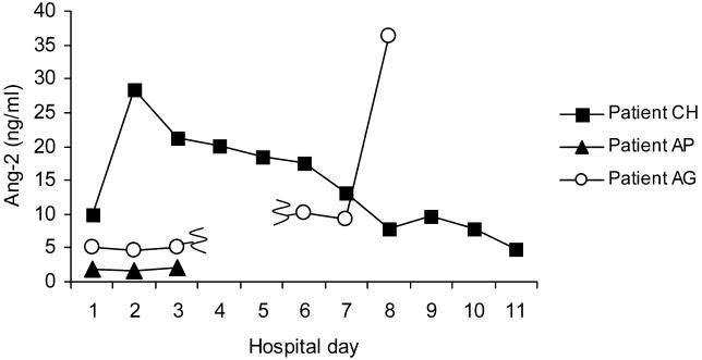
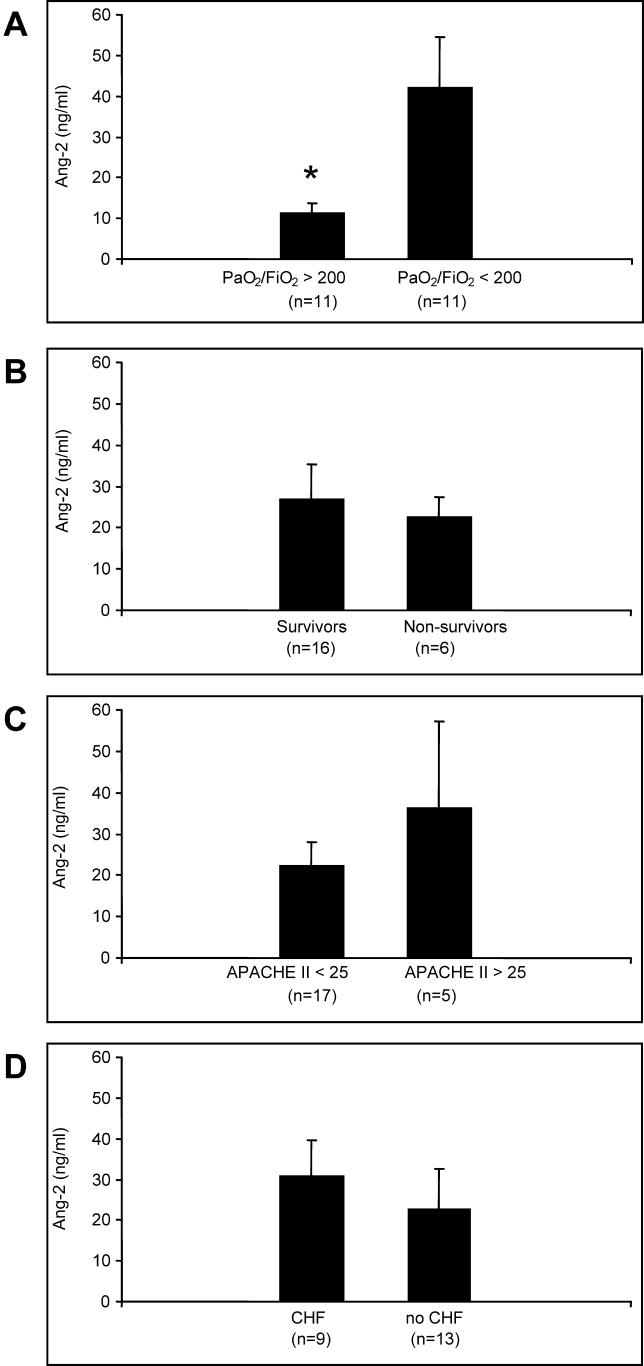
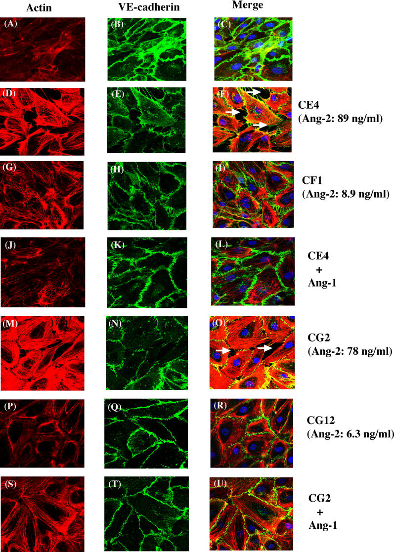
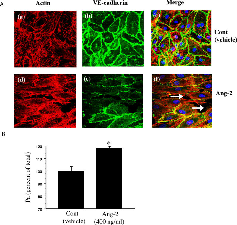
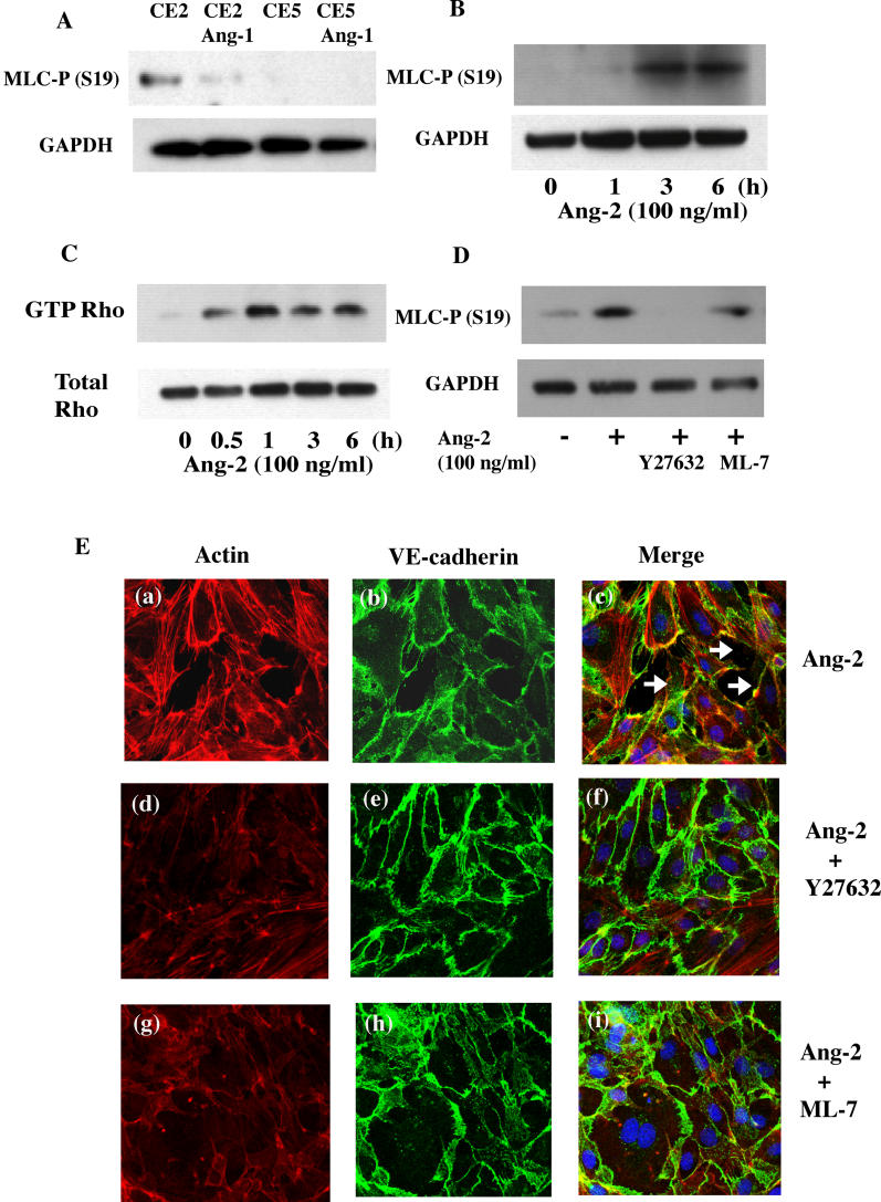
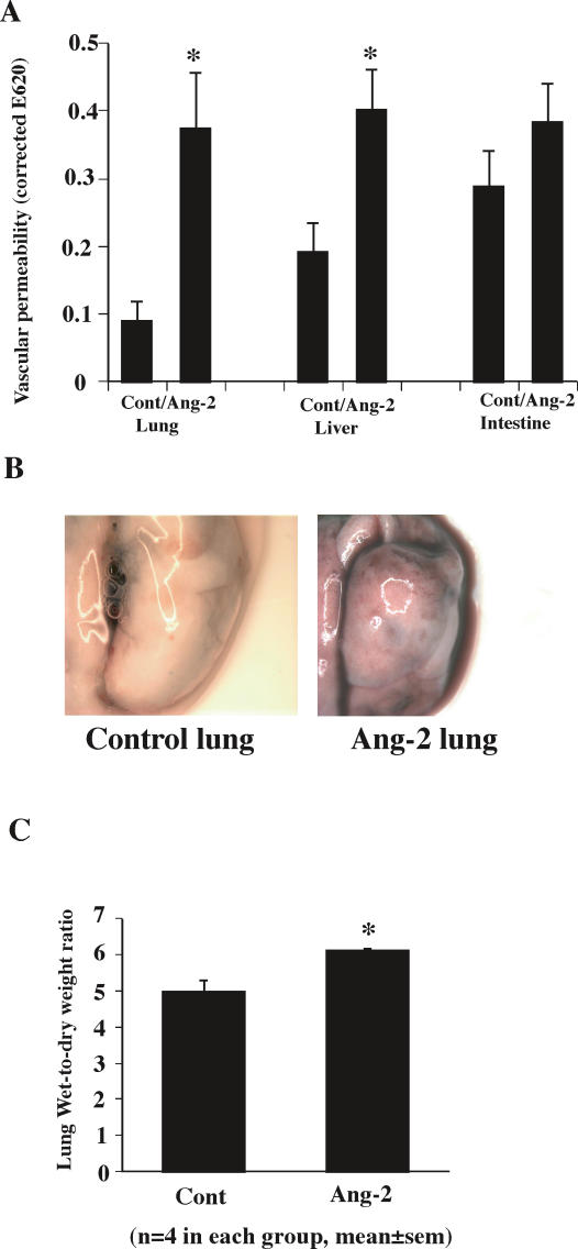
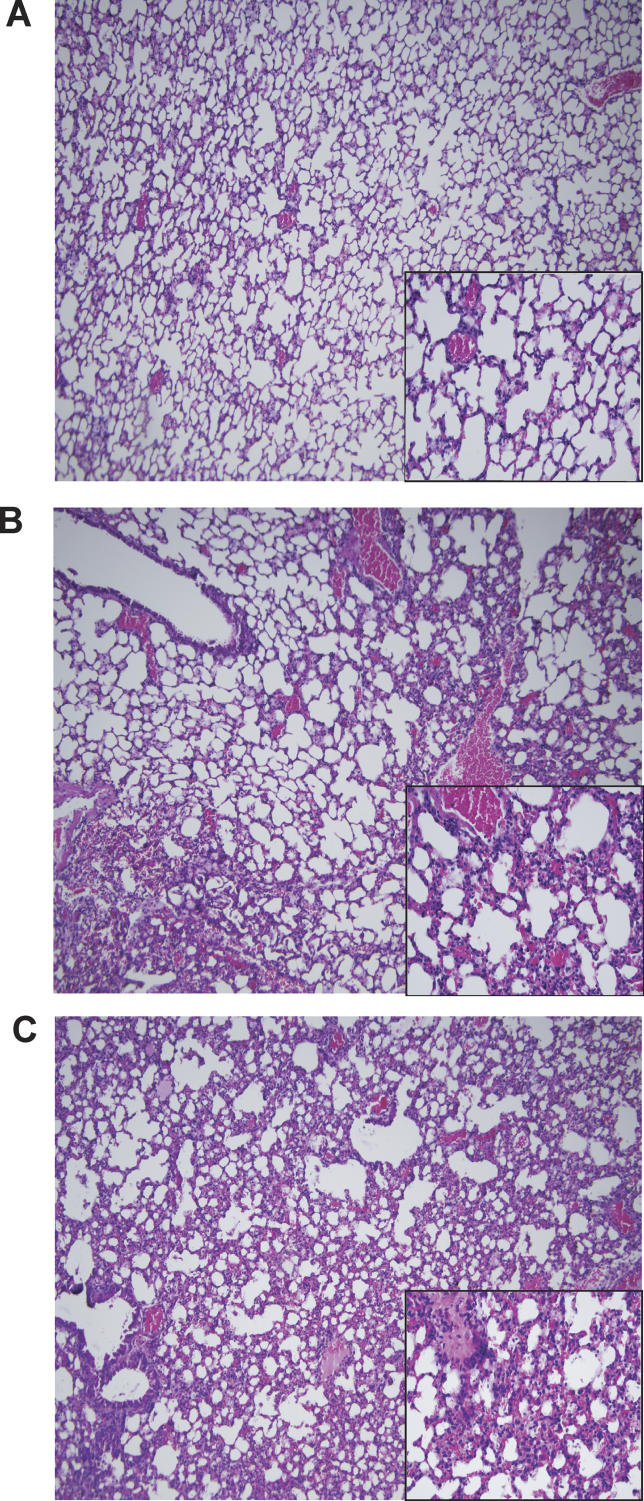
Methods and findings: We describe complementary human, murine, and in vitro investigations that implicate Ang-2 as a mediator of this process. We show that circulating Ang-2 is significantly elevated in humans with sepsis who have impaired oxygenation. We then show that serum from these patients disrupts endothelial architecture. This effect of sepsis serum from humans correlates with measured Ang-2, abates with clinical improvement, and is reversed by Ang-1. Next, we found that endothelial barrier disruption can be provoked by Ang-2 alone. This signal is transduced through myosin light chain phosphorylation. Last, we show that excess systemic Ang-2 provokes pulmonary leak and congestion in otherwise healthy adult mice.

Conclusions: Our results identify a critical role for Ang-2 in disrupting normal pulmonary endothelial function.


Conflict of interest statement

Competing Interests: The authors have declared that no competing interests exist.

Figures

Figure 1
Figure 1. Serum Ang-2 at Study Enrollment
Ang-2 was measured in serum specimens obtained prospectively from patients meeting criteria for sepsis (n = 22) and from randomly selected hospitalized patients (n = 29), used as a control, with a variety of illnesses ranging from infectious (e.g., pyelonephritis, aseptic meningitis, pneumonia) to cardiovascular (e.g., angina, syncope) and neurologic (e.g., stroke) diseases. Patients with sepsis were further subdivided into those with severe sepsis—defined by the presence of shock or multi-organ dysfunction (n = 17)—and those without severe sepsis (mild sepsis, n = 5). Individuals who were controls (marked Controls) and individuals with sepsis without shock or multi-organ dysfunction (marked Mild Sepsis) had low serum Ang-2 at enrollment (3.5 ± 0.6 ng/ml and 4.8 ± 1.5 ng/ml, respectively). Patients hospitalized with severe sepsis (marked Severe Sepsis) had significantly higher serum Ang-2 at enrollment (23.2 ± 9.1 ng/ml, p = 0.0071) compared with control patients. During the course of the hospitalization, only the severe sepsis group had higher peak Ang-2 (32.4 ± 8.7 ng/ml), whereas those patients in the control group and those patients with mild sepsis maintained stable Ang-2 < 10 ng/ml (unpublished data).
Figure 2
Figure 2. Temporal Trends of Circulating Ang-2 in Three Illustrative Hospitalized Patients
Patient CH (—▪—), a 74-y-old woman, was admitted to the medical intensive care unit with severe sepsis. She was treated with broad-spectrum antibiotics, initially required three vasoactive agents to manage shock, and was mechanically ventilated. Patient CH's nadir PaO2/FiO2 = 240 occurred on hospital day 2, correlating with her peak circulating Ang-2. Enterococcus was grown from her urine. She progressively convalesced and was extubated prior to discharge. Patient AP (—▴—), a 92-y-old woman, was admitted to the general medicine service from a nursing home for increased confusion over her baseline dementia. She had no evidence of sepsis, shock, or respiratory compromise—PaO2/FiO2 > 300. She was treated for a foot wound infection with two antibiotics and was discharged in stable condition back to the nursing home. Patient AG (—○—), a 77-y-old man, was first admitted to the general medicine service with hypotension following excessive fluid removal at hemodialysis—there was no evidence of infection, systemic inflammatory response, or respiratory compromise with PaO2/FiO2 > 300 (hospital days 1–3). However, 3 mo later (graphed as hospital days 6–8 for purposes of illustration), the same patient (—○—) was re-admitted to the intensive care unit following emergent right leg amputation for gangrene complicated by shock and inability to extubate. Nadir PaO2/FiO2 = 144 occurred on the same day as peak Ang-2 (depicted as hospital day 8), when he died despite full care.
Figure 3
Figure 3. Peak Circulating Ang-2 Correlates with Impaired Pulmonary Gas Exchange
(A) Impaired oxygenation of blood, as assessed by the nadir PaO2/FiO2 ratio, correlates with significant differences in circulating Ang-2, *p = 0.0195. (B) Circulating Ang-2 does not correlate with survival to discharge. Among the five patients who did not survive, medical care was withdrawn from three patients in accordance with family wishes; the remaining two died despite full measures. (C) APACHE II is a commonly used scoring system to rate overall severity of critical illness. Ang-2 does not differ significantly among individuals with high (more severe illness) or low (less severe illness) APACHE II scores. (D) History of congestive heart failure (defined by clinical documentation in medical record of measured ejection fraction = < 40%) does not correlate with significant differences in circulating Ang-2.
Figure 4
Figure 4. Serum from People with Sepsis Disrupts Endothelial Architecture and This Effect Resolves with Clinical Improvement, Correlates with Measured Ang-2, and Is Reversed by Ang-1
Ten percent FBS or 10% serum from one of two patients with sepsis was incubated with EC monolayers to assess effects on endothelial architecture. High Ang-2 serum (Patient CE4, Ang-2 = 89 ng/ml) induced thick actin stress fibers and intercellular gap formation (D–F), whereas low Ang-2 serum (CF1, Ang-2 = 8.9 ng/ml) did not (G–I). The gap-promoting effect of Patient CE4′s serum was reversed with addition of 100 ng/ml recombinant human Ang-1 (J–L) and was indistinguishable from control cells that exhibit thin actin fibers and no intercellular gaps (A–C). Serum was then taken from one patient (Patient CG), drawn on hospital day 2 (Patient CG2, Ang-2 = 78 ng/ml) and hospital day 16 (Patient CG12, Ang-2 = 6.3 ng/ml), and was added at 10% to HMVEC monolayers. Again, high-Ang-2 serum (CG2) induced gap formation and thick actin stress fibers (M–O), effects not seen in the serum of the same patient at discharge (CG12) (P–R) and effects that were reversed with the addition of 100 ng/ml Ang-1 (S–U). Arrows indicate intercellular gaps.
Figure 5
Figure 5. Ang-2 Alone Disrupts Endothelial Architecture at Physiologic Concentrations
(A) Control (vehicle) or recombinant human Ang-2 (100 ng/ml) was added to HMVEC monolayers. These cells were then fixed and stained for F-actin and VE-cadherin. Shown are healthy control cells (panels a–c) versus Ang-2 treated cells (panels d–f), which exhibit thick actin stress fibers and disrupted junctions, leaving intercellular gaps (arrows). (B) HMVECs were grown to confluence on Transwell membranes coated with fibronectin. Monolayers were treated with vehicle or Ang-2 (400 ng/ml in luminal chamber) plus FITC-albumin. Pa was calculated after 8 h as described in the Methods section. Pa values are expressed as percentage of control cells.*p < 0.01.
Figure 6
Figure 6. Ang-2 Disrupts Endothelial Architecture by Upregulating MLC-p in an MLCK- and Rho-Kinase–Dependent Fashion
(A) Serum was taken from two patients—Patient CE2 (Ang-2 = 77 ng/ml) and Patient CF5 (Ang-2 = 7.9 ng/ml)—and added at 20-fold dilution to 24-h serum-starved HMVECs. High Ang-2-serum (Patient CE2) caused MLC phosphorylation that was diminished by addition of Ang-1 (100 ng/ml), whereas low Ang-2-serum (CF5) did not induce MLC phosphorylation. (B) After 24-h serum starvation, Ang-2 (100 ng/ml) was added to HMVECs, and cells were lysed at the indicated times. MLC-p was determined by Western blot as described in the Methods section. MLC-p was elevated at 3 h and 6 h of stimulation. (C) After 24-h serum starvation, Ang-2 (100 ng/ml) was added to HMVECs, and cells were lysed at the indicated times. GTP-RhoA was pulled down and blotted as described in the Methods section. GTP-RhoA peaked at 30–60 min of Ang-2 stimulation. (D) After 24-h serum starvation, HMVECs were stimulated with Ang-2 (100 ng/ml) with or without 10 μM Y27632 (Rho-kinase inhibitor) or 10 μM ML-7 (MLCK inhibitor) for 5 h. MLC-p was determined by Western blot as described in the Methods section. Y27632 had a more potent inhibitory effect on MLC-p than equimolar ML-7. (E) HMVECs were grown to confluence and incubated for 5 h with Ang-2 (100 ng/ml) (panels a–c). HMVECs were also stimulated with Ang-2 (100 ng/ml) in the presence of 10 μM Y27632 (panels d–f) or 10 μM ML-7 (panels g–i). Cells were fixed and stained for F-actin and VE-cadherin as described in the Methods section. Shown are representative confocal fluorescence microscopy images (600×). Ang-2 provokes stress fibers within cells (panel a) and gap formation between cells (panel c, arrows). These changes are reversed by co-incubation with Y27632 or ML-7. F-actin, panels a, d, and g; VE-cadherin, panels b, e, and h; merge images, panels c, f, and i.
Figure 7
Figure 7. Tie-2 Knock-Down Inhibits MLC-p and Promotes Disrupted Endothelial Architecture, Replicating the Effects of Ang-2
(A) HMVECs were stimulated with Ang-2 (100 ng/ml) in 2.5% FBS EBM-2 for the indicated times, and phospho-Tie-2 was detected by immunoprecipitation and Western blot (upper) as described in Methods. Similar amounts of total Tie-2 were present in HMVECs harvested at each time point. Phospho-Tie-2 declined over time whereas total Tie-2 remained relatively constant (lower). (B) Negative control siRNA (left column) or a Tie-2-specific siRNA (right column) was transfected in HMVECs. Cells were then serum-starved for 24 h, after which decreased Tie-2 expression (right column, upper blot) and increased MLC phosphorylation (right column, middle blot) were verified with Tie-2 siRNA. (C) Phase contrast (200×) and fluorescence images (600×) of cells stained for F-actin and VE-cadherin after transfection of negative control siRNA (panels a–d) or Tie-2 specific siRNA (panels e–h). Tie-2-siRNA caused thick actin stress fibers and gap formation in HMVECs (panel h, arrows). Phase contrast images, panels a and e; F-actin, panels b and f; VE-cadherin, panels c and g; merge images, panels d and h.
Figure 8
Figure 8. Systemic Administration of Ang-2 Promotes Pulmonary Hyperpermeability and Water Accumulation
(A) After injection of vehicle or Ang-2 (10 μg, intraperitoneal), mice were injected in the retro-orbital sinus with Evans blue (2%, 50 μl); after sacrifice, intravascular Evans blue was washed out with PBS and vascular leakage was evaluated by measuring extravasated Evans blue. The amount of Evans blue in organ homogenates was spectrophotometrically quantified. Evans blue content significantly increased in the lung and liver of Ang-2–treated mice, indicating leakage out of the vasculature and impregnation within the tissue, *p < 0.01. (B) Representative photographs of lungs were taken after washout of intravascular Evans blue with PBS for 10 min. The lung from a control (vehicle intraperitoneal) mouse (left) appears blanched in contrast to the purple-tinted, congested lung from an Ang-2-treated mouse (right). (C) The lung W/D weight ratio was determined as described in the Methods section. Ang-2 treatment for 16 h increased lung W/D weight ratio, consistent with congestion due to water accumulation, *p < 0.01.
Figure 9
Figure 9. Systemic Ang-2 Administration Provokes Rapid and Progressive Pulmonary Congestion
Ang-2 was administered intraperitoneally (10 μg), and lung sections were assessed for histologic changes. Control lung is shown at 100× in (A). Note the thin alveolar septa, particularly in the inset (400×). (B) 3 h after Ang-2, there is noticeable expansion of alveolar septa with increase in cellularity, reduction in air space, and some leakage of cells into the alveolar space. (C) These changes are more advanced after 2 d of systemic Ang-2 administration (total dose 20 μg).

References

    1. Angus DC, Linde-Zwirble WT, Lidicker J, Clermont G, Carcillo J, et al. Epidemiology of severe sepsis in the United States: Analysis of incidence, outcome, and associated costs of care. Crit Care Med. 2001;29:1303–1310. - PubMed
    1. Martin GS, Mannino DM, Eaton S, Moss M. The epidemiology of sepsis in the United States from 1979 through 2000. N Engl J Med. 2003;348:1546–1554. - PubMed
    1. Brun-Buisson C, Doyon F, Carlet J. Bacteremia and severe sepsis in adults: A multicenter prospective survey in ICUs and wards of 24 hospitals. French Bacteremia-Sepsis Study Group. Am J Respir Crit Care Med. 1996;154:617–624. - PubMed
    1. Hudson LD, Milberg JA, Anardi D, Maunder RJ. Clinical risks for development of the acute respiratory distress syndrome. Am J Respir Crit Care Med. 1995;151:293–301. - PubMed
    1. Bernard GR, Artigas A, Brigham KL, Carlet J, Falke K, et al. Report of the American-European Consensus Conference on acute respiratory distress syndrome: Definitions, mechanisms, relevant outcomes, and clinical trial coordination. Consensus Committee. J Crit Care. 1994;9:72–81. - PubMed

Publication types

MeSH terms