

Breast papillomas: current management with a focus on a new diagnostic and therapeutic modality
- PMID: 16417642
- PMCID: PMC1395317
- DOI: 10.1186/1477-7800-3-1
Breast papillomas: current management with a focus on a new diagnostic and therapeutic modality
Abstract
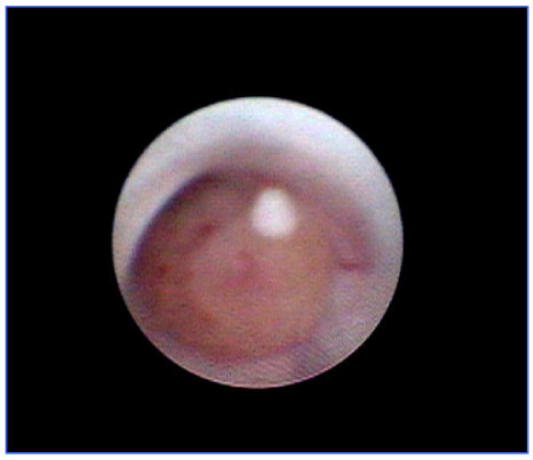
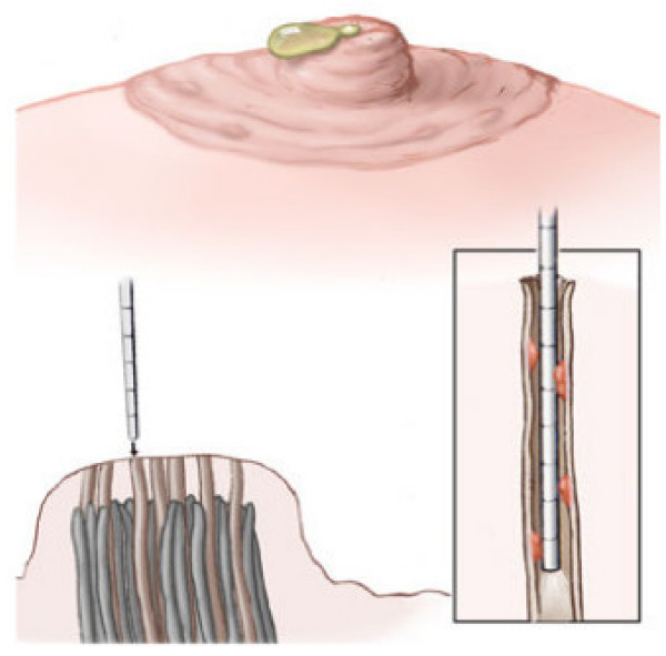
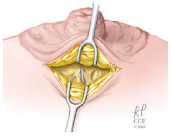
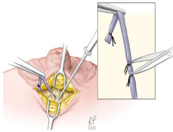
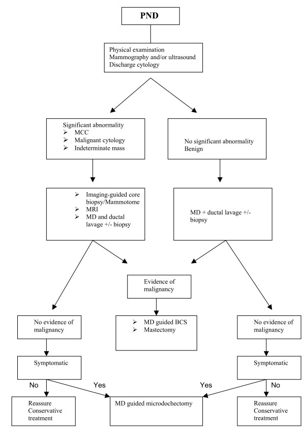
Breast papilloma is a term that describes an intraductal papillary configuration of the mammary epithelium on macroscopic or microscopic examination. It includes solitary intraductal papillomas, multiple papillomas, papillomatosis, and juvenile papillomatosis (JP).Recent advances in mammary ductoscopy (MD) have raised new possibilities in the diagnosis and treatment of breast papillomas. This technique represents an important diagnostic adjunct in patients with pathological nipple discharge (PND) by allowing direct visualisation and biopsy of intraductal lesions and guiding duct excision surgery. Treatment of breast papillomas often entails surgical duct excision for symptomatic relief and histopathological examination. Recently, more conservative approach has been adapted. MD-assisted microdochectomy should be considered the procedure of choice for a papilloma-related single duct discharge. Furthermore, there is increasing evidence that MD has the potential to reduce the number of duct excision procedures and minimise the extent of surgical resection. Imaging-guided vacuum-assisted core biopsy can be diagnostic and therapeutic for papillomas seen on mammography and/or ultrasound. Patients with multiple papillomas do have an increased risk of developing cancer and should be kept under annual review with regular mammography (preferably digital mammography) if treated conservatively. Magnetic resonance (MR) can be also used in surveillance in view of its high sensitivity. Because the risk is small, long term and affects both breasts, long-term follow-up is more appropriate than prophylactic mastectomy. Patients who prove to have solitary duct papilloma have insufficient increase in the risk of subsequent malignancy to justify routine follow-up.
Figures
References
-
- Hughes LE, Mansel RE, Webster DJT. Benign disorders and diseases of the breast: concept and clinical management. London: Bailliere Tindall; 1989. p. 42.
-
- Haagensen CD, Bodain C, Haagensen DE. Breast carcinoma risk and detection. Philadelphia: WB Saunders; 1981. p. 146.
LinkOut - more resources
Full Text Sources
Other Literature Sources
