. 2006;8(1):R7.
doi: 10.1186/bcr1371. Epub 2005 Dec 12.

CD24 staining of mouse mammary gland cells defines luminal epithelial, myoepithelial/basal and non-epithelial cells

Affiliations

CD24 staining of mouse mammary gland cells defines luminal epithelial, myoepithelial/basal and non-epithelial cells

Katherine E Sleeman et al. Breast Cancer Res. 2006.

Abstract

Introduction: Breast cancer is thought to arise in mammary epithelial stem cells. There is, therefore, a large amount of interest in identifying these cells. The breast is a complex tissue consisting of two epithelial layers (an outer myoepithelial/basal layer and an inner luminal epithelial layer) as well as a large non-epithelial component (fibroblasts, endothelial cells, lymphocytes, adipocytes, neurons and myocytes). The definitive identification of a mammary epithelial stem cell population is critically dependent on its purity. To date, this has been hampered by the lack of suitable markers to separate out the two epithelial layers, and to remove contaminating non-epithelial cells.

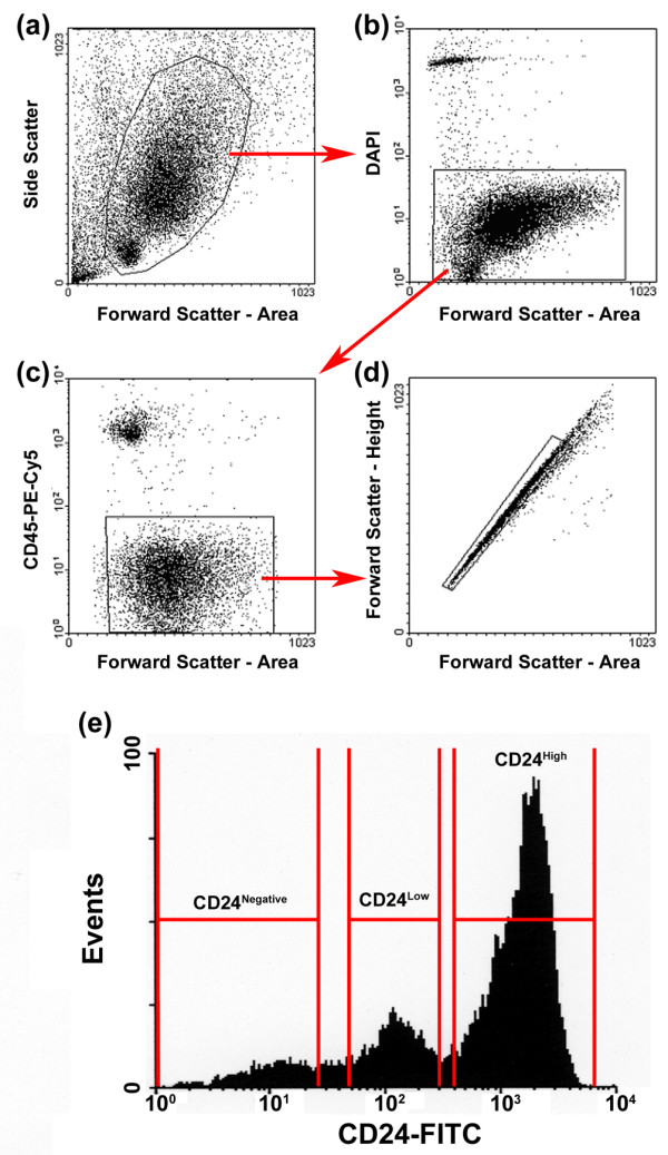
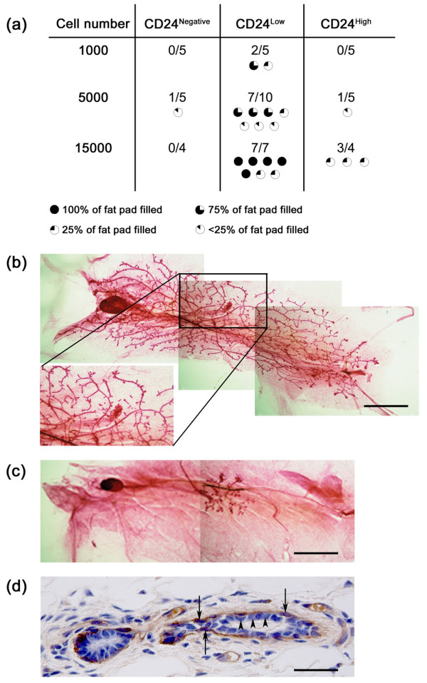
Methods: Mouse mammary glands were dissociated and stained with CD24. Cells were sorted into separate populations based on CD24 expression and assessed for luminal epithelial and myoepithelial/basal markers by direct fluorescent microscopy and real time PCR. The stem/progenitor potential of these cell populations was assessed in vivo by cleared mammary fat pad transplantation.

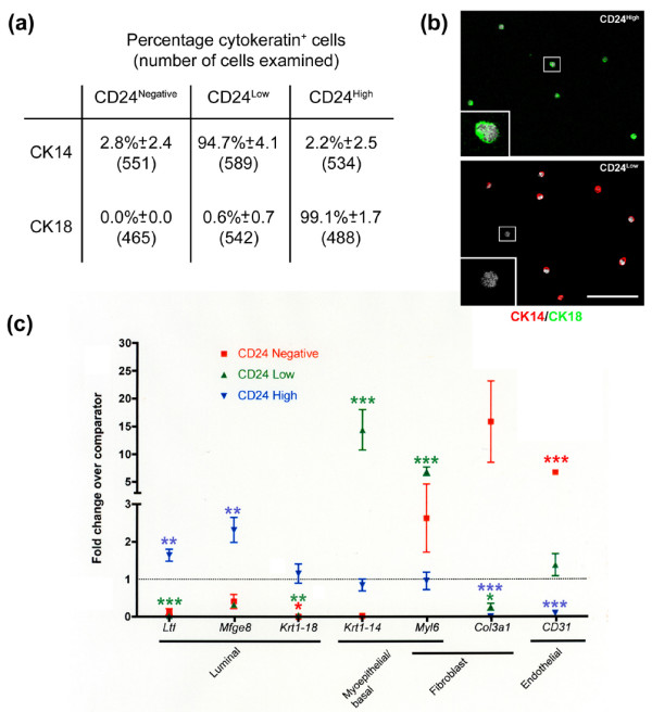
Results: Three populations of CD24 expressing cells were identified: CD24Negative, CD24Low and CD24High. Staining of these cells with cytokeratin markers revealed that these populations correspond to non-epithelial, myoepithelial/basal and luminal epithelial cells, respectively. Cell identities were confirmed by quantitative PCR. Cleared mammary fat pad transplantation of these cell populations revealed that extensive mammary fat pad repopulation capacity segregates with the CD24Low cells, whilst CD24High cells have limited repopulation capacity.

Conclusion: Differential staining of mammary epithelial cells for CD24 can be used to simultaneously isolate pure populations of non-epithelial, myoepithelial/basal and luminal epithelial cells. Furthermore, mammary fat pad repopulation capacity is enriched in the CD24Low population. As separation is achieved using a single marker, it will be possible to incorporate additional markers to further subdivide these populations. This will considerably facilitate the further analysis of mammary epithelial subpopulations, whilst ensuring high purity, which is key for understanding mammary epithelial stem cells in normal tissue biology and carcinogenesis.


Figures

Figure 1
Figure 1
Differential CD24 expression distinguishes between mammary gland subtypes. (a) Cells were sorted on the basis of forward scatter and side scatter. (b) Dead cells (DAPI bright) were excluded. (c) B lymphocytes and immature T lymphocytes are CD24+ and, therefore, CD45 staining was used to exclude all leukocytes. (d) Doublets and higher order cell clumps were excluded using a time-of-flight approach (boxed cells represent single cells). (e) Typical CD24 staining profile, indicating the three populations of cells: CD24High (69.9 ± 5.5%; n = 15), CD24Low (22.2 ± 5.6%; n = 15) and CD24Negative (6.3 ± 1.5%; n = 15). DAPI = 4',6-diamidino-2-phenylindole dihydrochloride; FITC, fluorescein isothiocyanate; PE-Cy5, phycoerythrin-Cy5.
Figure 2
Figure 2
Characterisation of CD24High, CD24Low and CD24Negative populations. (a) Cell populations sorted on the basis of CD24 expression were stained for cytokeratin (CK)8/18 or CK14. The mean percentage of CK14 and CK8/18 positive cells (±standard deviation) and the total number of cells counted is indicated for each population. Results from four independent sorts. (b) Cells were double stained with CK14 and CK8/18. Only CK18+/CK14- cells were observed in the CD24High population (top panel). The majority of CD24Low cells were CK14+, with occasional CK18-/CK14- cells (bottom panel, inset). No CK18+/CK14+ cells were observed. Scale bar = 75 μm. (c) Quantitative rtPCR reactions were carried out to determine fold changes in expression of a selection of genes with known luminal epithelial (Ltf, Mfge8, Krt1-18), myoepithelial/basal (Krt1-14, Myl6a) or non-epithelial (Myl6a, Procollagen 3a1 (Col3a1), CD31) distribution, compared to leucocyte-depleted, bulk mammary cells. The analysis was carried out on two independent cDNA syntheses from each of three independent sorts (CD24Low and CD24High) or on two independent cDNA syntheses from one sort and a single cDNA synthesis from a pool of two further sorts (CD24Negative). Significance levels are indicated by: *P < 0.05; **P < 0.01; ***P < 0.001.
Figure 3
Figure 3
Transplantation of CD24High, CD24Low and CD24Negative populations. Cell populations sorted on the basis of CD24 expression were transplanted into cleared mammary fat pads of syngeneic FVB mice. (a) Table indicates the number of cells of each population injected into each fat pad, the total number of fat pads injected, and the number of transplants that generated epithelial outgrowths. Failed clears were excluded from the analysis. The extent to which each outgrowth filled the host fat pad is indicated graphically. CD24Low cells showed the highest rate of successful transplantation and formed the most extensive outgrowths at each cell number studied. (b) Example of an outgrowth derived from transplantation of CD24Low cells that fills 100% of the host fat pad. The magnified boxed region highlights the point of origin of the outgrowth. Scale bar = 4.25 mm. (c) Example of an outgrowth derived from transplantation of CD24High cells which fills <25% of the host fat pad. Scale bar = 4.25 mm. (d) Section through CD24Low outgrowth stained with anti-α-isoform smooth muscle actin showing the positive outer myoepithelial layer (arrows) and the negative inner luminal epithelial layer (arrowheads). Scale bar = 40 μm.

References

    1. Boulanger CA, Smith GH. Reducing mammary cancer risk through premature cell senescence. Oncogene. 2001;20:2264–2272. doi: 10.1038/sj.onc.1204312. - DOI - PubMed
    1. Chepko G, Smith GH. Mammary epithelial stem cells: our current understanding. J Mammary Gland Biol Neoplasia. 1999;4:35–52. doi: 10.1023/A:1018752519356. - DOI - PubMed
    1. Reya T, Morrison SJ, Clarke MF, Weissman IL. Stem cells, cancer, and cancer stem cells. Nature. 2001;414:105–111. doi: 10.1038/35102167. - DOI - PubMed
    1. Smalley M, Ashworth A. Stem cells and breast cancer: A field in transit. Nat Rev Cancer. 2003;3:832–844. doi: 10.1038/nrc1212. - DOI - PubMed
    1. Alvi AJ, Clayton H, Joshi C, Enver T, Ashworth A, Vivanco MM, Dale TC, Smalley MJ. Functional and molecular characterisation of mammary side population cells. Breast Cancer Res. 2003;5:R1–8. doi: 10.1186/bcr563. - DOI - PMC - PubMed

Publication types