. 2006 Jan 24;103(4):945-50.
doi: 10.1073/pnas.0510164103. Epub 2006 Jan 17.

Somitic origin of limb muscle satellite and side population cells

Affiliations

Somitic origin of limb muscle satellite and side population cells

Jaclyn Schienda et al. Proc Natl Acad Sci U S A. .

Abstract

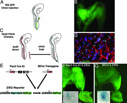
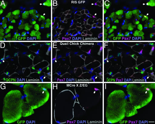
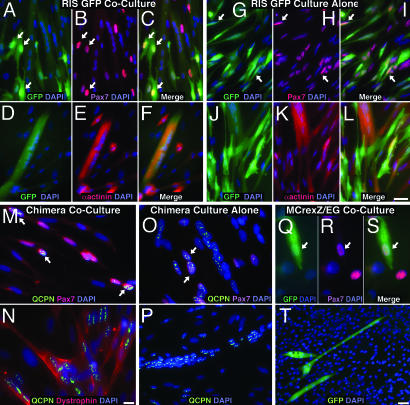
Repair of mature skeletal muscle is mediated by adult muscle progenitors. Satellite cells have long been recognized as playing a major role in muscle repair, whereas side population (SP) cells have more recently been identified as contributing to this process. The developmental source of these two progenitor populations has been considerably debated. We explicitly tested and quantified the contribution of embryonic somitic cells to these progenitor populations. Chick somitic cells were labeled by using replication-defective retroviruses or quail/chick chimeras, and mouse cells were labeled by crossing somite-specific, Pax3-derived Cre driver lines with a Cre-dependent reporter line. We show that the majority of, if not all, limb muscle satellite cells arise from cells expressing Pax3 specifically in the hypaxial somite and their migratory derivatives. We also find that a significant number of, but not all, limb muscle SP cells are derived from the hypaxial somite. Notably, the heterogeneity in the developmental origin of SP cells is reflected in their functional heterogeneity; somitically derived SP cells are intrinsically more myogenic than nonsomitically derived ones. Thus, we show that the somites, which supply embryonic and fetal myoblasts, are also an important source of highly myogenic adult muscle progenitors.


Figures

Fig. 1.
Fig. 1.
Methods for labeling somitic cells in chick and mouse. (A) Labeling of chick psm through injection of RIS-GFP. (B) GFP labeling of chick hindlimb muscle. (C) Somitic labeling through replacement of chick psm with quail psm. (D) QCPN+ myonuclei in chimeric hindlimb muscle. (Scale bar: 5 μm.) (E) Labeling of mouse Pax3+ cells and their progeny through Pax3CreKI or MCre;Z/EG mice. (F and G) GFP labeling of mouse hindlimb muscle in both crosses. Sections through quadriceps muscles were simultaneously stained for LacZ and visualized for GFP. (Scale bar: 0.5 mm for all sections.)
Fig. 2.
Fig. 2.
Somitic cells give rise to limb satellite cells. (AC) Section through P2–P3, RIS-GFP-labeled chick iliofibularis muscle with two GFP+, somitically derived Pax7+ satellite cells (arrows). (DF) Section through E18 quail/chick chimeric iliofibularis muscle with two QCPN+, somitically derived Pax7+ satellite cells (arrows). (GI) Section through 6-week-old mouse tibialis anterior muscle with one GFP+ Pax7+ satellite cell (arrow) derived from a hypaxial Pax3+ cell. DAPI+ nuclei are shown in blue (AI); Pax7+ satellite cells are shown in red (B, C, E, F, H, and I); basal lamina is highlighted with laminin shown in gray (B, DF, and H); GFP+ cells are shown in green (A, C, G, and I); and QCPN+ nuclei are shown in green (D and F). (Scale bar: 5 μm for all sections.)
Fig. 3.
Fig. 3.
Somitically derived limb muscle SP cells are myogenic in culture. (AF) GFP+ SP cells from hindlimb muscle of RIS-GFP chick in coculture differentiate into Pax7+ mononuclear cells (AC, arrows) or multinucleate, α-actinin+ myofibers (DF). (GL) GFP+ SP cells from hindlimb muscle of RIS-GFP chick cultured alone differentiate into Pax7+ mononuclear cells (GI, arrows) or multinucleate, α-actinin+ myofibers (JL). (M and N) QCPN+ SP cells from quail/chick chimeric hindlimb muscle in coculture differentiate into Pax7+ mononuclear cells (M, arrows) or multinucleate, dystrophin+ myofibers (N). (O and P) QCPN+ SP cells from quail/chick chimeric hindlimb muscle cultured alone differentiate into Pax7+ mononuclear cells (O, arrows) or multinucleate myofibers (P). (QT) GFP+ SP cells derived from MCre;Z/EG mice in coculture differentiate into Pax7+ mononuclear cells (QS, arrows) or multinucleate myofibers (T). DAPI+ nuclei are shown in blue; Pax7+ cells are shown in red (B, H, M, O, R, and S); α-actinin+ myofibers are shown in red (E and K); dystrophin+ myofiber is shown in red (N); GFP+ cells are shown in green (A, C, D, F, G, I, J, L, Q, S, and T); and QCPN+ cells are shown in green (MP). (Scale bars: AL, 0.025 μm; MO and QS, 0.0125 μm; P and T, 0.05 μm.)

References

    1. Bischoff, R. & Franzini-Armstrong, C. (2004) in Myology, eds. Engel, A. G. & Franzinin-Armstrong, C. (McGraw–Hill, New York), Vol. 1, pp. 66–86.
    1. Mauro, A. (1961) J. Biophys. Biochem. Cytol. 9, 493–495. - PMC - PubMed
    1. Charge, S. B. & Rudnicki, M. A. (2004) Physiol. Rev. 84, 209–238. - PubMed
    1. Asakura, A., Komaki, M. & Rudnicki, M. A. (2001) Differentiation 68, 245–253. - PubMed
    1. Wada, M. R., Inagawa-Ogashiwa, M., Shimizu, S., Yasumoto, S. & Hashimoto, N. (2002) Development (Cambridge, U.K.) 129, 2987–2995. - PubMed

Publication types

MeSH terms

Substances