. 2006 Jan 24;103(4):915-20.
doi: 10.1073/pnas.0508452103. Epub 2006 Jan 17.

HIV-1 protease flaps spontaneously open and reclose in molecular dynamics simulations

Affiliations

HIV-1 protease flaps spontaneously open and reclose in molecular dynamics simulations

Viktor Hornak et al. Proc Natl Acad Sci U S A. .

Abstract

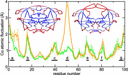
We report unrestrained, all-atom molecular dynamics simulations of HIV-1 protease that sample large conformational changes of the active site flaps. In particular, the unliganded protease undergoes multiple conversions between the "closed" and "semiopen" forms observed in crystal structures of inhibitor-bound and unliganded protease, respectively, including reversal of flap "handedness." Simulations in the presence of a cyclic urea inhibitor yield stable closed flaps. Furthermore, we observe several events in which the flaps of the unliganded protease open to a much greater degree than observed in crystal structures and subsequently return to the semiopen state. Our data strongly support the hypothesis that the unliganded protease predominantly populates the semiopen conformation, with closed and fully open structures being a minor component of the overall ensemble. The results also provide a model for the flap opening and closing that is considered to be essential to enzyme function.


Figures

Fig. 1.
Fig. 1.
The two experimentally determined conformations of HIV-PR. (A) Free HIV-PR with semiopen conformation of flaps (PDB ID code 1HHP). (B) Inhibitor-bound HIV-PR with closed flaps (PDB ID code 1HVR). Importantly, the handedness of the flaps changes in the two forms and is depicted above each structure. Color indicates distinct regions. Flaps: residues 43–58 and 43′-58′, red for free and blue for bound; flap tips: residues 49–52, yellow; flap elbow: residues 37–42, magenta; cantilever: residues 59–75, green; fulcrum: residues 10–23, orange; and interleaved β-strand motif forming the dimer interface: residues 1–4 and 96–99, blue/cyan.
Fig. 2.
Fig. 2.
Cα atomic fluctuations from simulation of inhibitor-bound (green line) and free (orange line) HIV-PR averaged for both monomers, with error bars reflecting the difference between monomers. Regions: a, N-terminal β-strand; b, fulcrum loop; c, flap elbow; d, flap tips; e, cantilever β-turn; f, Thr-80 loop; g, C-terminal β-strand. Also shown are crystal structures of bound (left) and unbound (right) protease with color indicating low (blue) to high (red) fluctuations in simulations. The most significant difference between the two simulations is a large increase in flap tip flexibility for the unbound simulation.
Fig. 3.
Fig. 3.
RMSD of flaps during simulation of bound and free HIV-PR, both starting from the closed state, overlapped on the crystal structures of semiopen (red line, PDB ID code 1HHP reference structure) and closed (blue line, PDB ID code 1HVR reference) HIV-PR. Low values for the blue line indicate that the flaps are in the closed state, and low values for the red line indicate semiopen flaps. (A) Inhibitor-bound HIV-PR flaps remain very close to the initial closed crystal structure (i.e., blue line remains at low RMSD values). (B) Unbound HIV-PR flaps that were initially closed (blue line has low initial RMSDs in the beginning) convert to semiopen, including reversal of handedness (low red values). The simulation later (≈35 ns) transiently samples the closed form before reverting back to semiopen.
Fig. 4.
Fig. 4.
Flaps RMSD and flap tips distance for free HIV-PR simulation started from a semiopen crystal structure. Snapshots (cartoon diagrams, side view) along the trajectory (cyan) are shown overlapped on the semiopen crystal structure (gray). Surface representations (top view) depict flap handedness and access to the active site, with the two flaps in green/orange and the catalytic Asp-25/25′ in yellow. The semiopen conformation is prevalent (low RMSDs for red line). Note that the transiently sampled closed structure (structure b) has the flap handedness characteristic of bound (closed) crystal structures, even though flaps do not become fully pulled into the active site in this simulations. Large flap openings are sampled (structures c–e), with flap tip distances reaching ≈30 Å and subsequently returning to the semiopen form (structures e–f).
Fig. 5.
Fig. 5.
Backbone amide generalized-order parameters (S2) averaged for both monomers, with error bars reflecting the difference. (A) Experimental data (black) (6) and simulation (red) for unliganded protease. General trends of flexibility are reproduced, although simulated values are lower. (B) Order parameters for inhibitor-bound HIV-PR from simulation (blue) and from two experiments of HIV-PR bound to DMP323 [black triangles (5) and black circles (18)]. The loss of flexibility in the flap tip region (residues 49–52) as compared with that in A is readily observed in data from simulations and from experiments.
Fig. 6.
Fig. 6.
Three forms of HIV-1 protease: closed (blue, PDB ID code 1HVR), semiopen (red, PDB ID code1HHP), and open (cyan, our model). We observe opening/closing wherein the flap elbows and exposed ends of fulcrum and cantilever move down and toward the terminal β-sheet dimer interface (indicated by yellow arrows). Examination of closed and semiopen crystal structures reveals differences in those regions that are smaller in magnitude but in agreement with the direction of changes seen during opening.

References

    1. Vondrasek, J. & Wlodawer, A. (2002) Proteins Struct. Funct. Genet. 49, 429–431. - PubMed
    1. Navia, M. A., Fitzgerald, P. M. D., McKeever, B. M., Leu, C. T., Heimbach, J. C., Herber, W. K., Sigal, I. S., Darke, P. L. & Springer, J. P. (1989) Nature 337, 615–620. - PubMed
    1. Wlodawer, A., Miller, M., Jaskolski, M., Sathyanarayana, B. K., Baldwin, E., Weber, I. T., Selk, L. M., Clawson, L., Schneider, J. & Kent, S. B. H. (1989) Science 245, 616–621. - PubMed
    1. Lapatto, R., Blundell, T., Hemmings, A., Overington, J., Wilderspin, A., Wood, S., Merson, J. R., Whittle, P. J., Danley, D. E., Geoghegan, K. F., et al. (1989) Nature 342, 299–302. - PubMed
    1. Nicholson, L. K., Yamazaki, T., Torchia, D. A., Grzesiek, S., Bax, A., Stahl, S. J., Kaufman, J. D., Wingfield, P. T., Lam, P. Y. S., Jadhav, P. K., et al. (1995) Nat. Struct. Biol. 2, 274–280. - PubMed

Publication types

LinkOut - more resources