. 2006 Jan 24;103(4):1000-5.
doi: 10.1073/pnas.0507350103. Epub 2006 Jan 17.

Stat5a/b are essential for normal lymphoid development and differentiation

Affiliations

Stat5a/b are essential for normal lymphoid development and differentiation

Zhengju Yao et al. Proc Natl Acad Sci U S A. .

Abstract

Cytokines that use the common gamma chain gammac are critical for lymphoid development and function. Mutations of the IL-7 receptor, gammac, or its associated kinase, Jak3, are the major cause of human severe combined immunodeficiency. Although activated by IL-7, Stat5a/b (Stat, signal transducer and activator of transcription) have been thought to play limited roles in lymphoid development. However, we now show that mice completely deficient in Stat5a/b have severely impaired lymphoid development and differentiation. Absence of Stat5 also abrogates T cell receptor gamma rearrangement and survival of peripheral CD8(+) T cells. Thus, deficiency of Stat5 results in severe combined immunodeficiency, similar in many respects to deficiency of IL-7R, gammac, and Jak3.


Figures

Fig. 1.
Fig. 1.
Stat5 deficiency results in a severe combined immunodeficiency phenotype. (A) Mice in which one Stat5 allele had been deleted by expression of MMTV-Cre (30) were intercrossed, and E18.5 fetuses were obtained for analysis. Peripheral blood smears and hematoxylin/eosin-stained sagittal sections of thymus and spleen were prepared from Stat5–/– fetuses (Right) and Stat5+/+ littermate controls (Left). The number of viable cells in thymi (B) and spleens (C) from E18.5 Stat5–/– fetuses (triangles) was determined and compared to E18.5 wild-type (Stat5+/+, squares), Stat5ΔN (inverted triangles), γc- (IL2rg–/–, diamonds) and IL-7R- (IL7r–/–, circles) deficient fetuses. Wild-type fetuses had 3.1 ± 0.6 × 10⁶ (mean ± SE) thymocytes (n = 15), whereas Stat5–/– fetuses had 0.05 ± 0.01 × 10⁶ thymocytes (n = 17, P < 0.001 compared to wild type). Stat5ΔN fetuses had 0.8 ± 0.3 × 10⁶ (n = 8, P < 0.05 compared to Stat5–/– fetuses). IL2rg–/– fetuses had 0.07 ± 0.01 × 10⁶ thymocytes and IL7r–/– fetuses had 0.2 ± 0.03 × 10⁶ thymocytes. Enumeration of splenocytes was as follows: wild type, 7 ± 1 × 10⁵; Stat5–/–, 0.3 ± 0.1 × 10⁵; Stat5ΔN, 1 ± 0.5 × 10⁵; IL2rg–/–, 5 ± 1 × 10⁵; IL7r–/–, 2 ± 0.3 × 10⁵.
Fig. 2.
Fig. 2.
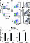
Defective B cell development in Stat5–/– fetuses. (A) Spleens from E18.5 Stat5+/+ and Stat5–/– fetuses were obtained and analyzed by flow cytometry with antibodies against CD19 and B220. (B) Fetal liver cells were obtained from E18.5 Stat5+/+, Stat5–/–, and Il7r–/– fetuses. B cells were assessed by staining with antibodies against CD19 and B220 (Left). B cell precursors were delineated by gating on B220+ cells and staining with antibodies against BP1 and CD24 (Right). (C) The expression of the B cell transcription factors Ebf and Pax5 in splenocytes and fetal liver cells was determined by quantitative PCR. Filled bars, Stat5+/+; open bars, Stat5–/–.
Fig. 3.
Fig. 3.
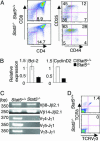
Altered T cell development in Stat5–/– fetuses. (A and B) Stat5–/– fetuses produce CD4+ and CD8+ SP T cells but have increased proportions of immature DN thymocytes. Cells were obtained from thymi of E18.5 Stat5+/+ or Stat5–/– fetuses. Single-cell suspensions were prepared and stained for surface expression of CD4, CD8, CD44, and CD25. (A) Expression of CD4 and CD8 is shown at Left. Gating on the CD4CD8 population, the proportion of CD25CD44+ (DN1), CD25+CD44+ (DN2), CD25+CD44 (DN3), and CD25CD44 (DN4) cells is depicted (Right) and summarized in Fig. 8. (B) Impaired expression of Bcl-2 and cyclin D2. RNA was isolated from thymocytes of Stat5+/+ and Stat5–/– E18.5 fetuses, and the levels of Bcl-2 and cyclin D2 were assessed by real-time PCR. Open bars, Stat5+/+; filled bars, Stat5–/–. (C and D) TCRγδ, but not αβ, rearrangement depends on Stat5 (C). Genomic DNA was isolated from thymi from wild-type and mutant mice. Products corresponding to V β8-J β2.1, V β14-J β2.1, Vγ4-Jγ1, Vγ5-Jγ1, and Vγ3-Jγ1 rearrangement were identified by PCR. (D) Thymocytes were stained with antibodies against TCRVγ3 and TCRδ and analyzed by flow cytometry.
Fig. 4.
Fig. 4.
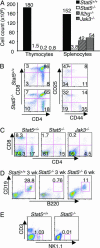
Severe disruption of lymphoid development in viable Stat5–/– mice. (A) Thymus and spleen cell counts for 3-week-old Stat5+/+ (filled bars), Stat5–/– (open bars), Il2rg–/– (diagonal hatched bars), and Jak3–/– (horizontal hatched bars) mice. (B) Thymocytes were stained for surface expression of CD4 and CD8 (Left). In addition, CD4/CD8 double-negative cells were gated and stained for surface expression of CD25 and CD44 to distinguish between four developmental stages within that population (Right; DN1, CD44+CD25; DN2, CD44+CD25+; DN3, CD44CD25+; DN4, CD44CD25). (CE) Splenocytes were stained for CD4 and CD8 (C), B220 and CD19 (D), and NK1.1 and CD3 (E).
Fig. 5.
Fig. 5.
Requirement for Stat5 in peripheral T cells. (A) Splenocytes from 6-week-old Stat5fl/–/CD4cre and Stat5fl/– mice were stained for CD3, CD4, and CD8 expression. (B and C) The expression of memory markers on CD4+ T cells (B; CD62L and CD44) and CD8+ T cells (C; CD44 and CD122) were assessed.
Fig. 6.
Fig. 6.
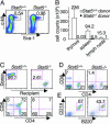
Stat5–/– fetuses have reduced numbers of lymphoid stem cells with poor repopulating function. (A) Fetal liver cells from E14.5 fetuses were stained for lineage negative Sca-1+ c-Kit+ (LSK) stem cells and analyzed by flow cytometry. Lineage negative cells were gated and analyzed for c-Kit and Sca-1 expression. Although the proportion of LSKs in Stat5–/– mice is greater than or equal to that in wild-type littermates, the absolute number of LSKs is reduced in Stat5–/– fetuses (Stat5+/+, 16.7 × 10⁴± 0.07 × 10⁴; Stat5–/–, 5.6 × 10⁴± 0.5 × 10⁴.(BE) Irradiated C57BL/6 rag2–/– CD45.1+ congenic mice were reconstituted with 2 × 10⁶ total fetal liver cells from CD45.2+ wild-type or Stat5–/– mice and housed in pathogen-free conditions on acidified water. Eight weeks after reconstitution, mice were killed; spleens, thymi, and lymph nodes were harvested, counted, and stained for various cell lineages along with antibodies against CD45.1 and CD45.2 to detect recipient and donor cells, respectively. (B) Donor-derived cell counts are shown. (C) The proportion of donor-derived thymocytes is shown in Upper. Donor-derived thymocytes were stained for CD4 and CD8 populations (Lower). (D and E) Donor-derived splenocytes were stained for T cell populations (D) or B cell populations (E). Stat5–/– fetal liver cells were unable to repopulate T and B cell lineages to any significant degree.

References

    1. Khaled, A. R. & Durum, S. K. (2002) Nat. Rev. Immunol. 2, 817–830. - PubMed
    1. Puel, A., Ziegler, S. F., Buckley, R. H. & Leonard, W. J. (1998) Nat. Genet. 20, 394–397. - PubMed
    1. Noguchi, M., Yi, H., Rosenblatt, H. M., Filipovich, A. H., Adelstein, S., Modi, W. S., McBride, O. W. & Leonard, W. J. (1993) Cell 73, 147–157. - PubMed
    1. Russell, S. M., Tayebi, N., Nakajima, H., Riedy, M. C., Roberts, J. L., Aman, M. J., Migone, T. S., Noguchi, M., Markert, M. L., Buckley, R. H., et al. (1995) Science 270, 797–800. - PubMed
    1. Macchi, P., Villa, A., Giliani, S., Sacco, M. G., Frattini, A., Porta, F., Ugazio, A. G., Johnston, J. A., Candotti, F., O'Shea, J. J., et al. (1995) Nature 377, 65–68. - PubMed

Publication types

MeSH terms