Effect of CT acquisition parameters in the detection of subtle hypoattenuation in acute cerebral infarction: a phantom study
- PMID: 16418353
- PMCID: PMC7976079
Effect of CT acquisition parameters in the detection of subtle hypoattenuation in acute cerebral infarction: a phantom study
Abstract
Background and purpose: We evaluated the effects of varying tube voltage, current per rotation, and section thickness on detectability of 2- and 4-Hounsfield unit (HU) differences on brain CT between normal and ischemic gray matter within 6 hours of ischemia onset, by using a low-contrast phantom.
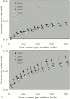
Methods: The phantom with an attenuation of 36 HU corresponding to normal gray matter contained 2 sets of spheres (34 HU and 32 HU) corresponding to the early CT signs of ischemic brain and complete infarction, respectively. The reproducibility of the CT numbers and the contrast-to-noise ratio (CNR), defined as the CT number difference between the background (36 HU) and the spheres (34 HU or 32 HU) divided by the SD of the background CT number were measured. Five radiologists rated the phantom images for detection of the low-contrast spheres by visual inspection.
Results: The CT numbers were reproducible within 1 HU with a tube current of > or =150 mAs at 120 kVp. The CNRs for the 34- and 32-HU spheres were positively correlated with the tube voltage, tube current per rotation, and the section thickness. A CNR of 1.0 was obtained for the 34-HU sphere when scanning was conducted with a section thickness of 10 mm at 120 kVp and 700 mAs, or 135kVp and 450 mAs, respectively. A significant improvement of the accuracy of detection was found with increasing tube current, tube voltage per rotation, and section thickness.
Conclusion: Our study indicated that the 2-HU hypoattenuation corresponding to the early CT sign of acute ischemic stroke can be detected by using appropriate parameter settings.
Figures


References
-
- Furlan A, Higashida R, Wechsler L, et al. Intra-arterial prourokinase for acute ischemic stroke. The PROACT II study: a randomized controlled trial: prolyse in acute cerebral thromboembolism. JAMA 1999;282:2003–11 - PubMed
-
- Hacke W, Kaste M, Fieschi C, et al. Intravenous thrombolysis with recombinant tissue plasminogen activator for acute hemispheric stroke: the European Cooperative Acute Stroke Study (ECASS). JAMA 1995;274:1017–25 - PubMed
-
- The NINDS rt-PA Stroke Study Group. Tissue plasminogen activator for acute ischemic stroke. N Engl J Med 1995;333:1581–88 - PubMed
-
- Arnold M, Schroth G, Nedeltchev K, et al. Intra-arterial thrombolysis in 100 patients with acute stroke due to middle cerebral artery occlusion. Stroke 2002;33:1828–33 - PubMed
Publication types
MeSH terms
LinkOut - more resources
Full Text Sources
Other Literature Sources
Medical
