Ruptured cavernous sinus aneurysms causing carotid cavernous fistula: incidence, clinical presentation, treatment, and outcome
- PMID: 16418380
- PMCID: PMC7976066
Ruptured cavernous sinus aneurysms causing carotid cavernous fistula: incidence, clinical presentation, treatment, and outcome
Abstract
Background and purpose: In this study, we present our experience with 11 patients with ruptured cavernous sinus aneurysms causing carotid cavernous fistulas (CCFs), to assess the incidence of ruptured cavernous sinus aneurysms causing CCFs and evaluate clinical presentations, treatments, and outcomes.
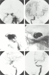
Patients and methods: During a 10-year period, 10 of 689 (1.5%) endovascular-treated ruptured aneurysms were ruptured cavernous sinus aneurysms causing CCF. One additional patient with a CCF died shortly before treatment of intracranial hemorrhage. All patients had audible pulsatile bruit. Exophthalmus, ocular motor palsy, and decreased vision correlated with venous drainage to the superior ophthalmic veins and intracerebral hemorrhage was associated with major cortical venous drainage in 2 patients.
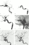
Results: Two low-flow CCFs closed spontaneously before treatment with resolution of symptoms; the aneurysms were subsequently treated. Eight CCFs were successfully occluded, 5 by coil occlusion of the aneurysm, one by occlusion of the aneurysm with a balloon, and 2 by simultaneous coil occlusion of the aneurysm and internal carotid artery. There were no complications of treatment. Visual acuity returned to normal in all but one patient, and ophthalmoplegia was cured in 6 of 8 patients. In 2 patients, a remaining abducens palsy was surgically corrected.
Conclusion: The incidence of CCF by a ruptured cavernous sinus aneurysm was 1.5%. CCF was the presenting symptom in 24.4% of treated symptomatic cavernous sinus aneurysms. Clinical symptoms correlate with venous drainage. Drainage to cortical veins may lead to intracranial hemorrhage. Endovascular treatment with coils is effective in occluding the fistula.
Figures


References
-
- Kasner SE, Liu GT, Galetta SL. Neuro-ophthalmologic aspects of aneurysms. Neuroimaging Clin N Am 1997;7:679–92 - PubMed
-
- Lee AG, Mawad ME, Baskin DS. Fatal subarachnoid hemorrhage from the rupture of a totally intracavernous carotid artery aneurysm: case report. Neurosurgery 1996;38:596–98 - PubMed
-
- van der Schaaf IC, Brilstra EH, Buskens E, et al. Endovascular treatment of aneurysms in the cavernous sinus: a systematic review on balloon occlusion of the parent vessel and embolization with coils. Stroke 2002;33:313–18 - PubMed
-
- Jafar JJ, Huang PP. Surgical treatment of carotid cavernous aneurysms. Neurosurg Clin N Am 1998;9:755–63 - PubMed
MeSH terms
LinkOut - more resources
Full Text Sources
Medical
