

MR diffusion tensor imaging and fiber tracking in 5 spinal cord astrocytomas
Affiliations
- PMID: 16418387
- PMCID: PMC7976075
Item in Clipboard
MR diffusion tensor imaging and fiber tracking in 5 spinal cord astrocytomas
AJNR Am J Neuroradiol.
2006 Jan.
Abstract
Spinal cord astrocytomas are rare neoplasms that can result in alteration of the spinal cord structural integrity, which can be assessed by using diffusion tensor imaging methods. Our objective was to visualize the deformation of the posterior spinal cord lemniscal and corticospinal tracts in 5 patients with low-grade astrocytomas compared with 10 healthy volunteers by using 3D fiber-tracking reconstructions.
Figures
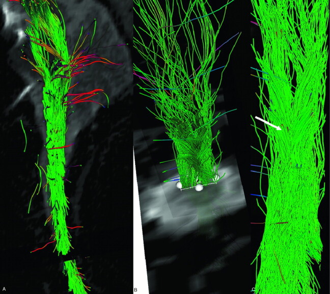
Fiber tracking performed on a volunteer’s cervical spinal cord focused on the posterior lemniscal tracts. Sagittal (A), axial (B), and coronal (C) views show tracts reconstructed over the b0 sequence. Note the decussating fibers (arrow).
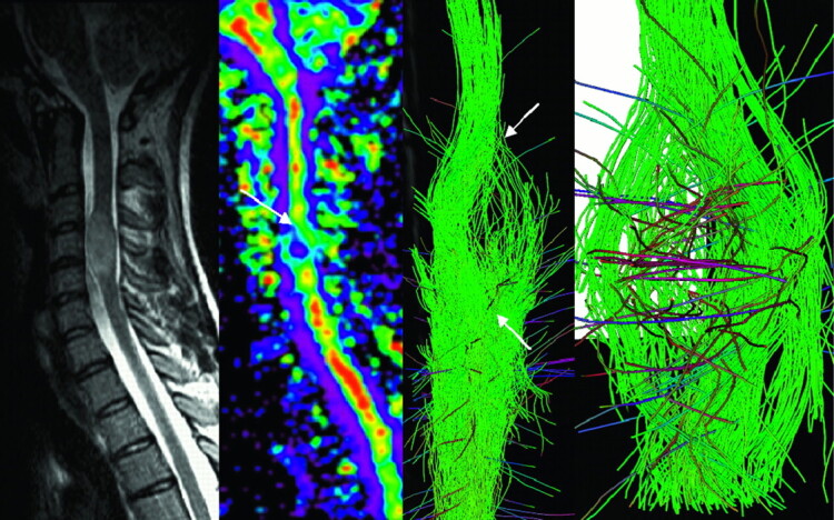
MR imaging of a spinal cord involvement due to a solid state astrocytoma. FA map and fiber tracking over b0 image show warped fibers around the tumor. Neither the vasogenic edema nor the cystic portion of the tumor was visible on the T2-weighted image, and boundaries of the lesion visible on both the FA map and 3D FT reconstructions (arrow) matched those on T2-weighted imaging.
References
-
- Stein BM, McCormick PC: Spinal intradural tumors. In: Wilkins RH, Rengachary SS, eds. Neurosurgery. New York: McGrawHill;1996. :1769–89
-
- Ries M, Jones R, Dousset V, et al. Diffusion tensor MRI of the spinal cord. Magn Reson Med 2000;44:884–92 - PubMed
-
- Basser P, Pierpaoli C. Microstructural and physiological features of tissues elucidated by quantitative diffusion tensor. MRI. J Magn Reson 1996;111:209–19 - PubMed
MeSH terms
LinkOut - more resources
Full Text Sources