. 2006 Jan 15;20(2):147-52.
doi: 10.1101/gad1367806.

Tissue-specific RNAi reveals that WT1 expression in nurse cells controls germ cell survival and spermatogenesis

Affiliations

Tissue-specific RNAi reveals that WT1 expression in nurse cells controls germ cell survival and spermatogenesis

Manjeet K Rao et al. Genes Dev. .

Abstract

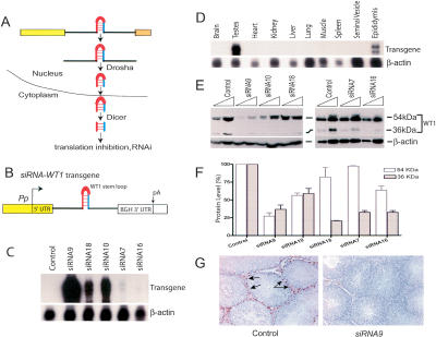
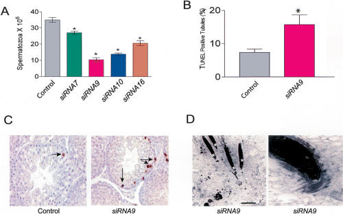
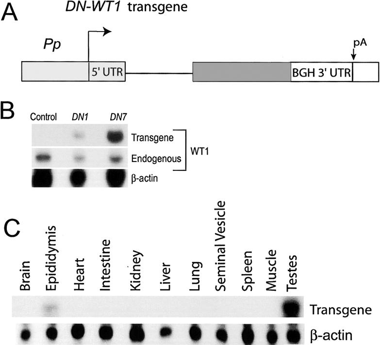
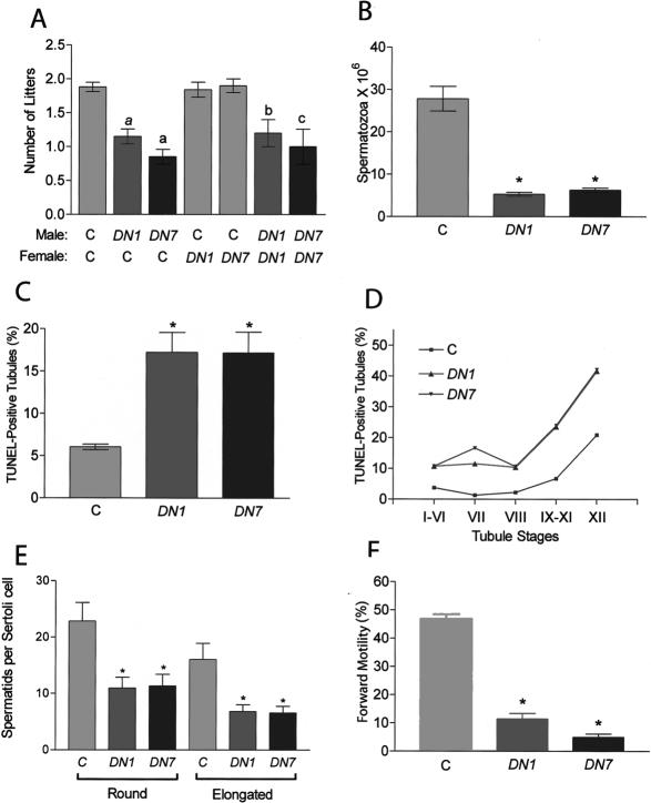
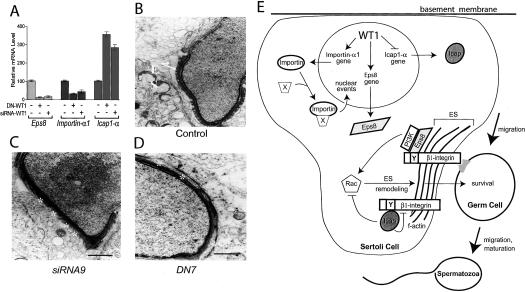
Using a novel tissue-specific RNA interference (RNAi) approach that mimics the principle by which naturally occurring microRNAs (miRNA) are made, we demonstrate that the Wilms' tumor 1 (WT1) transcription factor has an essential role in spermatogenesis. Mice depleted of WT1 in Sertoli nurse cells suffered from increased germ cell apoptosis, loss of adherens junctions, disregulation of adherence junction-associated genes, and impaired fertility. These effects were recapitulated in transgenic mice expressing a dominant-negative form of WT1 in Sertoli cells, demonstrating the validity of our RNAi approach. Our results indicate that the tumor suppressor WT1 promotes Sertoli cell-germ cell signaling events driving spermatogenesis.


Figures

Figure 1.
Figure 1.
WT1 silencing in siRNA-WT1 transgenic mice. (A) The canonical microRNA pathway (Tang 2005). (B) The siRNA-WT1 transgene (the WT1 stem-loop sequence is shown in Supplementary Fig. S1A). (UTR) Untranslated region; (BGH) bovine growth hormone; (pA) polyadenylation site. (C) Ribonuclease protection analysis of adult testes RNA (20 μg) annealed with transgene- and β-actin-specific probes. Transgene transcripts were detected with a 3′-UTR probe (Rao et al. 2003). The β-actin probe served as a loading control. (D) Transgene expression in various tissues from an adult siRNA9 mouse (the same pattern was also observed for the siRNA10 and siRNA16 lines; data not shown). (E) Western blot analysis of the indicated amounts of testicular lysate from control and transgenic mice using a WT1 polyclonal antibody (c19; Santa Cruz, Inc.; 1:1600 dilution). (F) Reduction in WT1 levels in siRNA-WT1 transgenic mice. Expression was quantified by densitometric analysis of three independent Western blots; means ± SEM. (G) Immunohistochemical analysis of testes from siRNA9 and control littermate adult mice using an anti-mouse WT1 monoclonal antibody (H6; DAKO, Inc.; 1:1000 dilution). Arrows indicate selected control Sertoli cell nuclei that stained positively for WT1. A reduction in WT1 immunostaining was also observed in siRNA10 mice testes.
Figure 2.
Figure 2.
Testes defects in siRNA-WT1 transgenic mice. (A) Caudal spermatozoa counts (means ± SEM). (*) P ≤ 0.001; n =6. (B) Quantitative analysis of TUNEL-positive tubules and cells (means ± SEM). (*) P ≤ 0.001; n = 1262 and 1052 tubules for control and siRNA9, respectively. (C) TUNEL analysis of apoptotic germ cells (arrows) in testes sections. (D) Transmission electron micrographs. The left panel shows a cluster of elongated spermatids deep within the seminiferous epithelium undergoing apoptosis (asterisks), and the right panel shows a high-magnification view of a step-14 spermatid with condensed chromatin, characteristic of an early stage of apoptosis.
Figure 3.
Figure 3.
Expression of the DN-WT1 transgene. (A) The DN-WT1 transgene. Solid bar is DN-WT1 coding region; see Figure 1 for definition of abbreviations. (B) RNase protection analysis of adult testes RNA (20 μg) from the DN1 and DN7 lines, performed with a WT1 probe that distinguishes between endogenous and transgene transcripts. Transgene transcripts were expressed at ∼1.2-fold and approximately twofold higher levels than endogenous WT1 transcripts in the testes of DN1 and DN7 mice, respectively (when corrected for [32P]UTP content in the protected bands). (C) RNase protection analysis of tissue RNA (20 μg) from an adult DN7 mouse annealed with the transgene-specific probe used in Figure 1C.
Figure 4.
Figure 4.
Testes defects in DN-WT1 transgenic mice. (A) Mean number of litters obtained from 60-d pairings of DN1 and DN7 mice and control littermates (labeled “C”). (a) P ≤ 0.001; (b) P < 0.05; (c) P ≤ 0.01; n = 25, 20, and 20 for control, DN1, and DN7, respectively. After a 5-d cohabitation, only 20% of DN1 males sired litters (n = 25) (data not shown), similar to the percentage for siRNA9 mice (27%; see text). (B) Caudal spermatozoa counts. (*) P ≤ 0.001; n =8 mice per group. (C) Percentage of TUNEL-positive tubules. (*) P ≤ 0.001; n = 628, 830, and 770 tubules for DN1, DN7, and control mice, respectively. (D) Percentage of TUNEL-positive tubules at different stages of the seminiferous epithelial cycle. In addition to having apoptotic spermatogonia and mitotic spermatocytes, both of which normally undergo apoptosis, the DN-WT1 mice had apoptotic round spermatids (data not shown). (E) Number of round and elongated spermatids in stage VII tubules. (*) P ≤ 0.05; n = 6 mice per group. (F) Percentage of sperm displaying forward motility. (*) P ≤ 0.001; n = 10 mice per group. The data in panels A-C, E, and F are means ± SEM.
Figure 5.
Figure 5.
Altered gene expression and disrupted adherens junctions in WT1-deficient mice. (A) The Eps8, Icap1-α, and importin-α1 genes are regulated in the same manner in both siRNA-WT1 and DN-WT1 mice testes. Real-time RT-PCR analysis was used to determine transcript expression relative to that of L19 mRNA (encodes a ubiquitously expressed ribosomal protein). (B-D) Transmission electron micrographs of testes from siRNA9 (siRNA-WT1), DN7 (DN-WT1), and control littermate mice. The control panel shows a normal step-10 spermatid embedded in a Sertoli cell; the ES junctional complex is present (arrows), and the acrosome extends over the apex and the dorsal curvature of the head. In contrast, the spermatid-Sertoli cell junctions in siRNA-WT1 and DN-WT1 mice often lacked the ES (asterisks). (E) Model of WT1 action in Sertoli cells. Eps8 is a signaling molecule that forms a molecular complex with Rac, phosphoinositide 3-kinase, and β1-integrin (see text). Because this complex is known to elicit cytoskeletal remodeling, we posit that by up-regulating Eps8, WT1 activates the actin remodeling events at the Sertoli cell membrane essential for migration of germ cells across its surface, as well as for germ cell survival and maturation. We also posit that these remodeling events are enhanced by WT1's ability to also inhibit the expression of Icap1-α, which is a negative regulator of Rac activation and β1-integrin avidity (see text). Lastly, WT1 increases the level of importin-α1, which we propose is necessary for the efficient nuclear import of proteins (designated by “×”) that control nuclear events essential for spermatogenesis.

References

    1. Armstrong, J.F., Pritchard-Jones, K., Bickmore, W.A., Hastie, N.D., and Bard, J.B. 1993. The expression of the Wilms' tumour gene, WT1, in the developing mammalian embryo. Mech. Dev. 40: 85-97. - PubMed
    1. Blume-Jensen, P., Jiang, G., Hyman, R., Lee, K.F., O'Gorman, S., and Hunter, T. 2000. Kit/stem cell factor receptor-induced activation of phosphatidylinositol 3′-kinase is essential for male fertility. Nat. Genet. 24: 157-162. - PubMed
    1. Bouvard, D., Vignoud, L., Dupe-Manet, S., Abed, N., Fournier, H.N., Vincent-Monegat, C., Retta, S.F., Fassler, R., and Block, M.R. 2003. Disruption of focal adhesions by integrin cytoplasmic domain-associated protein-1 α. J. Biol. Chem. 278: 6567-6574. - PubMed
    1. Calderwood, D.A., Fujioka, Y., de Pereda, J.M., Garcia-Alvarez, B., Nakamoto, T., Margolis, B., McGlade, C.J., Liddington, R.C., and Ginsberg, M.H. 2003. Integrin β cytoplasmic domain interactions with phosphotyrosine-binding domains: A structural prototype for diversity in integrin signaling. Proc. Natl. Acad. Sci. 100: 2272-2277. - PMC - PubMed
    1. Chang, D.D., Hoang, B.Q., Liu, J., and Springer, T.A. 2002. Molecular basis for interaction between Icap1 α PTB domain and β 1 integrin. J. Biol. Chem. 277: 8140-8145. - PubMed

MeSH terms