. 2006 Jan 15;20(2):253-66.
doi: 10.1101/gad.1360106.

Targeted deletion of a cis-regulatory region reveals differential gene dosage requirements for Pdx1 in foregut organ differentiation and pancreas formation

Affiliations

Targeted deletion of a cis-regulatory region reveals differential gene dosage requirements for Pdx1 in foregut organ differentiation and pancreas formation

Yoshio Fujitani et al. Genes Dev. .

Abstract

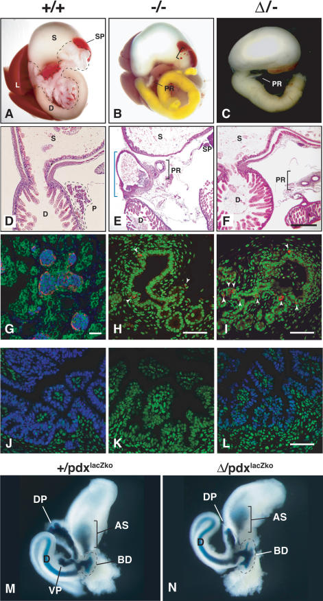
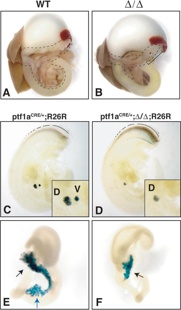
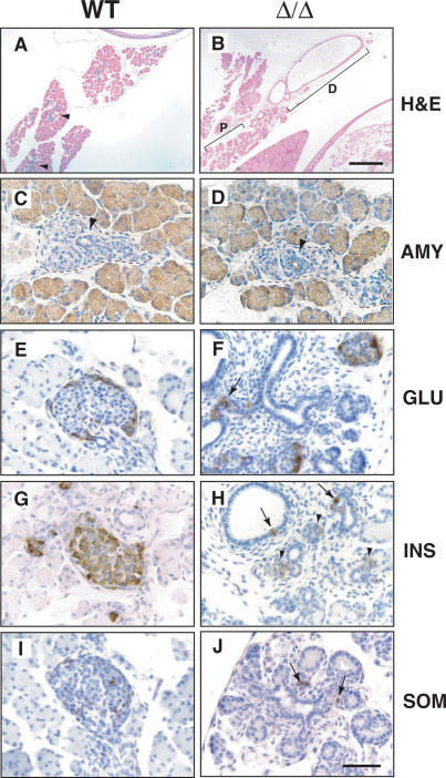
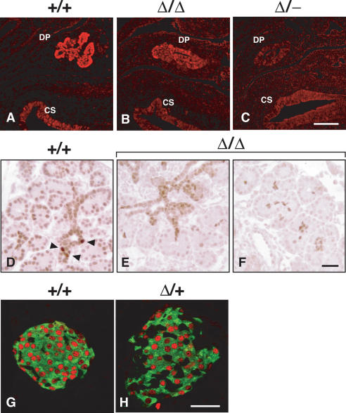
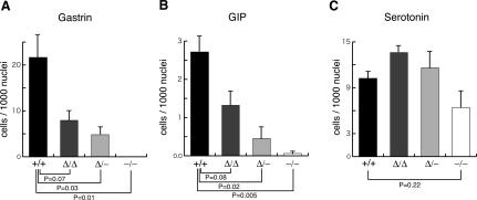
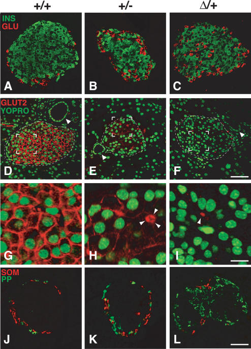
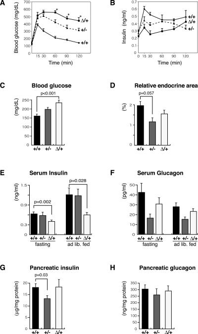
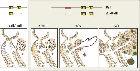
Pdx1 (IPF-1 in humans, which is altered in MODY-4) is essential for pancreas development and mature beta-cell function. Pdx1 is expressed dynamically within the developing foregut, but how its expression characteristics are linked to the various steps of organ specification, differentiation, and function is unknown. Deletion of a conserved enhancer region (Area I-II-III) from Pdx1 produced a hypomorphic allele (Pdx1(DeltaI-II-III)) with altered timing and level of expression, which was studied in combination with wild-type and protein-null alleles. Lineage labeling in homozygous Area I-II-III deletion mutants (Pdx1(DeltaI-II-III/DeltaI-II-III)) revealed lack of ventral pancreatic bud specification and early-onset hypoplasia in the dorsal bud. Acinar tissue formed in the hypoplastic dorsal bud, but endocrine maturation was greatly impaired. While Pdx1(-/-) (protein-null) mice have nonpancreatic abnormalities (e.g., distorted pylorus, absent Brunner's glands), these structures formed normally in Pdx1(DeltaI-II-III/DeltaI-II-III) and Pdx1(DeltaI-II-III/-) mice. Surprisingly, heterozygous (Pdx1(+/DeltaI-II-III)) mice had abnormal islets and a more severe prediabetic condition than Pdx1(+/-) mice. These findings provide in vivo evidence of the differential requirements for the level of Pdx1 gene activity in the specification and differentiation of the various organs of the posterior foregut, as well as in pancreas and gut endocrine cell differentiation.


Figures

Figure 1.
Figure 1.
Strategy for deleting Area I-II-III within Pdx1. (Top) Pdx1 genomic structure. Area I-II-III lies ∼2 kb 5′ of the transcription start site. Homologous recombination with the targeting vector generated the frt-neo allele; Flp deletion created the flox I-II-III allele; subsequent Cre deletion led to the ΔI-II-III allele. (Black boxes) Exons; (gray box) B-X external probe for Southern blot analysis (diagnostic band lengths pre/post-recombination indicated). (neoR) Neomycin resistance cassette; (TK) thymidine kinase cassette. Black and white arrowheads indicate LoxP and FRT sites, respectively. (B) BamHI; (Bs) BstEII; (P) PstI; (RV) EcoRV; (X) XbaI.
Figure 2.
Figure 2.
Characterization of Pdx1ΔI-II-III/- animals. (A-C) Gross analysis of pancreas differentiation in newborn Pdx+/+, Pdx+/-, and PdxΔI-II-III/- animals (labeled here as +/+, +/-, and Δ/-, respectively). (A) Pancreas tissue outlined. (D) Duodenum; (S) stomach; (SP) spleen; (L) liver. (B,C) (PR) Pancreatic rudiment derived from stunted outgrowth of dorsal pancreatic bud. The bracket in B indicates ectopic spleen tissue frequently observed in Pdx1-/- (-/-) or Pdx1ΔI-II-III/- (Δ/-) mutants. Yellow bile color in duodenum is often seen in -/- mutant animals. (D-F) Histological analysis of gastro-duodenal region (H&E stain). (D) Normal gastro-duodenal junction with a well-defined pylorus in P1 pup, with continuous lumen joining stomach and rostral duodenum. (E) This region is malformed in -/- littermates (blue bracket). In many -/- animals, the region forms a tortuous, sometimes blind-ended tube that lacks villi and is lined by cuboidal epithelium continuous with the pancreatic rudiment. (F) All Δ/- animals examined (n = 9) have a normal gastro-duodenal junction. (G) At P1, endocrine cell aggregations in +/+ animals represent immature islets (confocal immunofluroescence: peripheral glucagon-producing cells [red] surround insulin-producing cells [blue]; nuclei counterstained green by Yo-Pro-1). (H,I) Similar confocal immunofluorescence analysis—at higher magnification—of P1 dorsal bud-derived pancreatic rudiment in -/- and Δ/- mutants showed simple cuboidal epithelium similar to bile duct. Glucagon-producing cells (red, indicated by white arrowheads; nuclei green) were observed adjoining slight evaginations of the endodermal epithelium. (J-L) Immunofluorescence analysis, E16.5 tissues. A lower level of nuclear Pdx1 (blue; nuclei green) was found in the rostral duodenum of Δ/- animals compared with +/+ tissue (-/- tissue lacks signal). (M,N) E12.0 foregut organ development visualized with a lacZ knock-in allele of Pdx1 (Pdx1lacZko). X-gal staining labels (AS) antral stomach, (D) duodenum, (BD) common bile duct, and (DP) dorsal pancreas in both Pdx1lacZko and Δ-lacZko animals. The ventral pancreatic growth (VP) present in +/lacZko animals is undetected in Δ/lacZko animals, in which dorsal pancreatic growth is also stunted. Bars: D-F, 400 μm; G-L, 50 μm.
Figure 3.
Figure 3.
Reduced Pdx1 function leads to dysmorphic growth from the dorsal bud and loss of ventral bud specification. (A,B) Pancreas is outlined in newborn pups. Ventral pancreatic lobe (intraduodenal loop region) appears absent in Pdx1ΔI-II-III/ΔI-II-III (Δ/Δ) mutants. Bracket in B indicates cystic pancreatic epithelium with associated ectopic splenic tissue. (C-F) Heterozygous mice carrying a Ptf1aCRE knock-in allele (Ptf1aCRE/+) together with the Cre-activatable R26R reporter allele were used to assess dorsal (d) and ventral (v) bud pancreas specification at E10.5 and E14.5. (C,D) In contrast to normal pancreas specification (C), E10.5 Δ/Δ mutants (D) lack the ventral bud region-specific activation of Ptf1a expression and have a reduced dorsal bud. (E,F) At E14.5, the dorsal outgrowth (black arrows) is highly stunted compared with normal, and there is no evidence of ventral lobe-derived cells (blue arrow in normal situation in E). Dotted lines in C and D denote Ptf1a expression in developing spinal cord.
Figure 4.
Figure 4.
Distal dysmorphogenesis and proximal quasipancreatic differentiation in dorsal pancreas bud of Pdx1ΔI-II-III homozygous mice. All tissues are from P1 animals. (A,B) Distal region of dorsal bud tissue (D) differentiates as a simple dilated cystic epithelium (H&E staining), while gut-proximal tissue (P) undergoes quasipancreatic differentiation (cf. A), with a transitional zone between these regions. Blue hematoxylin-stained regions in A indicate endocrine compartment forming in newborn +/+ pancreas. (C-J) Immunohistochemical comparison of normal and gut-proximal pancreatic tissue of Pdx1ΔI-II-IIII-II-III (Δ/Δ) animals. Well-polarized acinar cells express almost normal levels of amylase (AMY) (C,D), and there are substantial numbers of disorganized glucagon-producing cells (GLU) (E,F), but only a few, weakly positive insulin-expressing cells (G,H; INS, arrowheads in H). (H) Some insulin-producing cells are located in or adjacent to ductal epithelium (arrows). (I,J) Somatostatin-producing cells (SOM) are found as small cell clusters or isolated cells in close proximity to ducts. Forming endocrine compartments are outlined in C and D. Ducts are denoted by black arrowheads. Bars: A-B, 400 μm; C-J, 50 μm.
Figure 5.
Figure 5.
Immunohistochemical assessment of Pdx1 protein expression levels with various combinations of Pdx1 deletion alleles. Tissue processing and immunostaining in parallel allowed comparison of cellular Pdx1 level; representative images are from serial section analysis of at least three embryos for each genotype. Confocal imaging and post-processing was identical for all samples; background is shown deliberately, so that the specific Pdx1 signal in the dorsal pancreas (DP) can be judged against it. (A-C) E12.0 tissues. Different combinations of mutant alleles affect Pdx1 levels in the early pancreas anlagen; signal intensity is similar in all cases in caudal stomach epithelium (CS). (D-F) E16.5 tissues. Embryos homozygous for the Pdx1ΔI-II-III allele (Δ/Δ) lack the focal high-level Pdx1-expressing cells (arrowheads) seen in normal tissue, with a significantly lower signal through the developing epithelial cords and acinar clusters. (G,H) Confocal immunofluorescence analysis on parallel-processed tissues from 4-wk-old mice from +/+ and Δ/+ adult islets (insulin, green; Pdx1, red). We noted an increased number of non-β cells (insulin-) in Δ/+ islets (Fig. 7, Supplementary Fig. 3). Bars: A-C, 50 μm; D-F, 50 μm; G,H, 50 μm.
Figure 6.
Figure 6.
Frequency of gastrin- and GIP-producing cells as a function of Pdx1 genotype. Sections analyzed were E18.5 for GIP- and serotonin-producing cells, and P7 for gastrin-producing cells. Results are from analyzing three mice of each genotype (see Materials and Methods for tissue sampling protocol). The Y-axis represents the number of gastrin-, GIP-, or serotonin-producing cells per 1000 epithelial nuclei; data expressed as mean ± SE (n = 3).
Figure 7.
Figure 7.
The Pdx1ΔI-II-III/+ genotype leads to severely compromised mature islet architecture and endocrine differentiation. All tissues analyzed were from 4-wk-old mice. (A-C) Pdx1ΔI-II-III/+ (Δ/+) mice show a reproducible increase and intra-islet scattering of glucagon cells (see also Supplementary Fig. 3) (D-F) β-Cell-selective expression of Glut2 is modestly decreased in Pdx1+/- (+/-) animals, but is absent in Δ/+ animals (arrowheads indicate ducts). (G-I) High magnification of bracketed region in D-F (arrowheads in H,I indicate autofluorescent erythrocytes in capillaries to show gain setting equivalence across the three panels; three erythrocytes clustered in H). (J-L) Frequency of PP cells is dramatically increased in Δ/+ animals; somatostatin cells are relatively unchanged in number and location (see also Supplementary Fig. 3). Bars: A-F, 50 μm; G-I, 12.5 μm; J-L, 50 μm.
Figure 8.
Figure 8.
Impaired glucose homeostasis in Pdx1ΔI-II-III/+ animals. Glucose clearance (A) and corresponding serum insulin levels (B) during IP-GTT (data from 8- to 11-wk-old female offspring from three litters resulting from crosses of Pdx1+/- [+/-] with Pdx1ΔI-II-III/+ [Δ/+] mice; N analyzed = 4 +/+, 5 +/-, and 5 Δ/+). (*) P < 0.05 compared with +/- mice. Note: In A, two mice showed 30 and 60 min off-scale measurments, which were assigned values of 600 mg/dL for the mean calculated here. (C) Serum glucose levels in +/+ (n = 7), +/- (n = 6) and Δ/+ littermates (n = 7) in ad lib. fed 10-wk-old mice. (D) Relative pancreatic endocrine area, 10-wk-old mice, as a percentage relative to the whole pancreas area (means ± SE, n = 3). (E) Serum insulin levels in +/+ (n = 10), +/- (n = 6), and Δ/+ (n = 10) adults (8-11 wk) in overnight fasting and ad lib. fed conditions. (F) Serum glucagon levels in +/+ (n = 10), +/- (n = 7), and Δ/+ (n = 10) adults (8-11 wk old) in overnight fasting and ad lib. fed conditions. (G,H) Total extractable pancreatic insulin and glucagon levels in +/+ (n = 6), +/- (n = 5), and Δ/+ (n = 7) adults (8-11 wk old) during ad lib. feeding.
Figure 9.
Figure 9.
Schematic summary: differential effects of Pdx1 dosage on pancreas and foregut epithelial development. The previous protein-null (Offield et al. 1996) and ΔI-II-III alleles are represented. Homozygous protein-null embryos frequently lack villi in the rostral-most duodenum (their variable presence indicated by lighter shading and dotted lines), with severe pyloric dysmorphology (light-orange plug). Brunner's glands are absent from the duodenal collar region adjacent to the pylorus. Not represented here is the enteroendocrine cell deficiency in stomach and duodenum (see text). One copy of the partially functional ΔI-II-III allele restores differentiation of the pylorus, Brunner's glands, and rostral duodenal villi, but the pancreas remains highly abrogated (Figs. 3, 4). Embryos homozygous for the ΔI-II-III allele exhibit quasi-pancreatic differentiation of a dorsal bud-derived pancreatic rudiment proximal to the gut tube, and a distal cystic epithelium (Fig. 4). (Blue dots) Lack of ventral bud specification as marked by Ptf1a-Cre;R26R lineage tracing (Fig. 3). The Δ/+ combination allows full-sized outgrowth of both dorsal and ventral buds, but the islets are highly abnormal (Fig. 7). (Yellow) Acini and Brunner's glands; (red) glucagon-producing α cells; (green) insulin-producing β cells.

References

    1. Ahlgren, U., Pfaff, S.L., Jessell, T.M., Edlund, T., and Edlund, H. 1997. Independent requirement for ISL1 in formation of pancreatic mesenchyme and islet cells. Nature 385: 257-260. - PubMed
    1. Ahlgren, U., Jonsson, J., Jonsson, L., Simu, K., and Edlund, H. 1998. β-Cell-specific inactivation of the mouse Ipf1/Pdx1 gene results in loss of the β-cell phenotype and maturity onset diabetes. Genes & Dev. 12: 1763-1768. - PMC - PubMed
    1. Andersen, F.G., Heller, R.S., Petersen, H.V., Jensen, J., Madsen, O.D., and Serup, P. 1999. Pax6 and Cdx2/3 form a functional complex on the rat glucagon gene promoter G1-element. FEBS Lett. 445: 306-310. - PubMed
    1. Bell, G.I. and Polonsky, K.S. 2001. Diabetes mellitus and genetically programmed defects in β-cell function. Nature 414: 788-791. - PubMed
    1. Ben-Shushan, E., Marshak, S., Shoshkes, M., Cerasi, E., and Melloul, D. 2001. A pancreatic β-cell-specific enhancer in the human PDX-1 gene is regulated by hepatocyte nuclear factor 3β (HNF-3β), HNF-1α, and SPs transcription factors. J. Biol. Chem. 276: 17533-17540. - PubMed

Publication types

MeSH terms

Substances