Review
. 2006 Jan;19(1):165-256.
doi: 10.1128/CMR.19.1.165-256.2006.

Real-time PCR in clinical microbiology: applications for routine laboratory testing

Affiliations
Review

Real-time PCR in clinical microbiology: applications for routine laboratory testing

M J Espy et al. Clin Microbiol Rev. 2006 Jan.

Erratum in

  • Clin Microbiol Rev. 2006 Jul;19(3):595

Abstract

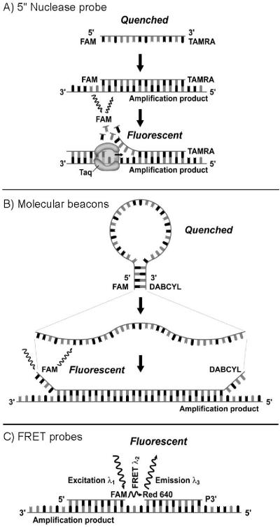
Real-time PCR has revolutionized the way clinical microbiology laboratories diagnose many human microbial infections. This testing method combines PCR chemistry with fluorescent probe detection of amplified product in the same reaction vessel. In general, both PCR and amplified product detection are completed in an hour or less, which is considerably faster than conventional PCR detection methods. Real-time PCR assays provide sensitivity and specificity equivalent to that of conventional PCR combined with Southern blot analysis, and since amplification and detection steps are performed in the same closed vessel, the risk of releasing amplified nucleic acids into the environment is negligible. The combination of excellent sensitivity and specificity, low contamination risk, and speed has made real-time PCR technology an appealing alternative to culture- or immunoassay-based testing methods for diagnosing many infectious diseases. This review focuses on the application of real-time PCR in the clinical microbiology laboratory.


Figures

FIG. 1.
FIG. 1.
Real-time probe technologies. (A) 5′ nuclease (TaqMan) probe. (B) Molecular beacon. (C) FRET hybridization probes. (Reprinted from reference with kind permission of Springer Science and Business Media.)
FIG. 2.
FIG. 2.
Melting curves obtained after PCR amplification of HSV DNA.
FIG. 3.
FIG. 3.
S.E.T.S. The inner tube will hold standard-sized swabs (approximately 5 to 8 mm in diameter, 10 of 25 mm in length) during spinning procedures. The collection tube is used for collection and storage of liquid from the swab. The screw cap is for tightly closing the collection tube.
FIG. 4.
FIG. 4.
Recovery template. The recovery template (internal control) has the same sequence as the PCR product except the probe region has been replaced with a sequence complementary to recovery template probes. FRET detection of the target DNA is with a probe labeled with the Red 640 dye in channel 2 of the LightCycler while the recovery template is detected with a probe labeled with the Red 705 dye in channel 3. A small amount of recovery template is added to the PCR and is amplified along with the target DNA by the same primers. Thus, the two reactions compete for the primers. Normally, the recovery template is amplified in all samples, including the negative control. If neither the recovery template nor target DNA is amplified, then it is assumed that inhibition of the PCR has occurred and the test for that sample is not valid. However, if target DNA is amplified but the recovery DNA template is not amplified, then it is assumed that the target DNA is present in a proportionally greater amount. In this situation, partial inhibition of the PCR may be present but the target DNA is successfully amplified or the recovery template may not be able to compete for primers and the recovery template signal may be weak or not present. When this occurs, the positive result is valid because the recovery template amplification result is unnecessary.
FIG. 5.
FIG. 5.
Work flow algorithm for processing specimens for the laboratory diagnosis of group A streptococcal infections by real-time PCR.

Similar articles

Cited by

References

    1. Abe, A., K. Inoue, T. Tanaka, J. Kato, N. Kajiyama, R. Kawaguchi, S. Tanaka, M. Yoshiba, and M. Kohara. 1999. Quantitation of hepatitis B virus genomic DNA by real-time detection PCR. J. Clin. Microbiol. 37:2899-2903. - PMC - PubMed
    1. Aberham, C., C. Pendl, P. Gross, G. Zerlauth, and M. Gessner. 2001. A quantitative, internally controlled real-time PCR assay for the detection of parvovirus B19 DNA. J. Virol. Methods 92:183-191. - PubMed
    1. Aberle, S. W., and E. Puchhammer-Stöckl. 2002. Diagnosis of herpesvirus infections of the central nervous system. J. Clin. Virol. 25(Suppl. 1):S79-85. - PubMed
    1. Aldea, C., C. P. Alvarez, L. Folgueira, R. Delgado, and J. R. Otero. 2002. Rapid detection of herpes simplex virus DNA in genital ulcers by real-time PCR using SYBR green I dye as the detection signal. J. Clin. Microbiol. 40:1060-1062. - PMC - PubMed
    1. Aliyu, S. H., M. H. Aliyu, H. M. Salihu, S. Parmar, H. Jalal, and M. D. Curran. 2004. Rapid detection and quantitation of hepatitis B virus DNA by real-time PCR using a new fluorescent (FRET) detection system. J. Clin. Virol. 30:191-195. - PubMed

MeSH terms

Substances