

Imaging findings of biliary hamartomas
- PMID: 16419165
- PMCID: PMC4320340
- DOI: 10.3748/wjg.v11.i40.6354
Imaging findings of biliary hamartomas
Abstract
Aim: To evaluate the imaging findings of biliary hamartomas (von Meyenburg complexes, VMCs) and discuss the differential diagnosis with other related diseases.
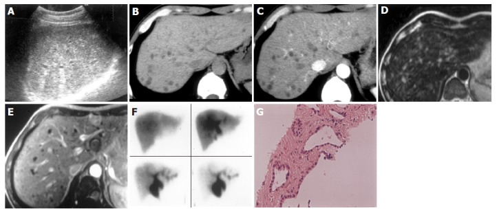
Methods: Imaging findings of biliary hamartomas on ultrasonography (US), computed tomography (CT), magnetic resonance imaging (MRI), MR cholangiopancreatography (MRCP)and hepatobiliary scintigraphy were retrospectively analyzed in six patients.
Results: On ultrasound images, five of the six cases showed multiple small hyper- and hypo-echoic lesions with comet-tail echoes, especially when magnified by US with the usage of zoom function. In all the six cases, multiple tiny hypodense lesions less than 10 mm in diameter were revealed as scattered throughout the liver with no enhancement on CT. These tiny lesions were demonstrated to be hyper- and hypo-intensity on T2- and TI-weighed images, respectively, in three patients who underwent MRI examinations. MRCP was performed in two patients, and clearly showed multiple tiny irregular- and round-shaped hyper-intensity lesions. MRCP and hepatobiliary scintigraphy showed normal appearances of intra- and extra-hepatic bile ducts in two and one patients, respectively.
Conclusion: Imaging modalities are useful in the diagnosis and differential diagnosis of VMCs. A correct diagnosis might be obtained when typical imaging findings are present even without a histological confirmation.
Figures


References
-
- Lev-Toaff AS, Bach AM, Wechsler RJ, Hilpert PL, Gatalica Z, Rubin R. The radiologic and pathologic spectrum of biliary hamartomas. AJR Am J Roentgenol. 1995;165:309–313. - PubMed
-
- Wei SC, Huang GT, Chen CH, Sheu JC, Tsang YM, Hsu HC, Chen DS. Bile duct hamartomas. A report of two cases. J Clin Gastroenterol. 1997;25:608–611. - PubMed
-
- Luo TY, Itai Y, Eguchi N, Kurosaki Y, Onaya H, Ahmadi Y, Niitsu M, Tsunoda HS. Von Meyenburg complexes of the liver: imaging findings. J Comput Assist Tomogr. 1998;22:372–378. - PubMed
-
- Cooke JC, Cooke DA. The appearances of multiple biliary hamartomas of the liver (von Meyenberg complexes) on computed tomography. Clin Radiol. 1987;38:101–102. - PubMed
-
- Mortelé B, Mortelé K, Seynaeve P, Vandevelde D, Kunnen M, Ros PR. Hepatic bile duct hamartomas (von Meyenburg Complexes): MR and MR cholangiography findings. J Comput Assist Tomogr. 2002;26:438–443. - PubMed
MeSH terms
LinkOut - more resources
Full Text Sources
