. 2005 Oct 28;11(40):6373-80.
doi: 10.3748/wjg.v11.i40.6373.

Effects of glucocorticoids on the growth and chemosensitivity of carcinoma cells are heterogeneous and require high concentration of functional glucocorticoid receptors

Affiliations

Effects of glucocorticoids on the growth and chemosensitivity of carcinoma cells are heterogeneous and require high concentration of functional glucocorticoid receptors

Yen-Shen Lu et al. World J Gastroenterol. .

Abstract

Aim: To determine how glucocorticoids (GCs) may affect the growth and chemosensitivity of common carcinoma cells.

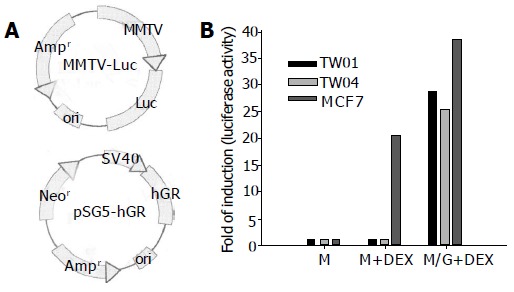
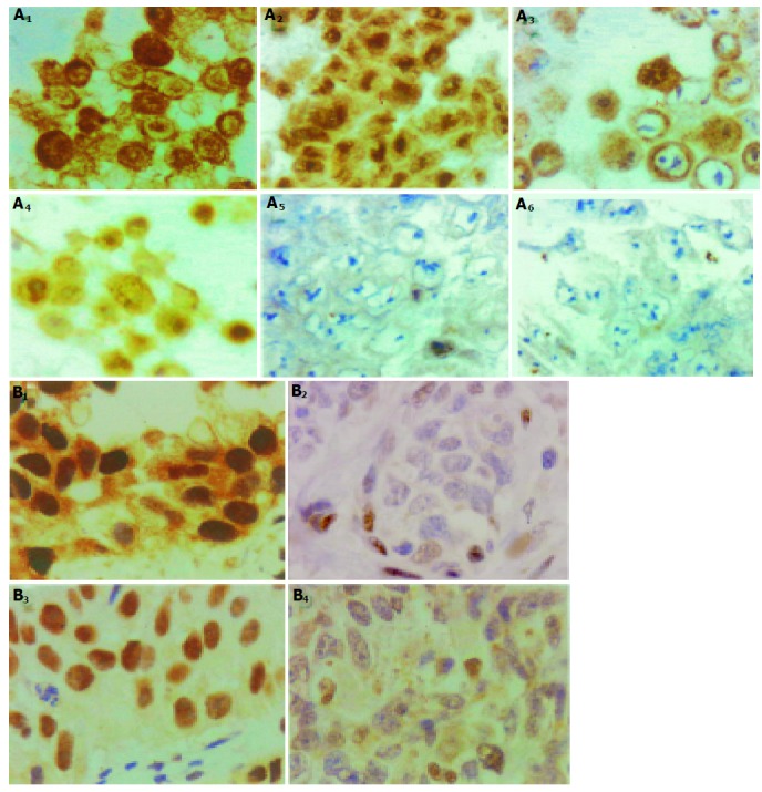
Methods: The effect of dexamethasone (DEX) on growth and chemosensitivity was assessed in 14 carcinoma cell lines. The function of GC receptors (GR) was assessed by MMTV reporter assay. Overexpression of GR was done by in vitro transfection and expression of a GR-expressing vector. Immunohistochemical stain of tissues and cells were done by PA1-511A, an anti-GR monoclonal antibody.

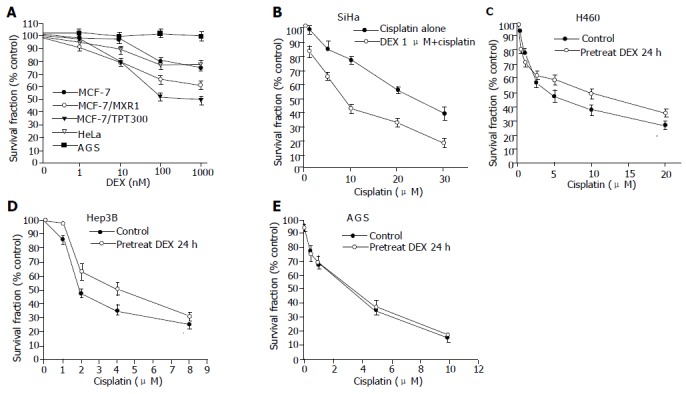
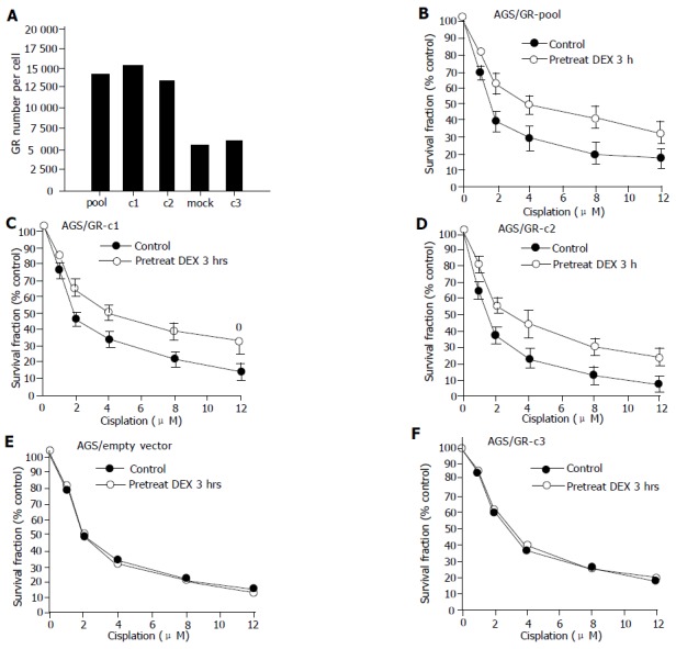
Results: DEX inhibited cell growth of four (MCF-7, MCF-7/MXR1, MCF-7/TPT300, and HeLa), increased cisplatin cytotoxicity of one (SiHa), and decreased cisplatin cytotoxicity of two (H460 and Hep3B) cell lines. The GR content of the seven cell lines affected by DEX was significantly higher than those of the seven cell lines unaffected by DEX (5.2+/-2.5 x 10(4) sites/cell vs 1.3+/-1.4 x 10(4) sites/cell, P = 0.005). Only two DEX-unresponsive cell lines (NPC-TW01 and NPC-TW04) contained high GR amounts in the range (1.9-8.1 x 10(4) sites/cell) of the seven DEX-responsive cell lines. The GR function of NPC-TW01 and NPC-TW04, however, was found to be impaired. The importance of high cellular amount of GR in mediating DEX susceptibility of the cells was further exemplified by GR dose-dependent drug resistance to cisplatin of AGS, a cell line with low GR content and was unaffected by DEX before transfection of GR-expressing vector. Immunohistochemical studies of human cancer tissues showed that 5 of the 45 (11.1%) breast cancer and 43 of the 85 (50.6%) non-small cell lung cancer had high GR contents at the ranges of the GC-responsive carcinoma cell lines.

Conclusion: The growth and chemosensitivity of human carcinomas with high GR contents may be affected by GC. However, in light of the heterogeneous and even contradictive effects of GC on these cells, routine examination of GR contents of human carcinoma tissues may not be clinically useful until other markers that help predict the ultimate effect of GC on individual patients are identified.


Figures

Figure 1
Figure 1
Effect of DEX on the growth and chemosensitivity in carcinoma cell lines. Cell numbers were measured by MTT assay and were plotted as a percentage of the control (cells not exposed to drugs); A: Growth of MCF-7, MCF-7/MXR1, MCF-7/TPT300, and HeLa cells were suppressed by DEX. Data of AGS represent the other 10 cell lines, growth of which was not affected by DEX; B: SiHa cells pretreated with DEX for 3 h were more sensitive to cisplatin; C and D: Pretreatment with DEX 1 mmol/L for 24 h diminished cisplatin cytotoxicity in H460 cells and Hep3B cells; E: Data of AGS represent the seven cell lines (AGS, N87, Caski, Hut 7, SNU1, NPC-TW01, and NPC-TW04), of which cytotoxicity of cisplatin was not affected by DEX. All values represent mean±SD of six separate wells.
Figure 2
Figure 2
Functional assay of the GR in NPC-TW01 and NPC-TW04 cells. NPC-TW01, NPC-TW04, and MCF-7 cells were transiently transfected with MMTV reporter plasmid (lanes M and M+DEX) or co-transfected with MMTV reporter plasmid and pS-hGR (lane M/G+DEX). The cells were then treated with 1 μmol/L DEX for 6 h (lanes M+DEX and M/G+DEX). Then the luciferase activity was assayed and represented in terms of folds of the induction activity of the control (lane M). All values represent mean±SD of three experiments(A,B).
Figure 3
Figure 3
Increased drug resistance to cisplatin in pS-hGR-transfected AGS. AGS cells were transfected with pS-hGR and MTT assay were performed. A: GR number measured by [3H]-labeled ligand binding assay. Pool: AGS/GR-pool; AGS cells transfected with pS-hGR, pooled cells. c1: AGS/GR-c1; AGS cells transfected with pS-hGR, single cell cloned, clone 1. c2: AGS/GR-c2; AGS cells transfected with pS-hGR, single cell cloned, clone 2. Mock: AGS/empty vector; AGS cells transfected with empty vector. c3: AGS/GR-c3; AGS cells transfected with pS-hGR, single cell cloned, clone 3; B-D: Pretreatment with DEX 1 μmol/L for 3 h diminished cisplatin cytotoxicity in AGS/GR-pool, AGS/GR-c1, and AGS/GR-c2 cells; E and F: Pretreatment with DEX 1 μmol/L for 3 h had no effect on the cisplatin cytotoxicity in AGS/empty vector cells and AGS/GR-c3. All values represent mean±SD of six separate wells.
Figure 4
Figure 4
A: Immunocytochemical stain for GR expression in representing carcinoma cell lines. (A1) and A2): SiHa cells, which had GR content about 8.1×10⁴/cell according to ligand binding assay; (A3) and (A4): HeLa cells, with GR content about 1.97×10⁴/cell; (A5) and (A6): N87 cells, with GR content about 5.0×10³/cell. (A2), (A4), and (A5): cells treated with DEX for 3 h before harvest. In (A1) and (A3), the GR immunoreactivity localized in cytoplasm. After DEX treatment, the immunoreactive GR translocalized to nuclei (A2) and (A4). The low GR content cancer cells N87 showed negligible immunoreactivity. B: Immunohistochemical stain for GR expression in human carcinoma tumor tissue samples. Non-small cell lung cancer tumor samples, with high GR expression (Ba), and low GR expression (B2). Breast cancer tumor samples, with high GR expression (B3), and low GR expression (B4).

References

    1. Frankfurt O, Rosen ST. Mechanisms of glucocorticoid-induced apoptosis in hematologic malignancies: updates. Curr Opin Oncol. 2004;16:553–563. - PubMed
    1. Wright AP, Zilliacus J, McEwan IJ, Dahlman-Wright K, Almlöf T, Carlstedt-Duke J, Gustafsson JA. Structure and function of the glucocorticoid receptor. J Steroid Biochem Mol Biol. 1993;47:11–19. - PubMed
    1. Schwartzman RA, Cidlowski JA. Glucocorticoid-induced apoptosis of lymphoid cells. Int Arch Allergy Immunol. 1994;105:347–354. - PubMed
    1. McEwan IJ, Wright AP, Gustafsson JA. Mechanism of gene expression by the glucocorticoid receptor: role of protein-protein interactions. Bioessays. 1997;19:153–160. - PubMed
    1. Rogatsky I, Trowbridge JM, Garabedian MJ. Glucocorticoid receptor-mediated cell cycle arrest is achieved through distinct cell-specific transcriptional regulatory mechanisms. Mol Cell Biol. 1997;17:3181–3193. - PMC - PubMed

Publication types