. 2006 Feb;4(2):459-68.
doi: 10.1111/j.1538-7836.2006.01723.x.

Function-blocking antithrombospondin-1 monoclonal antibodies

Affiliations

Function-blocking antithrombospondin-1 monoclonal antibodies

D S Annis et al. J Thromb Haemost. 2006 Feb.

Abstract

Background: Thrombospondin-1 (TSP-1) has been implicated in many different processes based in part on inhibitory activities of anti-TSP-1 monoclonal antibodies (mAbs).

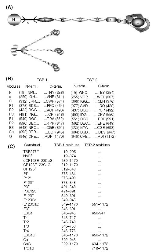
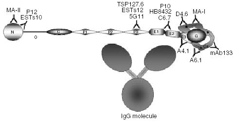
Objective: To map epitopes of 13 anti-TSP-1 mAbs to individual modules or groups of modules spanning TSP-1 and the closely related TSP-2 homolog.

Results: The mapping has led to assignment or reassignment of the epitopes of four mAbs, refinement of the epitopes of six mAbs, and confirmation of the epitopes of the remaining three mAbs. ESTs10, P12, and MA-II map to the N-terminal domain; 5G11, TSP127.6, and ESTs12 to the third properdin module; C6.7, HB8432, and P10 to epidermal growth factor (EGF)-like modules 1 and/or 2; and A6.1, mAb133, MA-I, and D4.6 to the calcium-binding wire module. A6.1, which recognizes a region of the wire that is identical in mouse and human TSP-1, reacts with TSP-1 from both species, and also reacts weakly with human TSP-2. Two other mouse antihuman TSP-1 mAbs, A4.1 and D4.6, also react with mouse TSP-1.

Conclusions: Consideration of previous literature and mapping of epitopes of inhibitory mAbs suggest that biological activities are present throughout TSP-1, including the EGF-like modules that have not been implicated in the past. Because the epitopes for 10 of the antibodies likely are within 18 nm of one another in calcium-replete TSP-1, some of the inhibitory effects may result from steric hindrance. Such seems to be the case for mAb133, which binds the calcium-binding wire but is still able to interfere with the activation of latent TGF-beta by the properdin modules.


Figures

Fig. 1
Fig. 1
Expression of thrombospondin modules and nomenclature. Modules or groups of modules spanning the human TSP-1 and -2 molecules were expressed in baculovirus. (A) Constructs that include the oligomerization domain are trimeric. The TSP monomer consists of an N-terminal module (N), an oligomerization domain (o), a procollagen module (C), three properdin or type I modules (P), three EGF-like or type II modules (E), several type III repeats or calcium-wire module (Ca), and a lectin-like globular module or G domain (G). This schematic is a representation of TSP in the calcium-replete form, showing the Ca-wire leaving the E3 module and looping around the G domain, forming the C-terminal globe. (B) The three N- and C-terminal residues for each module from TSP-1 and -2 are listed. The residue number is in parentheses. Numbering begins at the initiating methionine, with residue 19 being the first amino acid, and residues 1170 and 1172 from TSP-1 and -2 respectively, being the end of the mature proteins. (C) Tabulation of the recombinant modular arrays and the residues they encompass. The proteins are named based on their modular components. The exceptions are; TSP277, which is the first 277 residues (19–295) of TSP-1; and the E3Ca-1 truncations, Tr1, Tr2, Tr3, and Tr4, which include the E3 module and 1, 2, 3, or four calcium-binding motifs [25] from Ca. Modules derived from TSP-1 are followed by -1, and those derived from TSP-2 are followed by -2. All constructs are produced in coco with no thrombin cleavage site in the linker between the end of the TSP sequence and six his tag unless noted: produced in coco with a thrombin cleavage site in the linker (+), proteins not produced in coco with no tags or linkers (**), and proteins expressed in the GELEX system (*). P3-1 was produced in both coco and GELEX.
Fig. 2
Fig. 2
ESTs10 recognizes the N module, and C6.7 recognizes EGF domains 1 and/or 2. Approximate molar equivalents of TSP-derived proteins were resolved in reducing conditions on a 14% gel for ESTs10, and under non-reducing conditions on a 10% SDS–PAGE gel for C6.7. The proteins were transferred to PVDF membrane and immuno-blotted. (A) Western blot of ESTs10, lane (a) CP123E123CaG-1, (b) NoC-1 and (c) TSP277. (B) Western blot of C6.7, lane (a) NoC-1, (b) CP123-1, (c) P3E123-1, (d) E123-1, (e) E3CaG-1, (f) E3Ca-1, and (g) CP123E123CaG-1.
Fig. 3
Fig. 3
The epitope for A6.1 is in the 1st calcium-binding motif of Ca (residues 692–717). Approximate molar equivalents of TSP-derived proteins were resolved under the reducing conditions on a 14% SDS–PAGE gel, transferred to PVDF membrane and immuno-blotted. Western blot of A6.1, lane (a) E123-1, (b) E3Ca-1, (c) Tr1 (N700), (d) Tr2 (N700), (e) Tr3 (N700), (f) Tr4 (N700), and (g) Tr1 (S700).
Fig. 4
Fig. 4
Schematic representation of epitope placement along the TSP-1 molecule. The epitopes for MA-II, P12, and ESTs10 are located in the N module; 5G11, ESTs12, and TSP127.6 in the P3 module; C6.7, HB8432, and P10 in modules E12; A4.1 in the E3 module; and A6.1, mAb133, MAI, and D4.6 in the calcium-wire. The E123CaG portion of the molecule is drawn based on a partial structure of this region of hTSP-1 [4] and the structure of E123CaG from hTSP-2 [5]. The E3CaG complex is approximately 7 × 5.5 nm, with the G domain being approximately 4 × 3 nm, and the EGF-like modules 3 × 1.5 nm. The size of the properdin modules is based on the crystal structure published by Tan [35] and is approximately 5 × 1.2 nm. We have assumed the procollagen module to be similar in proportions to the properdins. The o domain is based on the structure of the coil-coil region of cartilage matrix protein [36] with a length of 5 nm. The N module is about 4 nm in diameter, and is based on the structure of the G modules of laminin [37]. A representation of an IgG molecule based on crystal structures and cryo-electron tomography [–40] is included to show the relative size compared to TSP-1. The Fc stem (larger globe) is approximately 6.2 × 7 nm, the Fab arms (smaller globes) are approximately 4.7 × 7 nm, and the distances between the three globes are 2–2.5 nm.

References

    1. Adams JC. Thrombospondins: multifunctional regulators of cell interactions. Annu Rev Cell Dev Biol. 2001;17:25–51. - PubMed
    1. Mosher DF, Huwiler KG, Misenheimer TM, Annis DS. Expression of recombinant matrix components using baculoviruses. Methods Cell Biol. 2002;69:69–81. - PubMed
    1. Calzada MJ, Annis DS, Zeng B, Marcinkiewicz C, Banas B, Lawler J, Mosher DF, Roberts DD. Identification of novel beta-1 integrin binding sites in the type 1 and type 2 repeats of thrombospondin-1. J Biol Chem. 2004;279:41734–43. - PubMed
    1. Kvansakul M, Adams JC, Hohenester E. Structure of a thrombospondin C-terminal fragment reveals a novel calcium core in the type 3 repeats. Embo J. 2004;23:1223–33. - PMC - PubMed
    1. Carlson CB, Bernstein AD, Annis DS, Misenheimer TM, Hannah BL, Mosher DF, Keck JL. Structure of the calcium-rich signature domain of human thrombospondin-2. Nat Struct Mol Biol. 2005;12:910–14. - PMC - PubMed

Publication types

MeSH terms