

Motor system activation after subcortical stroke depends on corticospinal system integrity
- PMID: 16421171
- PMCID: PMC3717515
- DOI: 10.1093/brain/awl002
Motor system activation after subcortical stroke depends on corticospinal system integrity
Abstract
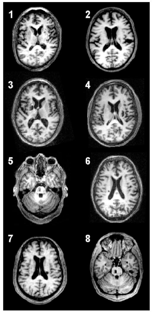
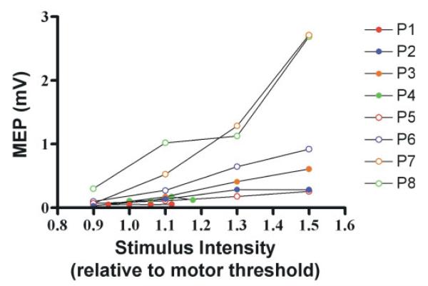
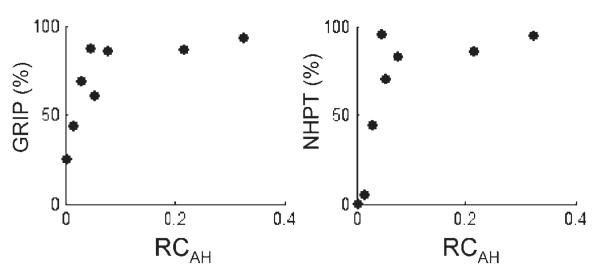
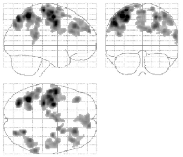
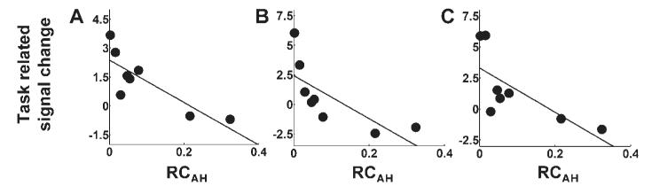
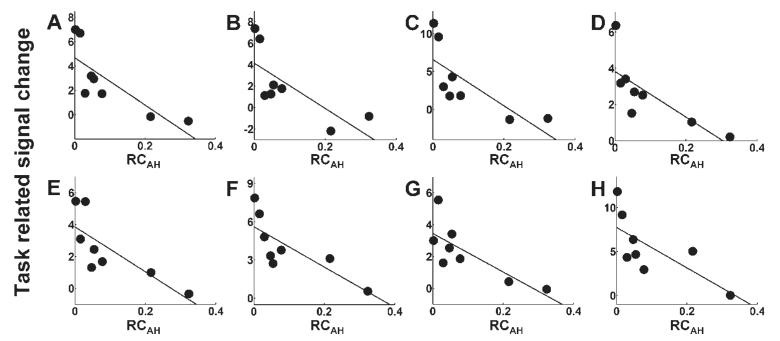
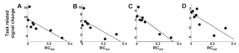
Movement-related brain activation patterns after subcortical stroke are characterized by relative overactivations in cortical motor areas compared with controls. In patients able to perform a motor task, overactivations are greater in those with more motor impairment. We hypothesized that recruitment of motor regions would shift from primary to secondary motor networks in response to impaired functional integrity of the corticospinal system (CSS). We measured the magnitude of brain activation using functional MRI during a motor task in eight chronic subcortical stroke patients. CSS functional integrity was assessed using transcranial magnetic stimulation to obtain stimulus/response curves for the affected first dorsal interosseus muscle, with a shallower gradient representing increasing disruption of CSS functional integrity. A negative correlation between the gradient of stimulus/response curve and magnitude of task-related brain activation was found in several motor-related regions, including ipsilesional posterior primary motor cortex [Brodmann area (BA) 4p], contralesional anterior primary motor cortex (BA 4a), bilateral premotor cortex, supplementary motor area, intraparietal sulcus, dorsolateral prefrontal cortex and contralesional superior cingulate sulcus. There were no significant positive correlations in any brain region. These results suggest that impaired functional integrity of the CSS is associated with recruitment of secondary motor networks in both hemispheres in an attempt to generate motor output to spinal cord motoneurons. Secondary motor regions are less efficient at generating motor output so this reorganization can only be considered partially successful in reducing motor impairment after stroke.
Figures
References
-
- Andersson JL, Hutton C, Ashburner J, Turner R, Friston K. Modeling geometric deformations in EPI time series. Neuroimage. 2001;13:903–19. - PubMed
-
- Benecke R, Meyer BU, Freund HJ. Reorganisation of descending motor pathways in patients after hemispherectomy and severe hemispheric lesions demonstrated by magnetic brain stimulation. Exp Brain Res. 1991;83:419–26. - PubMed
-
- Binkofski F, Fink GR, Geyer S, Buccino G, Gruber O, Shah NJ, et al. Neural activity in human primary motor cortex areas 4a and 4p is modulated differentially by attention to action. J Neurophysiol. 2002;88:514–9. - PubMed
-
- Boroojerdi B, Battaglia F, Muellbacher W, Cohen LG. Mechanisms influencing stimulus-response properties of the human corticospinal system. Clin Neurophysiol. 2001;112:931–7. - PubMed
