. 2006 Feb;133(4):697-709.
doi: 10.1242/dev.02241. Epub 2006 Jan 18.

The C. elegans Myt1 ortholog is required for the proper timing of oocyte maturation

Affiliations

The C. elegans Myt1 ortholog is required for the proper timing of oocyte maturation

Anna E Burrows et al. Development. 2006 Feb.

Abstract

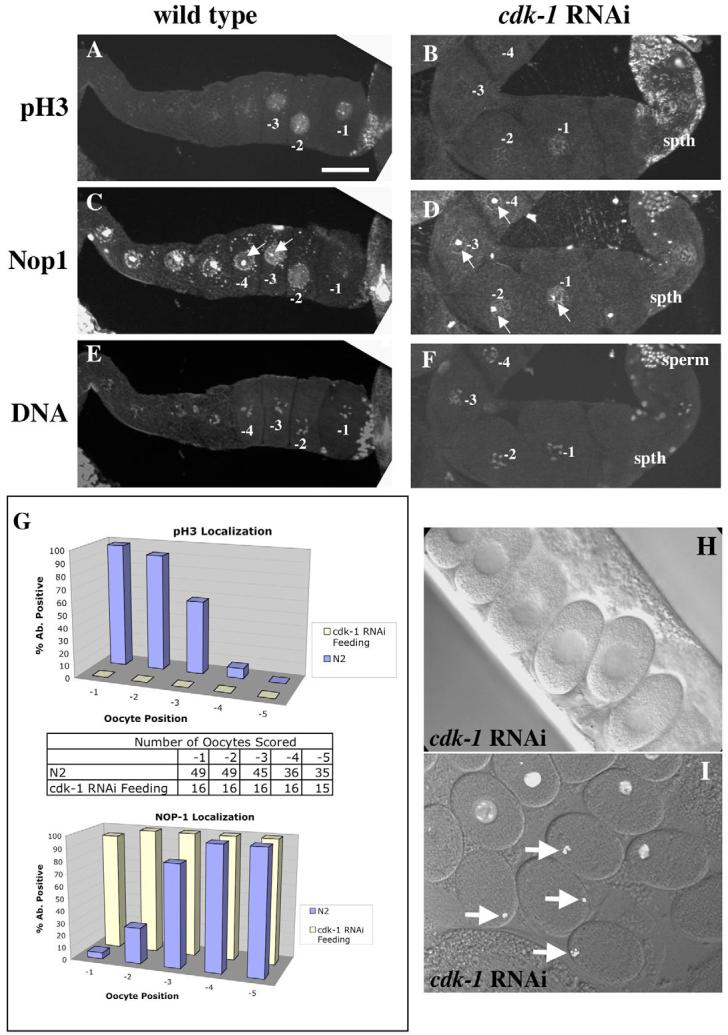
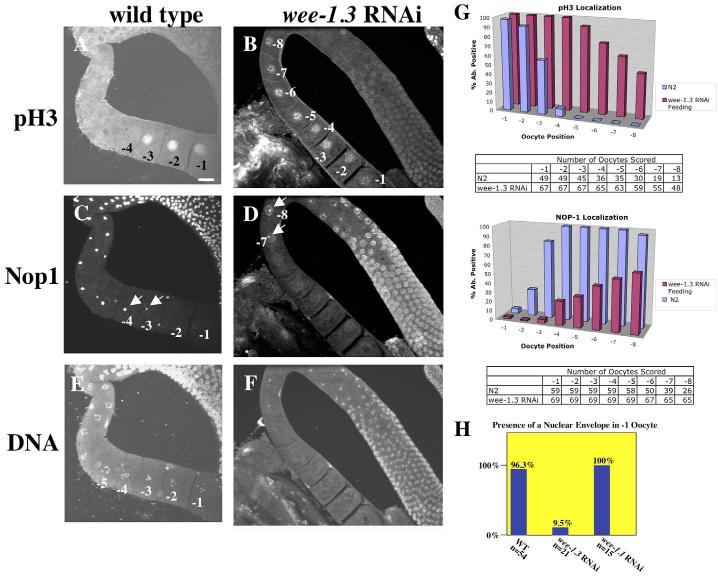
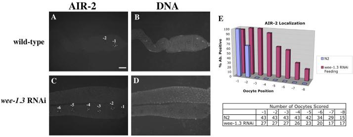
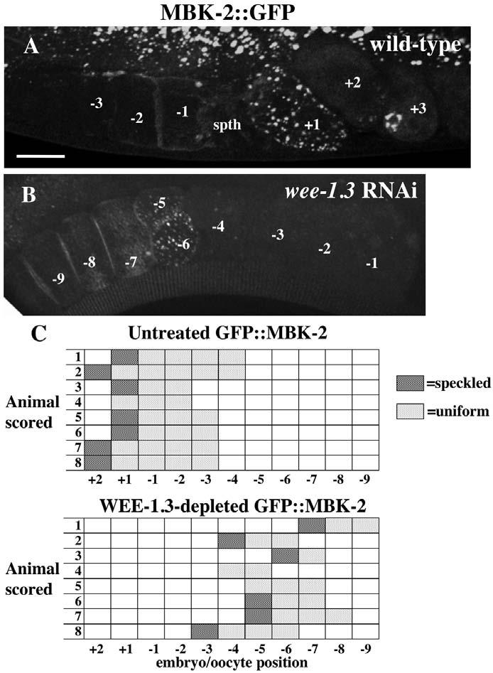
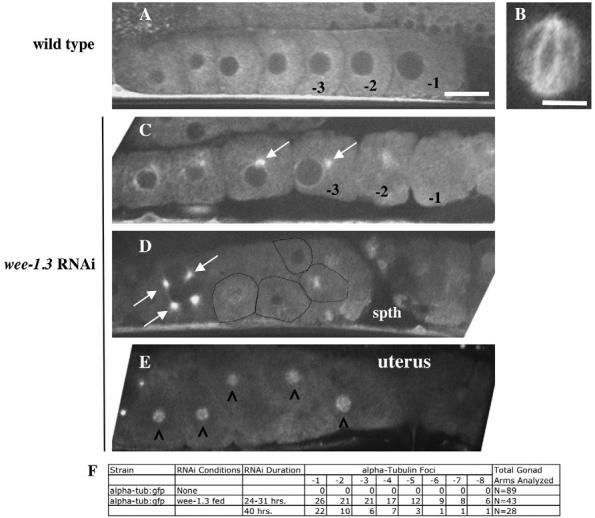
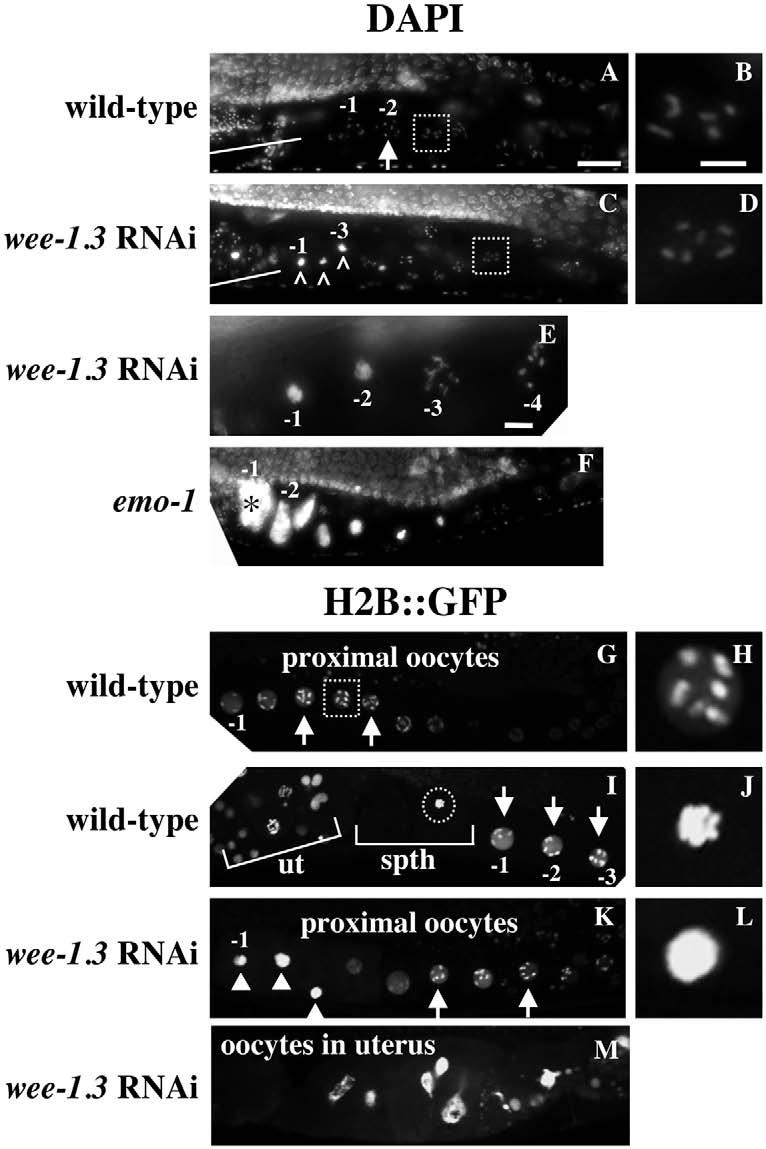
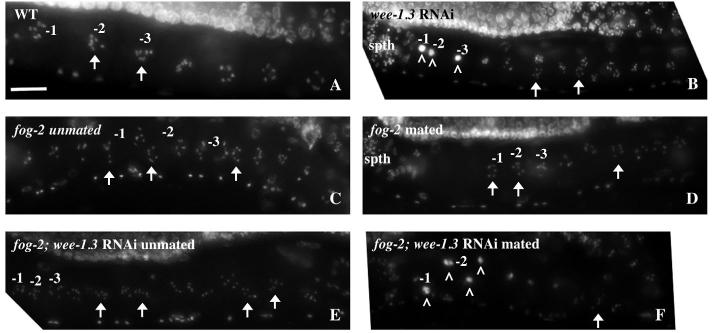
Maturation promoting factor (MPF), a complex of cyclin-dependent kinase 1 and cyclin B, drives oocyte maturation in all animals. Mechanisms to block MPF activation in developing oocytes must exist to prevent precocious cell cycle progression prior to oocyte maturation and fertilization. This study sought to determine the developmental consequences of precociously activating MPF in oocytes prior to fertilization. Whereas depletion of Myt1 in Xenopus oocytes causes nuclear envelope breakdown in vitro, we found that depletion of the Myt1 ortholog WEE-1.3 in C. elegans hermaphrodites causes precocious oocyte maturation in vivo. Although such oocytes are ovulated, they are fertilization incompetent. We have also observed novel phenotypes in these precociously maturing oocytes, such as chromosome coalescence, aberrant meiotic spindle organization, and the expression of a meiosis II post-fertilization marker. Furthermore, co-depletion studies of CDK-1 and WEE-1.3 demonstrate that WEE-1.3 is dispensable in the absence of CDK-1, suggesting that CDK-1 is a major target of WEE-1.3 in C. elegans oocytes.


Figures

Fig. 1
Fig. 1
CDK-1 depletion impairs oocyte maturation. (A-F) Wild-type (WT; A,C,E) and CDK-1-depleted (B,D,F) animals stained with pH3 antibodies (A,B), NOP1 antibodies (C,D), and TOTO-3 to visualize DNA (E,F). Scale bar: 20 μm. (G) Quantitation of pH3- and NOP1-positive nuclei by oocyte position in the proximal gonad. In between the graphs is a table showing the number of oocytes analyzed. N2 is the wild-type strain. (H) DIC image of one-cell arrested embryos in the uterus of a CDK-1-depleted adult. (I) One-cell arrested embryos dissected from the uterus of a histone H2B::GFP transgenic animal depleted of CDK-1. The four embryos marked by arrows show the congression of the oocyte chromosomes, similar to metaphase of meiosis I. Older embryos remain arrested as one-cell embryos but their chromosomes have decondensed. spth, spermatheca.
Fig. 2
Fig. 2
WEE-1.3 depletion causes precocious oocyte maturation. (A-F) Wild-type (A,C,E) and WEE-1.3-depleted (B,D,F) animals stained with pH3 antibodies (A,B), NOP1 antibodies (C,D), and TOTO-3 (E,F). Scale bar: 20 μm. (G) Quantitation of pH3- and NOP1-positive nuclei by oocyte position in the proximal gonad. Below each graph is a table showing the number of oocytes analyzed. (H) Quantitation of the presence of an intact NE in the −1 oocyte from adult hermaphrodites untreated (WT) or depleted of WEE-1.3 or WEE-1.1 (control). Animals were analyzed 16-20 hours post-injection. n, number of gonad arms analyzed.
Fig. 3
Fig. 3
WEE-1.3 depletion causes an increase in the number of oocytes that stain for AIR-2. (A-D) Wild-type (A,B) and WEE-1.3-depleted (C,D) animals stained with an AIR-2 antibody (A,C) and TOTO-3 (B,D). Scale bar: 20 μm. (E) Quantitation of AIR-2-positive nuclei by oocyte position in the proximal gonad. Below the graph is a table showing the number of oocytes analyzed.
Fig. 4
Fig. 4
GFP::MBK-2 aggregates in WEE-1.3-depleted oocytes. (A,B) Fluorescence images of wild-type GFP::MBK-2 (A) and WEE-1.3-depleted (B) oviducts. The speckled pattern is present in the +1 embryo in A and in the −5 and −6 embryos in B. Scale bar: 20 μm. (C,D) The position of each oocyte with a speckled GFP pattern and a uniformly cortical pattern is indicated for eight representative animals of each. n=19 for the total number of untreated GFP::MBK-2 animals, and n=22 for WEE-1.3 depleted. spth, spermatheca.
Fig. 5
Fig. 5
CDK-1 depletion suppresses the infertility of WEE-1.3-depleted animals. (A-D) DIC images of uteri from wild-type (A), CDK-1-depleted (B), WEE-1.3-depleted (C), and co-depleted (D) adult hermaphrodites. Wild-type animals have embryos of all stages (A), whereas CDK-1-depleted animals contain only one-cell arrested embryos (B). WEE-1.3-depleted animals have uteri containing unfertilized ‘mushy’ oocytes (C), whereas the co-depleted animals contain only one-cell arrested embryos (D). Embryos are ∼50 μm in length. (E,F) Quantitation of (E) pH3- and (F) NOP1-positive nuclei by oocyte position in the proximal gonad. Below each graph is a table showing the number of oocytes analyzed for each RNAi condition.
Fig. 6
Fig. 6
WEE-1.3 depletion results in the appearance of aberrant microtubules in developing oocytes. (A-E) Live α-tubulin::GFP animals were subjected to wee-1.3 RNAi. (A,B) Untreated animals. (A) Oocytes with typical cytoplasmic tubulin cytoskeletons. (B) A wild-type meiotic spindle; such a structure is normally observed in the oocyte upon fertilization in the spermatheca, or in the +1 embryo in the uterus, and is never observed in WEE-1.3-depleted oocytes in the spermatheca or uterus. (C-E) WEE-1.3-depleted oocytes in the oviduct (C,D) or the uterus (E). White arrows mark the perinuclear tubulin foci. Oocytes in D are outlined in black. (E) Tubulin clouds (marked by carets) form around the coalesced chromosomes of WEE-1.3-depleted oocytes that fail to be fertilized but nonetheless end up in the uterus. The spermatheca is to the right in each gonad shown. Scale bars: in A, 20 μm for A,C-E; 10 μm in B. (F) Quantitation of the number of oocytes with tubulin foci scored by oocyte position in the proximal gonad. spth, spermatheca.
Fig. 7.
Fig. 7.
Oocyte chromosomes do not maintain a diakinetic arrangement upon WEE-1.3 depletion. (A-F) Wild-type (A,B), WEE-1.3-depleted (C-E), and emo-1 (F) animals were DAPI stained to visualize oocyte chromosomes. Arrows and white boxes mark the normal diakinetic arrangement of chromosomes in growing oocytes; those marked with a white box are enlarged in B,D. White lines mark the spermatheca and the highly condensed sperm chromosomes. Carets (^) in C mark the most proximal oocyte chromosomes that have coalesced. (E) The −3 oocyte has stringy chromosomes and is flanked by normal diakinetic oocytes to the right and oocytes with coalesced chromosomes to the left. Asterisk in F marks an endoreplicating oocyte. (G-M) Images of live, untreated (G-J) or WEE-1.3-depleted (K-M) histone H2B::GFP transgenic animals. Diakinetic chromosomes are apparent in wild-type (G-I) and WEE-1.3-depleted (K) animals, and are marked with arrows and white boxes; the one in the white box in G is enlarged in H. The chromosomes of a wild-type fertilized oocyte congress during metaphase I (marked by a circle in I and enlarged in J). The coalesced chromosomes of WEE-1.3-depleted oocytes are marked by white arrowheads (K) and one is enlarged in L. (M) The uterus of a WEE-1.3-depleted animal in which the oocyte chromosomes have begun to endoreplicate. ut, uterus; spth, spermatheca. Scale bars: in A, 20 μm for A,C,F,G,I,K,M; in B, 5 μm for B,D,H,J,L; in E, 5 μm.
Fig. 8
Fig. 8
The chromosome coalescence phenotype of WEE-1.3-depleted oocytes is dependent on the presence of sperm. Diakinetic chromosomes (arrows) of oocytes in the proximal gonad are evident in DAPI-stained wild-type hermaphrodites (A), in unmated (C) or mated (D) fog-2 females, and in unmated WEE-1.3-depleted fog-2 females (E). Although the oocytes in fog-2 animals remain in diakinesis (C-E), the chromosomes of WEE-1.3-depleted fog-2 oocytes coalesce into one mass upon the introduction of sperm (F). This phenotype appears identical to that observed in wild-type hermaphrodites depleted of WEE-1.3 (B). The white carets (^) mark the oocyte chromosomes that have coalesced (B,F). The spermatheca (spth) is to the left in each panel. Scale bar: 20 μm.

References

    1. Albertson DG, Thomson JN. Segregation of holocentric chromosomes at meiosis in the nematode, Caenorhabditis elegans. Chrom. Res. 1993;1:15–26. - PubMed
    1. Aris JP, Blobel G. Identification and characterization of a yeast nucleolar protein that is similar to a rat liver nucleolar protein. J. Cell Biol. 1988;107:17–31. - PMC - PubMed
    1. Bishop JD, Schumacher JM. Phosphorylation of the carboxyl terminus of inner centromere protein (INCENP) by the Aurora B Kinase stimulates Aurora B kinase activity. J. Biol. Chem. 2002;277:27577–27580. - PMC - PubMed
    1. Bishop JD, Han Z, Schumacher JM. The Caenorhabditis elegans Aurora B kinase AIR-2 phosphorylates and is required for the localization of a BimC kinesin to meiotic and mitotic spindles. Mol. Biol. Cell. 2005;16:742–756. - PMC - PubMed
    1. Boxem M, Srinivasan DG, van den Heuvel S. The Caenorhabditis elegans gene ncc-1 encodes a cdc2-related kinase required for M phase in meiotic and mitotic cell divisions, but not for S phase. Development. 1999;126:2227–2239. - PubMed

Publication types

MeSH terms

LinkOut - more resources