. 2006 Apr;17(4):1971-84.
doi: 10.1091/mbc.e05-10-0956. Epub 2006 Jan 18.

A genetic dissection of Aip1p's interactions leads to a model for Aip1p-cofilin cooperative activities

Affiliations

A genetic dissection of Aip1p's interactions leads to a model for Aip1p-cofilin cooperative activities

Michael G Clark et al. Mol Biol Cell. 2006 Apr.

Abstract

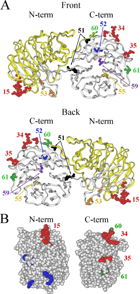
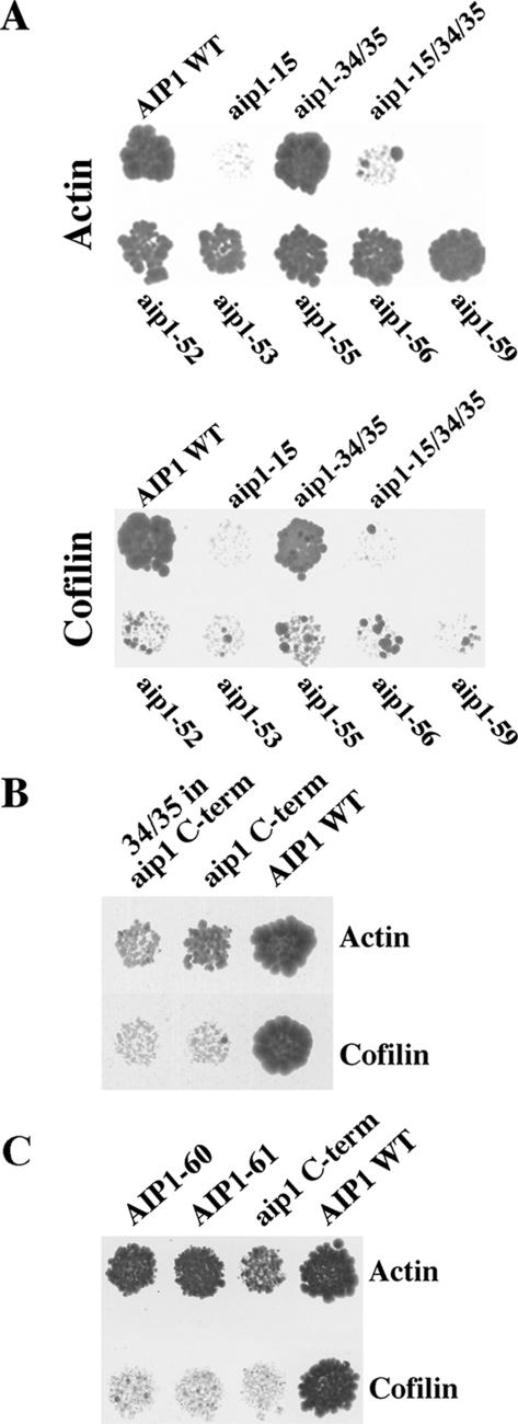
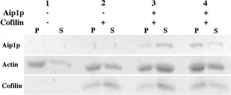
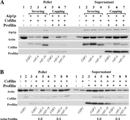
Actin interacting protein 1 (Aip1p) and cofilin cooperate to disassemble actin filaments in vitro and are thought to promote rapid turnover of actin networks in vivo. The precise method by which Aip1p participates in these activities has not been defined, although severing and barbed-end capping of actin filaments have been proposed. To better describe the mechanisms and biological consequences of Aip1p activities, we undertook an extensive mutagenesis of AIP1 aimed at disrupting and mapping Aip1p interactions. Site-directed mutagenesis suggested that Aip1p has two actin binding sites, the primary actin binding site lies on the edge of its N-terminal beta-propeller and a secondary actin binding site lies in a comparable location on its C-terminal beta-propeller. Random mutagenesis followed by screening for separation of function mutants led to the identification of several mutants specifically defective for interacting with cofilin but still able to interact with actin. These mutants suggested that cofilin binds across the cleft between the two propeller domains, leaving the actin binding sites exposed and flanking the cofilin binding site. Biochemical, genetic, and cell biological analyses confirmed that the actin binding- and cofilin binding-specific mutants are functionally defective, whereas the genetic analyses further suggested a role for Aip1p in an early, internalization step of endocytosis. A complementary, unbiased molecular modeling approach was used to derive putative structures for the Aip1p-cofilin complex, the most stable of which is completely consistent with the mutagenesis data. We theorize that Aip1p-severing activity may involve simultaneous binding to two actin subunits with cofilin wedged between the two actin binding sites of the N- and C-terminal propeller domains.


Figures

Figure 1.
Figure 1.
Aip1p mutagenesis reveals actin and cofilin binding footprints on Aip1p. (A) Aip1p is shown from a front (top) and back (bottom) view with residues changed by aip1 mutant alleles highlighted. Residues involved in the actin interaction are highlighted in red. The region expressed by the aip1-56 truncation mutant, which interacts with actin but not cofilin, is colored in yellow. Cofilin-specific alleles are shown in orange, black, blue, purple, and brown. (B) Sides views of the Aip1p N-terminal (left) and C-terminal (right) propellers depict analogous actin binding domains. Red residues represent loss-of-function cluster charged-to-alanine mutations involved in the actin interaction. Green residues represent randomly selected gain-in-function mutants that apparently increased the Aip1p-actin two-hybrid interaction. Blue residues represent the S. cerevisiae equivalents of site-directed mutants generated in C. elegans AIP1 (UNC-78) that are defective for actin filament disassembly in vitro (Mohri et al., 2004).
Figure 2.
Figure 2.
Two-hybrid analysis of aip1 mutants. (A) Yeast two-hybrid interactions between mutants of Aip1p and actin (top) or cofilin (bottom) were measured based on activation of expression of the HIS3 reporter (growth on minimal media containing 100 mM 3-AT [top] and 50 mM 3-AT [bottom]). (B) The C-terminal propeller of aip1p (aip1 C-term) interacts by yeast two-hybrid with actin but not with cofilin (10 mM 3-AT). This interaction is severely impaired by addition of the aip1-34/35 mutations. (C) Two spontaneously generated mutants on the aip1 C-term (aip1-60 and aip1-61) increase the two-hybrid interaction with actin.
Figure 3.
Figure 3.
Molecular docking of the Aip1p-cofilin complex. (A) The predicted protein complex between Aip1p (green) and cofilin (orange) as the result of molecular docking studies. The contacting interfaces of the two proteins are rendered as a transparent surface to illustrate their complementarities. (B) The specific residues found to affect Aip1p-cofilin binding have been labeled and rendered as spheres. Yellow residues represent mutations that disrupt cofilin's two-hybrid interaction with Aip1p but not actin. Purple residues represent mutations that disrupt Aip1p's two-hybrid interaction with cofilin but not actin. This figure was created using VMD (Humphrey et al., 1996).
Figure 4.
Figure 4.
Biochemical analysis reveals a severing defect for the aip1-15 mutation. Polymerized actin filaments (2.5 μM) were incubated with or without cofilin and GST-Aip1p (lane 3) or GST-aip1-15p (lane 4) for 10 min. The ratios of actin/cofilin and actin/Aip1p were 1:1 and 20:1, respectively. After high-speed centrifugation (360,000 × g), the extent of filament disassembly was determined based on the amount of actin that remained in the pellet fraction (F-actin) versus the supernatant fraction (G-actin), as detected by SDS-PAGE and SYPRO Ruby staining.
Figure 5.
Figure 5.
Biochemical capping assays demonstrate that cofilin-specific disassembly defects are Aip1p independent. In all cases, actin filament disassembly was measured by high-speed centrifugation (360,000 × g) followed by SDS-PAGE and SYPRO Ruby staining to compare the relative amounts of actin in the pellet (F-actin) versus actin in the supernatant (G-actin). (A) The cof1p mutants were tested for actin filament-severing and -capping defects in the presence of Aip1p. Polymerized actin filaments (2 μM) were incubated with or without cofilin, Aip1p, and profilin (where applicable). In lanes 2-4 (severing assay), the ratios of actin/cofilin and actin/Aip1p were 1:1 and 20:1, respectively. In lanes 5-7 (capping assay), the ratios of actin/cofilin, actin/Aip1p, and actin/profilin added were 5:1, 20:1, and 1:2, respectively. Lanes 2 and 5 contained wild-type cofilin. Lanes 3 and 6 contained cof1-4p. Lanes 4 and 7 contained cof1-19p. These data show that cof1-4p and cof1-19p have different defects depending on the method used to measure actin filament dynamics. (B) cof1p mutants were tested for actin filament-capping defects without Aip1p to determine whether the defects observed in 6A are Aip1p dependent. Polymerized actin filaments (2 μM) were incubated with or without cofilin and profilin. The actin/cofilin ratio is 5:1. The ratios for actin/profilin added were 1:2 (lanes 4-6) or 2:1 (lanes 7-9). Lanes 4 and 7 contained wild-type cofilin. Lanes 5 and 8 contained cof1-4p. Lanes 6 and 9 contained cof1-19p. These data show that under these conditions differences in assembly at the barbed end are Aip1p independent but cofilin dependent and differentially effected by cof1-4p versus cof1-19p. cof1-4p is more defective in the capping assay than cof1-19p, whereas cof1-19p is more defective than cof1-4p for Aip1p-induced severing.
Figure 6.
Figure 6.
Physical and genetic interactions among cortical patch-localized genes and gene products. Dotted lines represent physical interactions between gene products. Dashed lines represent synthetic sick genetic interactions between null alleles or site-directed mutant alleles (cofilin). Solid lines represent synthetic lethal genetic interactions. Data obtained from the S. cerevisiae Genome Database (Balakrishnan, 2005) and this work.

References

    1. Amatruda, J. F., Cannon, J. F., Tatchell, K., Hug, C., and Cooper, J. A. (1990). Disruption of the actin cytoskeleton in yeast capping protein mutants. Nature 344, 352-354. - PubMed
    1. Amatruda, J. F., Gattermeir, D. J., Karpova, T. S., and Cooper, J. A. (1992). Effects of null mutations and overexpression of capping protein on morphogenesis, actin distribution and polarized secretion in yeast. J. Cell Biol. 119, 1151-1162. - PMC - PubMed
    1. Amberg, D. C., Basart, E., and Botstein, D. (1995). Defining protein interactions with yeast actin in vivo. Nat. Struct. Biol. 2, 28-35. - PubMed
    1. Amberg, D. C., Burke, D. J., and Strathern, J. N. (2005). Methods in Yeast Genetics: A Cold Spring Harbor Laboratory Course Manual, Cold Spring Harbor, NY: Cold Spring Harbor Laboratory Press.
    1. Baggett, J. J., and Wendland, B. (2001). Clathrin function in yeast endocytosis. Traffic 2, 297-302. - PubMed

Publication types

MeSH terms