Comparative Study
. 2006 Jan 18;26(3):841-50.
doi: 10.1523/JNEUROSCI.3523-05.2006.

Selective attenuation of afferent synaptic transmission as a mechanism of thalamic deep brain stimulation-induced tremor arrest

Affiliations
Comparative Study

Selective attenuation of afferent synaptic transmission as a mechanism of thalamic deep brain stimulation-induced tremor arrest

Trent R Anderson et al. J Neurosci. .

Abstract

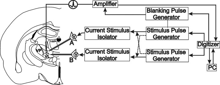
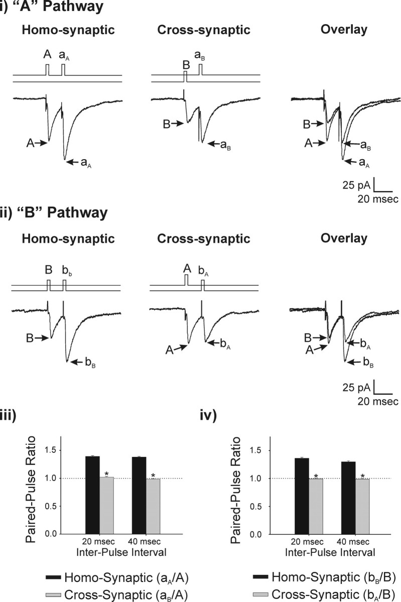
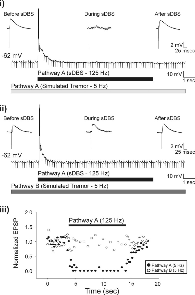
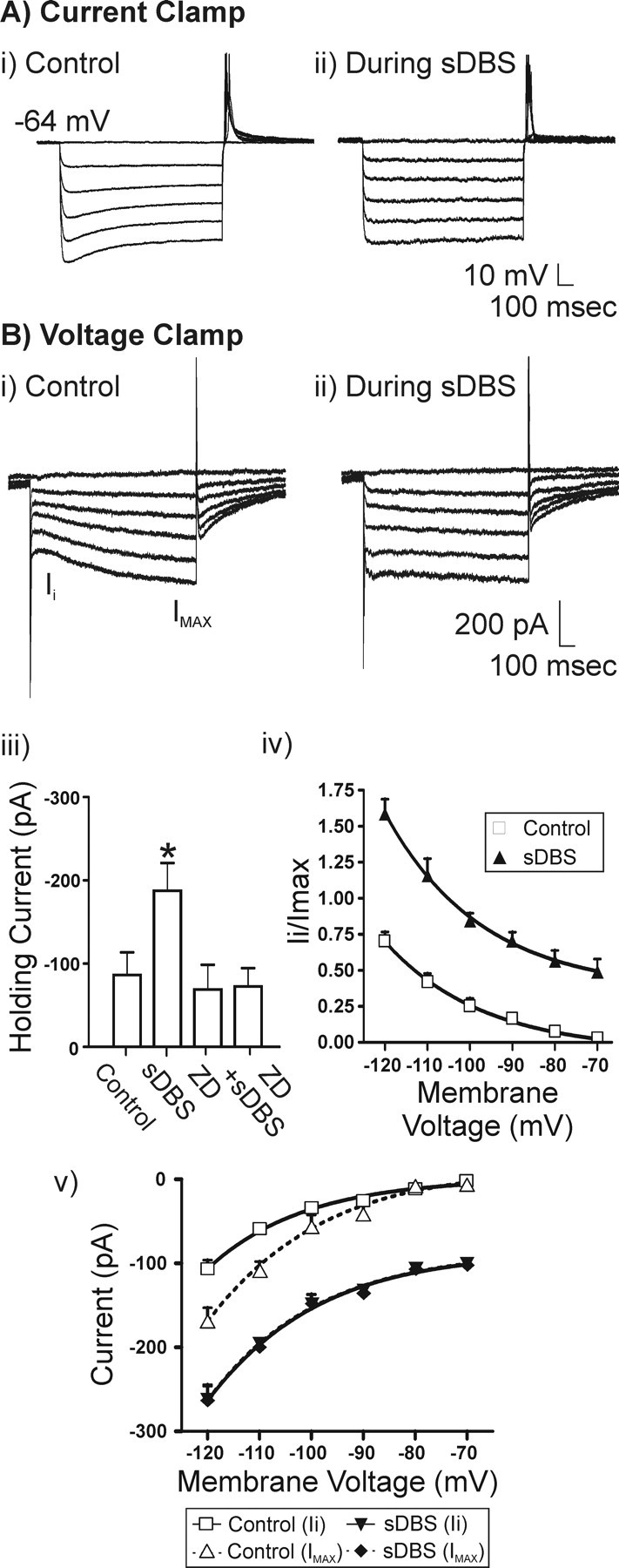
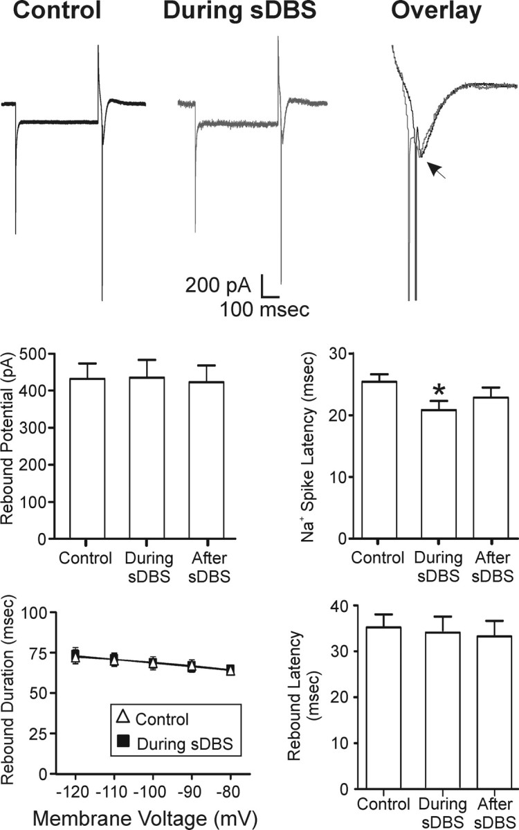
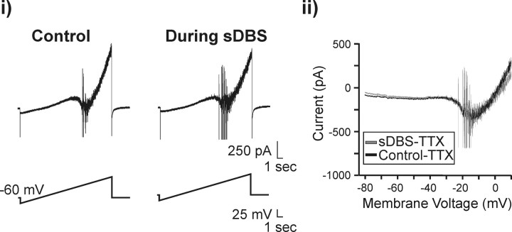
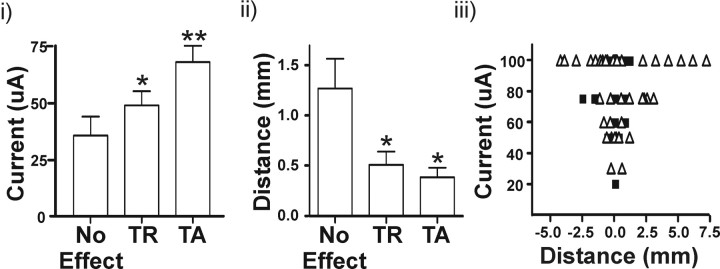
Deep brain stimulation (DBS) of the ventrolateral thalamus stops several forms of tremor. Microelectrode recordings in the human thalamus have revealed tremor cells that fire synchronous with electromyographic tremor. The efficacy of DBS likely depends on its ability to modify the activity of these tremor cells either synaptically by stopping afferent tremor signals or by directly altering the intrinsic membrane currents of the neurons. To test these possibilities, whole-cell patch-clamp recordings of ventral thalamic neurons were obtained from rat brain slices. DBS was simulated (sDBS) using extracellular constant current pulse trains (125 Hz, 60-80 micros, 0.25-5 mA, 1-30 s) applied through a bipolar electrode. Using a paired-pulse protocol, we first established that thalamocortical relay neurons receive converging input from multiple independent afferent fibers. Second, although sDBS induced homosynaptic depression of EPSPs along its own pathway, it did not alter the response from a second independent pathway. Third, in contrast to the subthalamic nucleus, sDBS in the thalamus failed to inhibit the rebound potential and the persistent Na+ current but did activate the Ih current. Finally, in eight patients undergoing thalamic DBS surgery for essential tremor, microstimulation was most effective in alleviating tremor when applied in close proximity to recorded tremor cells. However, stimulation could still suppress tremor at distances incapable of directly spreading to recorded tremor cells. These complementary data indicate that DBS may induce a "functional deafferentation" of afferent axons to thalamic tremor cells, thereby preventing tremor signal propagation in humans.


Figures

Figure 1.
Figure 1.
Experimental methods. Schematic representation of whole-cell patch-clamp recording setup in thalamic rat brain slices. Left, Recordings were made from thalamic relay neurons in the VL thalamus. Extracellular stimulation (A or B) was selectively delivered through one (or combination) of two stimulating electrodes (NEX-100) placed within the VL. Right, Flow chart detailing experimental design enabling independent stimulus pulse generation along an individual or across electrodes.
Figure 2.
Figure 2.
Selective activation of independent thalamic afferents. A test for independence was conducted by comparing the paired-pulse ratio obtained from homosynaptic or cross-synaptic stimulation. Cells were held at –60 mV. i, The stimulation protocol is illustrated above each voltage-clamp recording. The effect of a homosynaptic prepulse (A) on a second homosynaptic pulse (aA) is shown. Similarly, the effect of cross-synaptic prepulse (B) on the A pathway (aB) is shown (n = 18). ii, A similar experimental protocol was used to test pathway B (n = 15). Summary data of the resulting paired-pulse ratio is seen for pulses delivered at 20 and 40 ms apart for pathway A (iii) and pathway B (iv).
Figure 3.
Figure 3.
sDBS selectively inhibits extracellular simulated tremor. Tremor was mimicked in the slice by extracellularly evoking EPSPs at 5 Hz. sDBS applied homosynaptically during simulated tremor resulted in complete inhibition of the tremor signal (i) but had no effect on tremor generated along the cross-synaptic pathway (ii; n = 5). Insets show expanded views of single EPSPs before, during, and after sDBS. The effects of sDBS on the amplitude of the homosynaptic (closed circles) and cross-synaptic (open circles) tremor EPSPs as a function of their respective controls are plotted over time (iii). Note that the tremor stimuli were delivered at a subharmonic frequency of the sDBS train to ensure noninterference when delivered homosynaptically down the same electrode. To confirm successful delivery, the tremor stimulation artifacts were not blanked.
Figure 4.
Figure 4.
sDBS-mediated modulation of Ih. A, Representative current-clamp recording and V–I relationship (20 pA for each step) obtained during control (i) and sDBS (ii)(n = 5). B, Representative voltage-clamp recording and I–V relationship (10 mV for each step) obtained from another neuron. i, In control, Ih is seen as a slowly activating inward current on the hyperpolarizing steps (n = 6). ii, During extracellular sDBS, the time dependency of Ih activation is nearly absent as the current is activated tonically with the onset of sDBS and consequently has reached steady state before application of the hyperpolarizing test pulse. All data are derived from ZD7288 (20 μm)-sensitive current and obtained after subtracting ZD7288-resistant currents from the controls (n = 6). iii, A comparison of holding current before or during sDBS. Cells were held at –70 mV. Note that sDBS increased the holding current from control levels (p < 0.001), but this sustained inward current was blocked by ZD7288. iv, Ih activation curve obtained based on the ratio of Ii versus Imax (control step to –120 mV). v, I–V relationship for instantaneous (Ii) and steady-state (Imax) current. Imax was obtained at the end of each 1 s voltage step. During sDBS, a significant increase in instantaneous and steady-state current was observed over control (p < 0.001). However, there was no statistical difference between instantaneous and steady-state current that developed during sDBS. This indicates a tonic activation of Ih induced by sDBS.
Figure 5.
Figure 5.
Lack of modulatory effects of sDBS on IT. Intracellular voltage steps (–50 mV, 500 ms) were delivered repeatedly once per second in control or during extracellular sDBS (n = 7). There was no statistical difference in the peak amplitude, onset latency, or duration of the rebound potential. However, there was a decrease in the latency of Na+ spike onset (p < 0.05). The arrow indicates peak of rebound potential. Action potentials were truncated in overlay for clarity.
Figure 6.
Figure 6.
Effect of sDBS on the persistent Na+ current. i, A slow voltage ramp command (5 mV/s) was applied from –80 to +10 mV resulting in a slow inward current (INap). This current was unaltered during sDBS (n = 4). ii, Overlay of TTX (0.3 μm)-sensitive INap current in control or during sDBS. The INap current was obtained by subtracting the current evoked in the presence of TTX from that obtained under normal aCSF. Action potentials were truncated for clarity.
Figure 7.
Figure 7.
Alleviation of tremor is related with stimulation proximity to tremor cells. Thalamic microelectrode stimulation was performed in eight patients before implantation of the DBS macroelectrode. We identified 36 tremor cells and applied microstimulation at multiple sites at various current intensities. i, At stimulation sites <1 mm away from a tremor cell, increasing the stimulation current intensity increased the effectiveness of tremor stoppage. ii, At a constant stimulation intensity of 50 μA, better tremor suppression was achieved at stimulation sites closer to recorded tremor cells. iii, The relationship between current and distance for tremor reduction (open triangles) and tremor arrest (closed squares) are plotted. Note that tremor suppression was observed at distances >2 mm from recorded tremor cells. However, at these distances, low-current intensity-induced tremor reduction was not observed, suggesting the absence of other nearby tremor cells. TR, Tremor reduction; TA, tremor arrest. *p < 0.05 from control (no effect); **p < 0.05 from control (no effect) and TR.

References

    1. Anderson ME, Postupna N, Ruffo M (2003) Effects of high-frequency stimulation in the internal globus pallidus on the activity of thalamic neurons in the awake monkey. J Neurophysiol 89: 1150–1160. - PubMed
    1. Anderson T, Hu B, Pittman Q, Kiss ZH (2004a) Mechanisms of deep brain stimulation: an intracellular study in rat thalamus. J Physiol (Lond) 559: 301–313. - PMC - PubMed
    1. Anderson T, Hu B, Pittman Q, Kiss ZH (2004b) The effects of deep brain stimulation of rat ventral thalamus are age and stimulation parameter dependent. Soc Neurosci Abstr 30: 753.13.
    1. Anderson T, Hu B, Kiss ZH (2005) The “roadblock” to tremor signal propagation: a selective action of deep brain stimulation on afferent synaptic transmission. Soc Neurosci Abstr 31: 988.12.
    1. Arai A, Lynch G (1998) AMPA receptor desensitization modulates synaptic responses induced by repetitive afferent stimulation in hippocampal slices. Brain Res 799: 235–242. - PubMed

Publication types