Comparative Study
. 2006 Jan 18;26(3):851-61.
doi: 10.1523/JNEUROSCI.3292-05.2006.

Calcium calmodulin-stimulated adenylyl cyclases contribute to activation of extracellular signal-regulated kinase in spinal dorsal horn neurons in adult rats and mice

Affiliations
Comparative Study

Calcium calmodulin-stimulated adenylyl cyclases contribute to activation of extracellular signal-regulated kinase in spinal dorsal horn neurons in adult rats and mice

Feng Wei et al. J Neurosci. .

Abstract

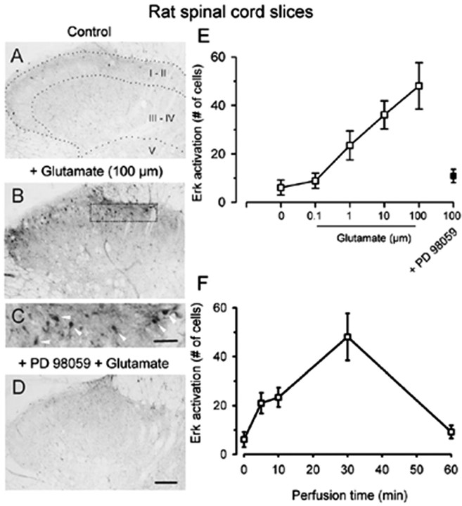
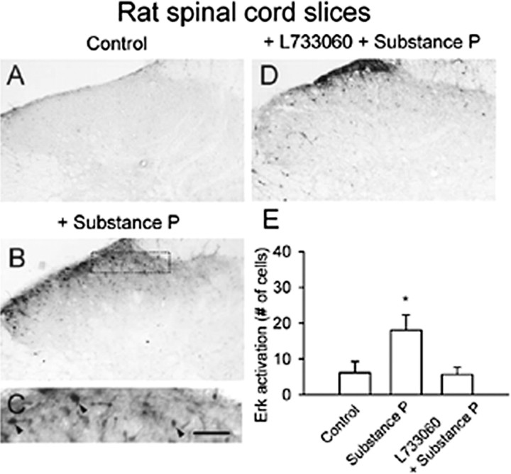
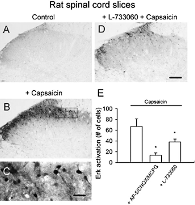
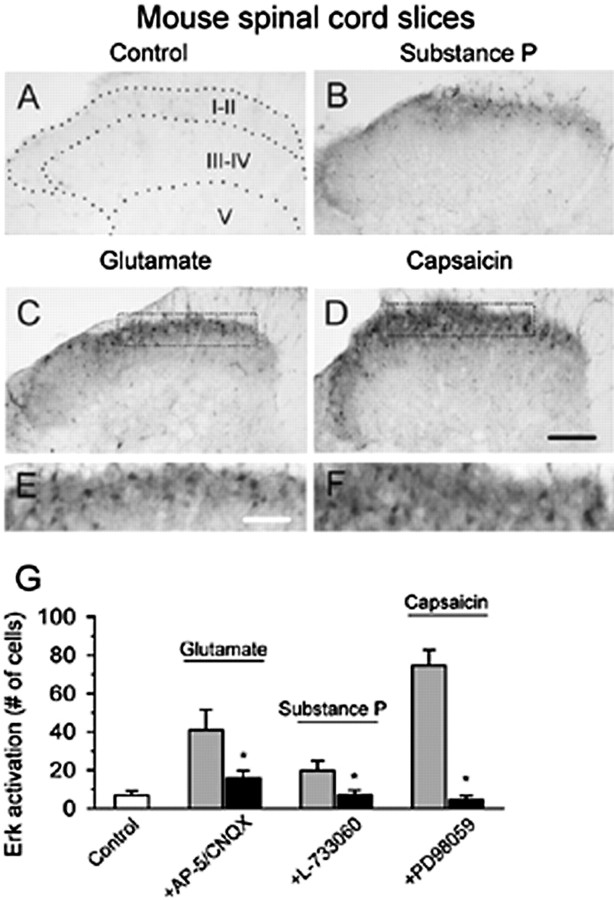
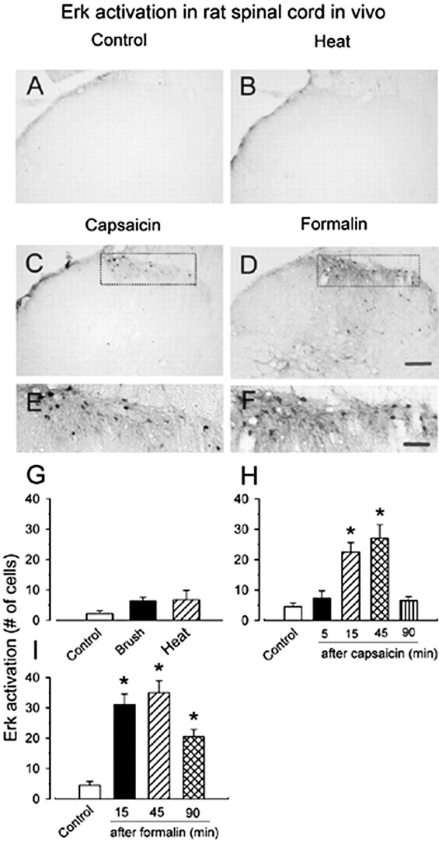
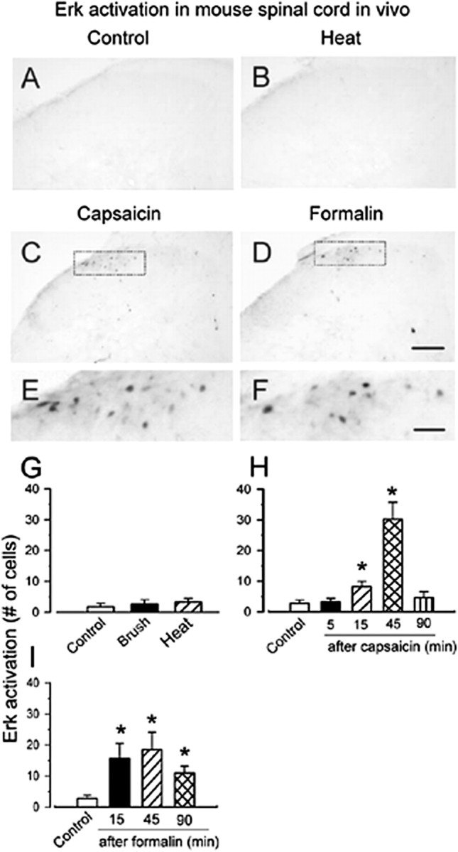
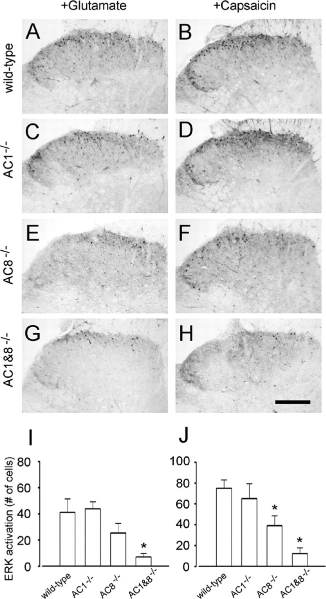
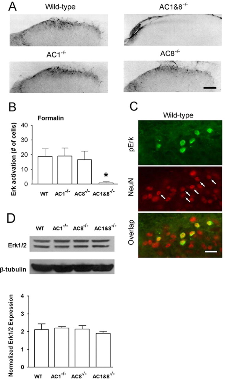
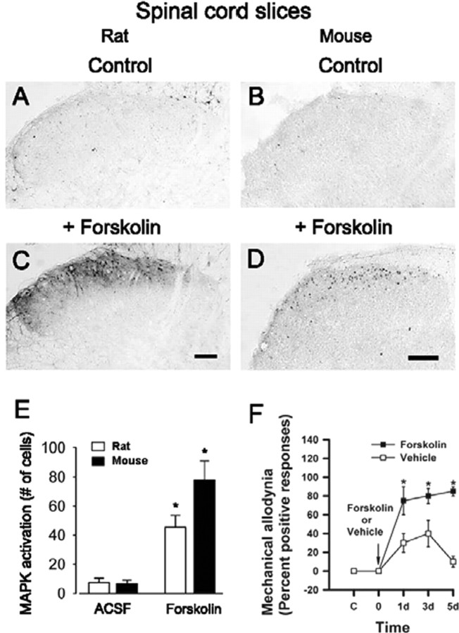
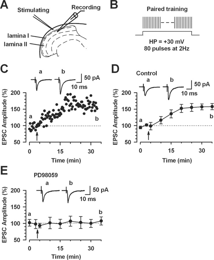
The extracellular signal-regulated kinase (Erk) cascades are suggested to contribute to excitatory synaptic plasticity in the CNS, including the spinal cord dorsal horn. However, many of their upstream signaling pathways remain to be investigated. Here, we demonstrate that glutamate and substance P (SP), two principal mediators of sensory information between primary afferent fibers and the spinal cord, activate Erk in dorsal horn neurons of both adult rat and mouse spinal cord. In genetic knock-out mice of calcium calmodulin-stimulated adenylyl cyclase subtypes 1 (AC1) and 8 (AC8), activation of Erk in dorsal horn neurons were significantly reduced or blocked, either after peripheral tissue inflammation or by glutamate or SP in spinal cord slices. Our studies suggest that AC1 and AC8 act upstream from Erk activation in spinal dorsal horn neurons and the calcium-AC1/AC8-dependent Erk signaling pathways may contribute to spinal sensitization, an underlying mechanism for the development of persistent pain after injury.


Figures

Figure 1.
Figure 1.
Dose- and time-dependent activation of Erk by glutamate in adult rat spinal cord slices. Bath application of high-dose glutamate (100 μm for 30 min) induced increases in pErk immunoreactivity in dorsal horn neurons (B), especially in the superficial layer (C, arrowheads), compared with sham treatment (A). This activation was completely inhibited by pretreatment with the MEK inhibitor PD98059 (D). E, Pooled data demonstrate a dose-dependent activation of Erk by bath application of glutamate (30 min; open squares). Erk activation was blocked by the MEK inhibitor PD98059 (filled squares). F, Erk activation depended on the perfusion time of glutamate (100 μm; 5, 10, 30, and 60 min). Scale bars: (in D) A, B, D, 100 μm; (in C) C, 40 μm. Error bars indicate SEM.
Figure 2.
Figure 2.
Pretreatment with glutamate receptor antagonists reduced glutamate-induced enhancement of pErk immunoreactivity in rat superficial dorsal horn. Pretreatment (10 min) with AP-5 (D), CNQX (E), and MCPG (F) decreased the number of immunopositive cells for pErk after glutamate exposure (100 μm; 30 min) (C). G, No obvious effect of the glutamate receptor antagonists on activation of Erk was observed (for a representative sample, see B) compared with that of vehicle (A). H, Summary data of the effect of different antagonists on the attenuation of glutamate-induced phosphorylation of Erk in superficial dorsal horn neurons. *Significant difference compared with application of glutamate alone; p < 0.05. Scale bar: F (for A–F), 100 μm. Error bars indicate SEM.
Figure 3.
Figure 3.
Activation of Erk by SP in adult rat spinal cord slices. Basal expression of pErk in ACSF-perfused slices (A). Bath application of SP (10 μm for 30 min) increased pErk immunoreactivity in dorsal horn neurons (B). High magnification of the middle part of the superficial dorsal horn in B showing labeled neurons (C, arrowheads). This activation was inhibited by pretreatment with the NK1 receptor antagonist L-733060 (D). E, Summary data of pErk immunoreactivity in superficial dorsal horn. *Significant difference compared with control; p < 0.05. Scale bar: (in C) A, B, D, 100 μm; C, 40 μm. Error bars indicate SEM.
Figure 4.
Figure 4.
Activation of spinal Erk by capsaicin in rat spinal cord slices. Compared with control slices (A), bath application of capsaicin (1 μm for 30 min) increased intensity of pErk immunoreactivity in dorsal horn neurons. B, A high-magnification image of the medial superficial dorsal horn in B is provided (C). Immunostaining of pErk was observed in cell bodies and their proximal dendrites. The Erk activation in superficial dorsal horn was significantly decreased by the NK1 receptor antagonist L-733060 (D, E) or a combination of the three glutamate receptor antagonists (E). *Significant difference compared with application of capsaicin alone; p < 0.05. Scale bar: (in D) A, B, D, 100 μm; (in C) C, 50 μm. Error bars indicate SEM.
Figure 5.
Figure 5.
Representative activation of Erk in the dorsal horn from mouse spinal slices. Erk activation in the dorsal horn of spinal slices treated with bath application of SP (10 μm for 30 min)(B), glutamate (100 μm for 30 min) (C), or capsaic in (1 μm for 30 min) (D) is shown. Higher magnifications of the superficial dorsal horn in C and D are provided in E and F, respectively. G, Summary data showed that the combination of AP-5 with CNQX attenuates the increase in activation of Erk by glutamate; SP-induced increase of Erk activation is completely blocked by the NK1 receptor antagonist L-733060 and pretreatment with the MEK inhibitor PD98059 blocked capsaicin-induced Erk activation. *Significant difference between groups with and without antagonists; p < 0.05. Scale bars: (in D) A–D, 100 μm; (in E) E, F, 40 μm. Error bars indicate SEM.
Figure 6.
Figure 6.
Activation of Erk by noxious stimulation in rat spinal dorsal horn. Representative pErk immunostaining in ipsilateral spinal dorsal horn from control and stimulated rats is shown. A, Saline-treated control. B, Forty-five minutes after brief noxious thermal stimulation (55°C; 12 s) on a hindpaw skin. C, Hindpaw injection of capsaicin (100 μg for 15 min) produced spinal Erk activation in the medial half of the superficial dorsal horn, in which additional enlargement (E) showed a group of labeled neurons in this region. No obvious increase in pErk immunoreactivity was observed in deep dorsal horn. D, Hindpaw injection of formalin (5%; 50 μl; 45 min) induced substantial increases in pErk immunoreactivity in the superficial and deep dorsal horn. F, High magnification of the medial part of the superficial dorsal horn in D showing that a majority of labeled neurons have labeled dendrites located in this region. Graphical representations of change in average number of pErk-labeled neurons in the laminas I–II of ipsilateral spinal cord section after somatosensory stimuli in A–F is shown in G–I. G, Forty-five minutes after repeated brush or noxious heat (55°C) on the hindpaw for 12 s. Different time points after hindpaw injection of capsaicin (H) or formalin (I) into the plantar surface of the left hindpaw are shown. *p < 0.05; compared with control animals. Scale bars: (in D) A–D, 100 μm; (in F) E, F, 40 μm. Error bars indicate SEM.
Figure 7.
Figure 7.
Activation of Erk by sensory stimulation in mouse spinal dorsal horn. Representative pErk immunostaining in ipsilateral spinal dorsal horn from control and stimulated mice are shown. A, Control. B, Brief noxious heat (55°C; 12 s) did not increase in pErk immunoreactivity. A similar pattern of Erk activation was observed at 45 min after injection of capsaicin (10 μg) (C) and 5% formalin (10 μl) (D). E, F, A higher magnification image of the superficial dorsal horn in C and D respectively, with obviously labeled neurons. Graphical representations of changes in the average number of pErk-labeled neurons in the laminas I–II of the ipsilateral spinal cord section after somatosensory stimuli in A–F are shown in G–I. G, Forty-five minutes after repeated brushing or noxious heat (55°C) on the hindpaw for 12 s. Different time points after subcutaneous injection of capsaicin (H) or formalin (I) into the plantar surface of the left hindpaw, respectively, are shown. *p < 0.05; compared with control animals. Scale bars: (in D) A–D, 100 μm; (in F) E, F, 30 μm. Error bars indicate SEM.
Figure 8.
Figure 8.
Activation of Erk by glutamate and capsaicin in genetic mice spinal cord slices. Bath application of glutamate (100 μm for 30 min; A, C, E, G) or capsaicin (1 μm for 30 min; B, D, F, H) induced expression of pErk in superficial dorsal horn from WT (A, B), AC1 (C, D), or AC8 knock-out (E, F) but not AC1&8 DKO mice (G, H). I, J, Summary data showing variation of pErk expression in different genetic knock-out mice. *p < 0.05, compared with WT groups. Scale bar: (in H) A–H, 100 μm. Error bars indicate SEM.
Figure 9.
Figure 9.
Erk expression in the spinal dorsal horn of AC1, AC8, and AC1&8 DKO mice. Different patterns of pErk expression in AC1&8 DKO mice compared with WT mice. A, pErk immunoreactivity in dorsal horn of knock-out mice. Scale bar, 50 μm. The value n = 6 mice per group. B, Graphical representation of the number of immunoreactive cells at 45 min after injection of formalin (n = 6 mice per group; *p < 0.05). C, Double immunostaining showed Erk activation in superficial dorsal horn neurons of WT mice labeled by NeuN (arrows; n = 4 mice) at 15 min after hindpaw formalin injection. Scale bar, 20 μm. D, Representative Western blots of Erk expression in dorsal horn homogenates in AC1, AC8, AC1&8 DKO, and WT mice. The normalized Erk expression between all groups was unchanged (n = 3 mice per group; p > 0.05). Error bars indicate SEM.
Figure 10.
Figure 10.
Activation of Erk by forskolin in rat and mouse spinal cord slices. Forskolin-induced pErk immunoreactivity in dorsal horn neurons in spinal cord slices from adult rats (C) and mice (D) compared with the control (A and B, respectively) is shown. Pooled data from rats and mice are shown in E. F, Pretreatment with an intrathecal injection of forskolin (12 nmol) 30 min before the unilateral hindpaw injection of CFA in AC1&8 DKO mice rescued mechanical allodynia (n = 4 mice in each group). Behavioral responses to the von Frey stimulation (filament No 2.44) on the dorsum of the ipsilateral hindpaw are plotted against time. Mechanical allodynia was tested on days 1, 3, and 5. Asterisks indicate significant differences compared with control; *p < 0.05; **p < 0.001. Scale bars, 100 μm. Error bars indicate SEM.
Figure 11.
Figure 11.
Activation of Erk for the induction of LTP in the superficial dorsal horn neurons. A, Diagram of a slice showing the placement of a whole-cell patch-clamp recording and a stimulation electrode in the superficial dorsal horn of the spinal cord. B, Schematic illustrating the induction protocol consisting of 80 pulses at 2 Hz while holding at +30 mV (paired training). C, LTP was induced by paired training in the superficial dorsal horn neurons. D, Summary result of the LTP experiments under control conditions (n = 9 neurons). EPSC responses were averaged over 5 min intervals. E, The MEK inhibitor PD98059 (50 μm) in the intracellular solution completely blocked LTP induction (n = 7 neurons). EPSC responses were averaged over 5 min intervals. C–E, Traces show averages of six EPSCs 3 min before (a) and 25 min after (b) the paired training (arrow). The dashed line indicates the mean basal synaptic responses. Error bars indicate SEM.

References

    1. Adwanikar H, Karim F, Gereau IV RW (2004) Inflammation persistently enhances nocifensive behaviors mediated by spinal group I mGluRs through sustained ERK activation. Pain 111: 125–135. - PubMed
    1. Bliss TV, Collingridge GL (1993) A synaptic model of memory: long-term potentiation in the hippocampus. Nature 361: 31–39. - PubMed
    1. Chetkovich DM, Sweatt JD (1993) nMDA receptor activation increases cyclic AMP in area CA1 of the hippocampus via calcium/calmodulin stimulation of adenylyl cyclase. J Neurochem 61: 1933–1942. - PubMed
    1. Coderre TJ, Katz J, Vaccarino AL, Melzack R (1993) Contribution of central neuroplasticity to pathological pain: review of clinical and experimental evidence. Pain 52: 259–285. - PubMed
    1. Dubner R, Basbaum AI (1994) Spinal dorsal horn plasticity following tissue or nerve injury. In: Textbook of pain, Ed 3 (Wall PD, Melzack R, Bonica JJ, eds), pp 230–233. New York: Churchill Livingstone.

Publication types

MeSH terms

Substances

LinkOut - more resources