Comparative Study
. 2006 Jan 18;26(3):882-92.
doi: 10.1523/JNEUROSCI.3162-05.2006.

Multiple Eph receptors and B-class ephrins regulate midline crossing of corpus callosum fibers in the developing mouse forebrain

Affiliations
Comparative Study

Multiple Eph receptors and B-class ephrins regulate midline crossing of corpus callosum fibers in the developing mouse forebrain

Shannon W Mendes et al. J Neurosci. .

Abstract

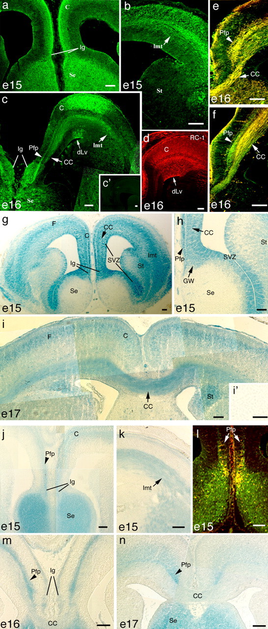
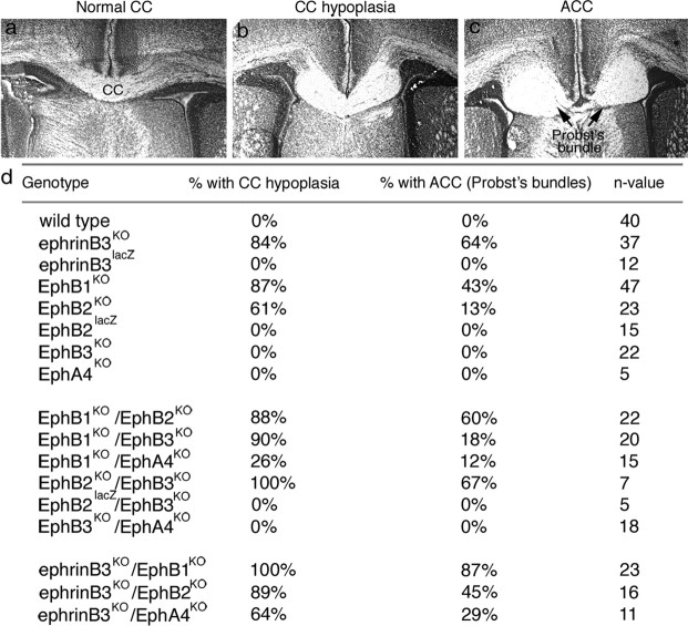
Agenesis of the corpus callosum (CC) is a rare birth defect that occurs in isolated conditions and in combination with other developmental cerebral abnormalities. Recent identification of families of growth and guidance molecules has generated interest in the mechanisms that regulate callosal growth. One family, ephrins and Eph receptors, has been implicated in mediating midline pathfinding decisions; however, the complexity of these interactions has yet to be unraveled. Our studies shed light on which B-class ephrins and Eph receptors function to regulate CC midline growth and how these molecules interact with important guideposts during development. We show that multiple Eph receptors (B1, B2, B3, and A4) and B-class ephrins (B1, B2, and B3) are present and function in developing forebrain callosal fibers based on both spatial and temporal expression patterns and analysis of gene-targeted knock-out mice. Defects are most pronounced in the combination double knock-out mice, suggesting that compensatory mechanisms exist for several of these family members. Furthermore, these CC defects range from mild hypoplasia to complete agenesis and Probst's bundle formation. Further analysis revealed that Probst's bundle formation may reflect aberrant glial formations and/or altered sensitivity of CC axons to other guidance cues. Our results support a significant role for ephrins and Eph receptors in CC development and may provide insight to possible mechanisms involved in axon midline crossing and human disorder.


Figures

Figure 1.
Figure 1.
Developmental timing of the CC, Pfp, and midline glial guideposts is normal in CD1 mice. Few anti-β-tubulinIII (β-tub)-labeled callosal fibers crossed the midline at E15 (a) and E16 (b). c, Cortex; s, septum. c, By E17, many CC fibers have crossed the midline and project to the contralateral cortex. Anti-NF (145 kDa)-labeled Pfp fibers at E16 (d) and E17 (e) are shown. f, High-magnification image of anti-NF-labeled Pfp fibers at the midline. High-magnification image of anti-β-tub-labeled CC fibers (g) and anti-NF-labeled Pfp fibers (h) at E17 are shown. i, Callosal axon growth is regulated by glial guideposts, including Ig, GW, and mzg as visualized using anti-GFAP at E18. a′, DiI tracing of cortical fibers at E15 show CC axons projecting to the midline but not crossing the midline (dashed line represents midline). b′, DiI tracing of cortical fibers at E16 shows that CC fibers have crossed the midline. c′, No primary control. Scale bars, 100 μm.
Figure 2.
Figure 2.
B-class ephrins are expressed in the developing forebrain. At E15, ephrinB1 is expressed in forebrain regions corresponding to the cortex (C), septum (Se), Ig, and lateromedial tract (lmt) (a, b), whereas at E16, ephrinB1 is also expressed in regions that include the CC, Pfp, Ig, C, Se, and unique regions of the dLv (c). d, RC-1 is also expressed in the dLv, similar to ephrinB1 expression. e, At E16, ephrinB1 (green) colabels withβ-tubulinIII (red) in both the CC and Pfp fibers (yellow). f, EphrinB1 (green) colabels with Tag-1 (red) in CC (yellow) and not Pfp fibers. EphrinB2 expression was visualized by examining β-gal expression in the ephrinB2LacZ mice. g, h, At E15, ephrinB2 expression is localized to regions corresponding to the C, CC, GW, Ig, Pfp, striatum (St), and subventricular zone (SVZ) but absent in the Se. F, Forebrain. j, k, By E17, ephrinB2 expression localized to the C, CC, GW, and St (i). EphrinB3 expression was visualized using ephrinB3lacZ mice. At E15, ephrinB3 was expressed in the Pfp, Ig, Se, and lmt. l, Anti-β-galactosidase antibodies (green) colabeled with GFAP (red) in the Ig (yellow). m, n, At E16 and E17, ephrinB3 expression is maintained in the Pfp, Ig, and Se, but little to no expression is observed in the CC. c′ and i′ are no primary and wild-type tissue controls, respectively. Scale bars, 100 μm.
Figure 3.
Figure 3.
Eph receptors are expressed in the developing forebrain. EphB1 (a) and EphB3 (b) are expressed specifically in the regions of the CC. EphB2 expression was visualized by examining β-gal expression in the EphB2lacZ mice. c, e, At E15 and E16, EphB2lacZ expression was detected in areas corresponding to GW, CC, septum (Se), and regions of the subventricular zone (SVZ). d, f, High-magnification images of EphB2 expression in the GW, CC, and SVZ regions. g, EphA4 was expressed in the regions of the CC, GW, Ig, striatum (St), and SVZ at E15. h, High-magnification image of EphA4 expression at the midline in the CC, GW, and Ig. i, EphA4 (green) and GAP-43 (red) are coexpressed in the CC but not in the GW or Ig at E16. j–l, EphA4 (green) colabels with GFAP (red) in the GW and Ig but not the CC at E16 and E17. l, High-magnification image of EphA4 (green) and GFAP (red) in the GW. m–o represent control anti-EphB1, anti-EphB3, and anti-EphA4 immunoreactivity on EphB1KO, EphB3KO, and EphA4KO tissues, respectively. Scale bars, 100 μm.
Figure 4.
Figure 4.
Frequency of CC Probst's bundle and hypoplasia formation in single and double knock-out ephrin/Eph receptor mice. a, Representative Nissl-stained coronal section of a normal CC phenotype. b, Nissl-stained section showing a representative CC hypoplasia phenotype from knock-out tissue. c, Representative Nissl-stained section exemplifies complete ACC with >90% of axons not crossing the midline and the formation of Probst's bundles. d, Frequencies of CC hypoplasia or ACC in single and combination mutant mice at P1 compared with WT CC phenotypes.
Figure 5.
Figure 5.
Pathfinding defects in the CC of ephrinB3KO P1 mice results in aberrant glial cells localized to Probst's bundles. a, DiI tracing of CC fibers (red) in WT tissue colabeled with GFAP (green) shows crossing of callosal fibers in the developing forebrain. b, High-magnification image of DiI tracing shows callosal fibers (red) avoiding the GW and Ig in WT mice. c, DiI tracing of CC fibers (red) in ephrinB3KO mice colabeled with GFAP (green)-expressing cells in the pathway of CC fibers. d, High-magnification image of CC fibers (red) in ephrinB3KO mice shows close association of GFAP (green)-expressing glial cells. Confocal images of DiO-labeled CC axons (e, green) and GFAP-expressing glia (f, red) show regions of axon-cell contact (yellow, arrowhead) (g). Expression of the radial glia markers RC-1 (h) and RC-2 (i) shows normal expression in the guideposts in WT mice. Localization of RC-1- (j) and RC-2- (l) positive cells in the region of Probst's bundle formation in ephrinB3KO tissues is shown. High-magnification images of RC-1- (k) and RC-2- (m) positive radial glial cells in Probst's bundles are shown. Scale bars, 100 μm.
Figure 6.
Figure 6.
Probst's bundles include Pfp fibers in EphB1KO mice. a, Neurofilament-145 kDa-expressing (green) Pfp fibers colabeled with GFAP-expressing (red) guideposts (including GW, Ig, and mzg) in WT mice. b, Neurofilament-expressing (green) Pfp fibers colabeled with GFAP expressing (red) guideposts (including GW, Ig, and mzg) in EphB1KO mice. c, High-magnification image showing NF-labeled Pfp axons extending through and around the Probst's bundle. Scale bars, 100 μm.
Figure 7.
Figure 7.
Cortical neurons have reduced neurite outgrowth when grown on GW feeder cells, where EphB1 interacts with ephrinBs to promote neurite outgrowth. Cortical neurons transfected with eGFP markers allow measurements of neurite length (a, between arrowheads) after 3 d of growth on GW feeder layers that express both GFAP (b) and RC-2 (c). Scale bars, 100μm. d, Average (Avg) neurite length of WT and EphB1KO cortical neurons are reduced when grown on WT glial wedge feeder (Wtgw) cells compared with WT cortical glial feeder (Wtctx) cells. WT and EphB1KO cortical neurons grown on Wtctx cells have similar mean neurite length, whereas EphB1KO cortical neurons grown on Wtgw cells shows significantly shorter neurites then WT cortical neurons grown on Wtgw cells. e, WT cortical neurons grown on dishes coated with ephrinB1-Fc and ephrinB2-Fc showed significantly enhanced neurite outgrowth compared ephrinB3-Fc or Fc fragments, which are not observed in EphB1KO cortical neurons. Error bars indicate mean ± SEM. **p < 0.01; ***p < 0.001.
Figure 8.
Figure 8.
a, Summary of expression of B-class ephrins and Eph receptors superimposed with a schematic coronal cross section showing CC, Pfp, and midline guideposts [GW, Ig, glial sling (GS), and mzg] during development.b, Schematic representation of CC and Pfp growth patterns in the ephrinB3 and EphB1 knock-out mice. c, Schematic representation of a cortical fiber interacting with the GW guidepost. Callosal axons contain both reverse and forward signaling mediated by ephrins and Eph receptors after interactions with either EphB2 or ephrinB3 in guideposts, respectively.

References

    1. Barinaga M (1995) Receptors find work as guides. Science 269: 1668–1670. - PubMed
    1. Bergemann AD, Zhang L, Chiang MK, Brambilla R, Klein R, Flanagan JG (1998) Ephrin-B3, a ligand for the receptor EphB3, expressed at the midline of the developing neural tube. Oncogene 16: 471–480. - PubMed
    1. Birgbauer E, Oster SF, Severin CG, Sretavan DW (2001) Retinal axon growth cones respond to EphB extracellular domains as inhibitory axon guidance cues. Development 128: 3041–3048. - PubMed
    1. Blits-Huizinga CT, Nelersa CM, Malhotra A, Liebl DJ (2004) Ephrins and their receptors: binding versus biology. IUBMB Life 56: 257–265. - PubMed
    1. Brambilla R, Bruckner K, Orioli D, Bergemann AD, Flanagan JG, Klein R (1996) Similarities and differences in the way transmembrane-type ligands interact with the Elk subclass of Eph receptors. Mol Cell Neurosci 8: 199–209. - PubMed

Publication types

LinkOut - more resources