Comparative Study
. 2006 Jan 18;26(3):934-47.
doi: 10.1523/JNEUROSCI.3656-05.2006.

In vivo imaging of preferential motor axon outgrowth to and synaptogenesis at prepatterned acetylcholine receptor clusters in embryonic zebrafish skeletal muscle

Affiliations
Comparative Study

In vivo imaging of preferential motor axon outgrowth to and synaptogenesis at prepatterned acetylcholine receptor clusters in embryonic zebrafish skeletal muscle

Jessica A Panzer et al. J Neurosci. .

Abstract

Little is known about the spatial and temporal dynamics of presynaptic and postsynaptic specializations that culminate in synaptogenesis. Here, we imaged presynaptic vesicle clusters in motor axons and postsynaptic acetylcholine receptor (AChR) clusters in embryonic zebrafish to study the earliest events in synaptogenesis in vivo. Prepatterned AChR clusters are present on muscle fibers in advance of motor axon outgrowth from the spinal cord. Motor axon growth cones and filopodia are selectively extended toward and contact prepatterned AChR clusters, followed by the rapid clustering of presynaptic vesicles and insertion of additional AChRs, hallmarks of synaptogenesis. All initially formed neuromuscular synapses contain AChRs that were inserted into the membrane at the time the prepattern is present. Examination of embryos in which AChRs were blocked or clustering is absent showed that neither receptor activity or receptor protein is required for these events to occur. Thus, during initial synaptogenesis, postsynaptic differentiation precedes presynaptic differentiation, and prepatterned neurotransmitter clusters mark sites destined for synapse formation.


Figures

Figure 1.
Figure 1.
Location of prepatterned AChR clusters in myotomal muscle. Presynaptic vesicles in motor axons and terminals were labeled with an antibody against SV2 (green), and postsynaptic AChR clusters were labeled with rhodamine αBTX (red) in 24 hpf embryos. Except where indicated, all images are oriented so that rostral is to the left and dorsal is at the top. The dashed line in each panel indicates the edge of the spinal cord. Higher-magnification views of the boxed regions in A are shown in B–D. A, In rostral segments (left), primary motor axons have grown past the choice point and have begun to extend along the medial surface of the myotome. In middle segments, motor axons have just reached the choice point. In the most caudal segments (right), motor axons have not yet exited the spinal cord. Prepatterned AChR clusters are visible throughout the dorsal and ventral extent of the myotome and along the lateral myosepta. Scale bar, 10 μm. B, In rostral segments, as motor axons form the initial neuromuscular synapses at the choice point (bracket) and extend beyond it along the medial surface of the myotome, some prepatterned AChR clusters dorsal and ventral to the choice point have disappeared. C, In middle segments, as motor axons enter the myotome, prepatterned AChR clusters are reduced in number, smaller, and more punctate in appearance. D, In caudal segments, prepatterned AChR clusters are elongated and diffuse along each muscle fiber, well in advance of outgrowing motor axons. Scale bar, 10μm.
Figure 2.
Figure 2.
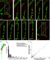
Dynamics of prepatterned AChR clusters. RhodamineαBTX-labeled AChRs (red) imaged intransgenic embryo expressing GFPinneuronstovisualizemotoraxons(HuC:GFP;green) from 20–26 hpf at 1 h intervals (supplemental movie 1, available at www.jneurosci.org as supplemental material). A, Time-lapse images of prepatterned AChR clusters. The first movie plane was taken at time 0:00, before motor axon outgrowth from the spinal cord. Boxed regions in the first panel are shown at higher magnification in B and D. Prepatterned AChR clusters are initially elongated and diffuse and coalesce over time (e.g., compare panels 1:00 and 3:00). The final panel is postimaging immunostaining for presynaptic vesicles (SV2; green) that demonstrates that motor axons have grown to and synapses have been established at the choice point (bracket) by 6:00. Scale bar, 10μm. B, Higher magnification of boxed region in A. As motor axons exit the spinal cord and innervate the choice point, many prepatterned AChR clusters disappear (disappearing cluster indicated by arrow). In this small region, many prepatterned AChRs disappear over the 6 h imaging interval (compare cluster marked with an arrow from panels 0:00 to 6:00). However, throughout the myotome, 20% of prepatterned AChR clusters disappear over a 6 h imaging interval. Scale bar, 10μm. C, Line scan (8 μm in length) through the cluster marked with an arrow in B shows that cluster fluorescence intensity (arbitrary units) and length decrease gradually over the 6 h imaging interval. D, Higher magnification of boxed region in A. In this small region, two of the four prepatterned AChR clusters present at the first views are incorporated into synapses (compare cluster marked with an arrow in panels 0:00–6:00) as evidenced by the appearance of a GFP+ axon over the AChR cluster at panel 5:00 (asterisk) that persists to 6:00. However, throughout the myotome, 30% of prepatterned AChR clusters are incorporated into synapses. D′, Postimaging immunostaining demonstrating an SV2+ vesicle cluster formed at this AChR cluster. D″, D\m='\\m='\\m='\, Single-channel images of the boxed region in D′, demonstrating colocalization of the SV2+ vesicle cluster (indicated by dashed line) with the AChR cluster. Scale bar, 10μm. E, Line scan (11μm) through the AChR cluster marked with an arrow in D shows that cluster width gradually decreases and that fluorescence persists throughout the 6 h imaging interval. F, Quantification of AChR cluster length at 0:00 and 6:00, demonstrating that prepatterned AChR clusters coalesce and become significantly reduced in length over time (n = 4 fish, 19 myotome segments; Kolmogorov–Smirnov test, p < 0.001). G, Quantification of AChR cluster intensity at 0:00 and 6:00, determined by measuring the average pixel intensity along a line scan, demonstrating that prepatterned AChR clusters significantly increase in fluorescence over time (n = 4 fish, 19 myotome segments; Kolmogorov–Smirnov test, p < 0.001). H, Quantification of the fate of prepatterned AChR clusters. Of all prepatterned AChR clusters, 20% disappeared, 30% become synaptic during the 6 h imaging interval, and 50% remain nonsynaptic during the imaging interval (n = 4 fish, 14 myotome segments). Error bars indicate SE.
Figure 3.
Figure 3.
Motor axon growth cones preferentially extend toward prepatterned AChR clusters. A–C, A primary motor neuron, MiP, expressing VAMP-GFP (green) and AChR clusters labeled with rhodamine αBTX (red) from a ∼24 hpf embryo were imaged for >4 h at intervals of 20 min (supplemental movie 2, available at www.jneurosci.org as supplemental material). The dashed line indicates the edge of the spinal cord. In this series of images, the position of AChRs is fixed while a single motor axon growth cone advances across the field of view. In the supplemental movie (available at www.jneurosci.org as supplemental material), AChR clusters near the bottom of the frame appear to move because the embryo is growing. Because we did not attempt to focus on this region during image capture, these clusters come in and out of the field of view on several occasions. A, At the start of time-lapse imaging, MiP has extended to the choice point (bracket), and a dorsal axon branch is growing toward prepatterned AChR clusters (arrow). The boxed region is shown at higher magnification in B. B, Time-lapse images of the MiP growth cone extending toward, contacting, and then extending beyond, prepatterned AChR clusters. In some cases, VAMP-GFP+ clusters of presynaptic vesicles accumulate over prepatterned AChR clusters (panel 0:40, asterisks). In panels 3:00–4:20, the growth cone turns 65° toward prepatterned AChR clusters at the dorsal edge of the myotome and lateral myosepta (arrowhead). Scale bar, 10 μm. C, At the end of time-lapse imaging, the MiP axon has reached the dorsal edge of the myotome and has just contacted AChR clusters located at the dorsal edge of the myosepta. The boxed region is the same as in A. Scale bar, 10μm. D, To quantify axon and growth cone outgrowth with respect to prepatterned AChR clusters, the position of a growth cone was determined from a point in its geometric center (arrow). The closest AChR cluster in advance of the growth cone was defined as a potential target AChR cluster (red oval). Three angles were then measured: angle 1, the angle between the actual growth cone trajectory (black line) and a straight trajectory toward the AChR cluster (red dashed line); angle 2, the angle between the actual trajectory and the initial trajectory if the growth cone were to grow in a straight line (blue dashed line); and angle 3, the angle between the initial trajectory and the target AChR cluster (n = 9 22–30 hpf embryos, 9 motor neurons, 38 growth cone out growth events). E, The distribution of angles 1 and 2. Sixty-three percent of growth conesare extended with an angle ≤10°, and 95% are extended at an angle of ≤30°, with respect to a prepatterned AChR cluster (black bars). Growth cones were observed to turn toward a prepatterned AChR cluster over a wide range of angles, from 10 to 130° (gray bars). F, Angle 2 was plotted against angle 3, and their relationship was analyzed by linear regression. A significant correlation exists between the location of a prepatterned AChR cluster with respect to the initial trajectory of the motor axon (angle 3) and the angle (angle 2) that the growth cone eventually turns toward that cluster (r = 0.91).
Figure 4.
Figure 4.
Motor axon filopodia preferentially extend toward and contact prepatterned AChR clusters. A–C, A primary motor neuron, RoP, expressing VAMP-GFP (green) and prepatterned AChR clusters labeled with rhodamine αBTX (red) from a ∼24 hpf embryo imaged for >5 h at intervals of 10 min (supplemental movie 3, available at www.jneurosci.org as supplemental material). Because of focal plane drift, the leftmost AChR cluster in supplemental movie 3 (available at www.jneurosci.org as supplemental material) briefly disappears for the first five frames at the beginning of the movie. A, The growth cone pauses at the choice point (bracket) and forms a rostrally extending branch (panel 5:00). Two areas containing prepatterned AChRs are present (panel 0:00: rostral area, arrowhead; caudal and ventral area, arrow). Scale bar, 5 μm. B, Many filopodia are extended toward the caudal area containing prepatterned AChR clusters (panel 0:00, arrow). Filopodia contact this AChR cluster twice (panels 0:10 and 3:30, asterisk) and retract. Scale bar, 5 μm. C, Filopodia are also extended toward the rostral area containing prepatterned AChR clusters (panel 3:20, arrowhead). A filopodia contacts an AChR cluster (panel 4:00, asterisk) and then retracts (panel 4:10), contacts the cluster again (panel 4:20, asterisk), and persists until the end of the imaging session. Scale bar, 5 μm. D, Quantification of filopodial extension. The top panel illustrates a segment of a motor axon with three filopodia (green) and a prepatterned AChR cluster (red) off to the side of the axon. In the bottom panel, the dashed line indicates the shortest distance between the base of each filopodia at the axon and the AChR cluster. The solid line indicates the actual direction of filopodial extension. The angle between these two lines was measured for each filopodia. Scale bar, 5 μm. E, Distribution of the angle of filopodial extension with respect to prepatterned AChR clusters for the RoP axon shown in A–C. The location of prepatterned AChR clusters was aligned at 0° (red dot). Each line represents one filopodia, the length of the line represents filopodial length, and the angle represents the angle of filopodial extension with respect to a prepatterned AChR cluster. The majority of filopodia are extended at an angle ≤30° with respect to prepatterned AChR clusters. F, Distribution of the angle of filopodial extension with respect to a control area 15 μm away from the axon. Filopodia are extended randomly if an AChR cluster is not present. G, Summary plot of cumulative percent of angles of filopodial extension with respect to prepatterned AChR clusters (gray line; n = 6 22–30 hpf embryos, 6 motor neurons, 230 filopodia) or control areas (black line; n = 167 filopodia) for all filopodia from all motor neurons imaged (asterisk indicates significant difference, Kolmogorov–Smirnov test, p < 0.0001).
Figure 5.
Figure 5.
Filopodia are preferentially extended from synapses. Examples of filopodia extended from primary motor neurons expressing VAMP-GFP (green) and prepatterned AChR clusters labeled with rhodamine αBTX (red) from ∼22–30 hpf embryos. A–A″, Filopodia are extended from VAMP-GFP+ clusters of presynaptic vesicles (A) that are apposed to AChR clusters (A′) and are thussynapses(A″).B–B″,FilopodiaareextendedfromVAMP-GFP+vesicleclusters(B) that are not apposed to AChR clusters (B′, B″). C–C″, Relatively few filopodia are extended from axon regions lacking VAMP-GFP+ clusters. Scale bar, 10 μm. D, Quantification of the percentage of filopodia extended from VAMP-GFP+ vesicle clusters (90%), from synapses (63%), or from the axon (10%).
Figure 6.
Figure 6.
Postsynaptic AChR clusters precede presynaptic vesicle clusters during initial neuromuscular synaptogenesis. Primary motor neurons (CaP) expressing VAMP-GFP (green) and prepatterned AChR clusters labeled with rhodamine αBTX (red) from a ∼24 hpf embryos imaged for 2–5 h at intervals of 20 min. A, At the beginning of the imaging session, a prepatterned AChR cluster is present in advance of the growth cone. The boxed region is shown at higher magnification in B. Scale bar, 10μm. B, Time-lapse images of the growth cone extending toward, contacting, and then extending beyond the AChR cluster. In panels 1:20 and 1:40, clusters of VAMP-GFP+ vesicles have accumulated over the AChR cluster. Scale bar, 10 μm. C, D, The fluorescence intensity of postsynaptic AChRs (C) and presynaptic VAMP-GFP (D) are displayed as bars, the height and color of which are proportional to intensity (white, high; purple, low). This analysis shows that VAMP-GFP gradually accumulates over the AChR cluster. E, At the beginning of the imaging session, a prepatterned AChR cluster is present in advance of the growth cone (arrow). The boxed region is shown at higher magnification in F and G. Scalebar,10μm.F, G, Time-lapse images of the growth cone contacting and growing past a prepatterned AChR cluster and the subsequent appearance of an AChR cluster beneath a filopodia. AChR labeling alone is shown in G for clarity. The cluster that was present in advance of the growth cone is marked in F, panel 3:00, with an arrow. A VAMP-GFP+ cluster is subsequently induced over this AChR cluster (panel 4:00, asterisk). In addition, a cluster subsequently appears beneath a filopodia (F, G, panel 3:20, arrowhead). This AChR cluster becomes larger and increases in intensity over time (G, panel 4:40). A VAMP-GFP+ cluster appears at this site (panel 3:40) and persists throughout the imaging period (F, panel 4:40, open arrowhead). Scale bar, 10 μm.
Figure 7.
Figure 7.
Insertion of new AChRs and redistribution of prepatterned AChRs during initial neuromuscular synaptogenesis. All AChRs were optically saturated with rhodamine αBTX at 20 hpf, and 2, 4, or 7 h later, embryos were fixed and newly inserted AChRs were labeled with Cy5 αBTX. The location of existing (old; red) AChRs and receptors inserted since the initial rhodamine αBTX labeling (new; blue) was compared with the location of motor axons and terminals visualized after immunostaining for SV2 (green). A–A′, Prepatterned AChRs present before motor axon outgrowth at 20 hpf. B–B″, In embryos relabeled with Cy5 αBTX 4 h after initial labeling of old AChRs with rhodamine αBTX, all prepatterned AChRs have coalesced and some have disappeared, consistent with immunostaining and in vivo imaging observations. The majority of synaptic AChR clusters were composed of both old (B′) and new (B″) AChRs. The majority of nonsynaptic sites (B, arrows) were composed of only old AChRs. Few if any AChR clusters are composed of only newly inserted AChRs. C–C″, In embryos relabeled with Cy5 αBTX 7 h after initial labeling, old AChRs labeled with rhodamine αBTX were colocalized with new AChRs in clusters beneath presynaptic nerve terminals throughout the myotome. Scale bar, 10 μm. D, Quantification of the percentage of AChR clusters containing old, new, or both old and new receptors in synaptic and nonsynaptic sites (n = 6 22–24 hpf embryos, 170 clusters). Error bars indicate SE.
Figure 8.
Figure 8.
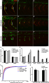
AChR activity or AChR clusters are not required for motor axon outgrowth or neuromuscular synaptogenesis. A–C, Embryos (24 hpf) in which SV2+ presynaptic axons and terminals (green) and rhodamine αBTX labeled AChRs (red) were analyzed in rostral (A, B, C), middle (A′, B′, C′), and caudal (A″, B″, C″) segments. Brackets indicate the location of the first neuromuscular synapses formed at the choice point. A, Motor axon outgrowth, prepatterned AChRs, and initial neuromuscular synaptogenesis in wild-type, unmanipulated control embryos (n = 4 24 hpf embryos, 8 myotome segments). B, Motor axon outgrowth, prepatterned AChRs, and initial neuromuscular synaptogenesis in embryos in which AChRs were blocked with rhodamine αBTX from 12 to 24 hpf (n = 4 24 hpf embryos, 8 myotome segments). The dorsal and ventral extent of motor axon outgrowth, prepatterned AChRs, and initial synaptogenesis are similar to control embryos. C, Motor axon outgrowth and formation of SV2+ presynaptic vesicle clusters in sop embryos (n = 7 24 hpf embryos, 14 myotome segments). Although AChRs are absent, the dorsal and ventral extent of motor axon outgrowth is similar to normal siblings (n = 6 24 hpf embryos, 12 myotome segments) (data not shown). Presynaptic vesicle clusters are present all along the length of axons, but clusters at the choice point (bracket) are enlarged. Scale bar, 10 μm. D, Quantification of motor axon outgrowth, normalized to myotome width, for rostral and middle myotomes in control and αBTX-blocked embryos, sop mutants, and normal siblings. Mean values ± SEM are not significantly different (Student's t test). E, Quantification of SV2+ cluster number and total area in control and αBTX-blocked embryos, sop mutants, and normal siblings, and of AChR cluster and synapse number and total area in control and αBTX-blocked embryos. Number and total area measurements were normalized to axon length. Mean values ± SEM are not significantly different between control and αBTX-blocked embryos or between sop mutants and normal siblings (Student's t test). F, Cumulative percentage of the area of individual SV2+ clusters in control and αBTX-blocked embryos, sop mutants, and normal siblings. Although the mean values for total area are not different among these groups (E), the distribution of the area of individual SV2+ clusters is shifted toward larger values in αBTX-blocked compared with control embryos (asterisks indicate significant differences, Kolmogorov–Smirnovtest, p = 0.002) and in sop mutants compared with normal siblings (p < 0.0001), probably reflecting the enlarged presynaptic SV2+ cluster area at three to four choice point synapses in each myotome. G, Quantification of prepatterned AChR cluster number and total area from caudal segments of control and αBTX-blocked embryos, normalized to myotome width. Mean values ± SEM are not significantly different (Student's t test).

References

    1. Anderson MJ, Cohen MW (1977) Nerve-induced and spontaneous redistribution of acetylcholine receptors on cultured muscle cells. J Physiol (Lond) 268: 757–773. - PMC - PubMed
    1. Aoki C, Venkatesan C, Go CG, Mong JA, Dawson TM (1994) Cellular and subcellular localization of NMDA-R1 subunit immunoreactivity in the visual cortex of adult and neonatal rats. J Neurosci 14: 5202–5222. - PMC - PubMed
    1. Apel ED, Glass DJ, Moscoso LM, Yancopoulos GD, Sanes JR (1997) Rapsyn is required for MuSK signaling and recruits synaptic components to a MuSK-containing scaffold. Neuron 18: 623–635. - PubMed
    1. Arber S, Burden SJ, Harris AJ (2002) Patterning of skeletal muscle. Curr Opin Neurobiol 12: 100–103. - PubMed
    1. Bloch RJ (1988) Molecular events in synaptogenesis: nerve-muscle adhesion and postsynaptic differentiation. Am J Physiol 254: C345–C364. - PubMed

Publication types

Substances

LinkOut - more resources