. 2005 Nov;52(11):1992-2002.
doi: 10.1109/tuffc.2005.1561668.

Influence of lipid shell physicochemical properties on ultrasound-induced microbubble destruction

Affiliations

Influence of lipid shell physicochemical properties on ultrasound-induced microbubble destruction

Mark A Borden et al. IEEE Trans Ultrason Ferroelectr Freq Control. 2005 Nov.

Abstract

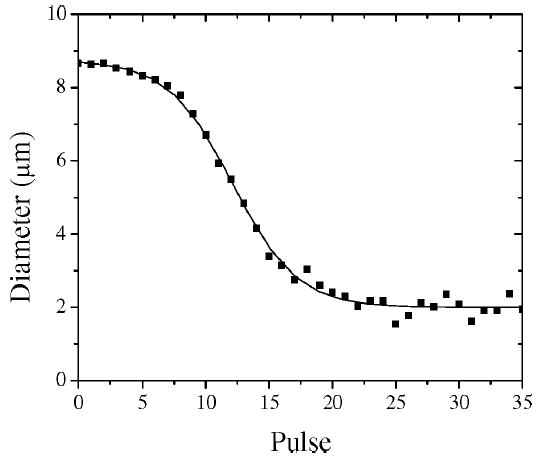
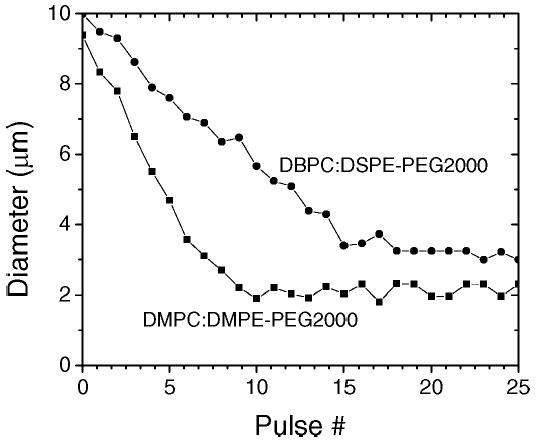
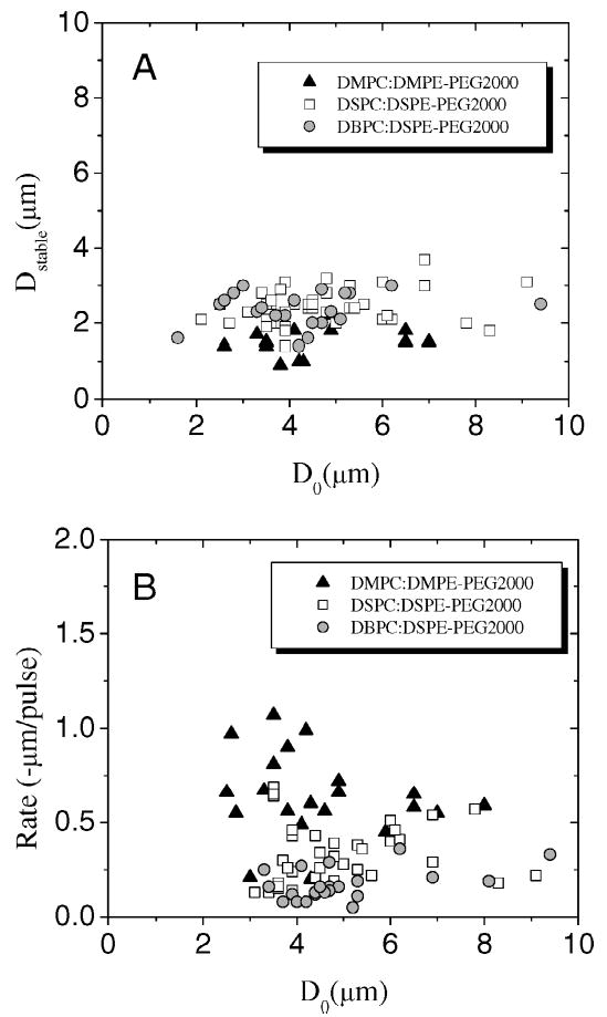
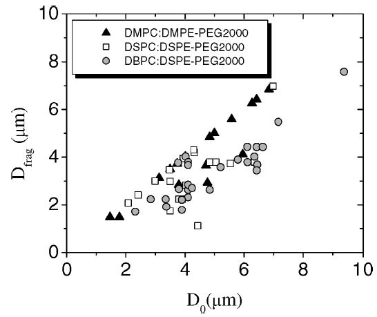
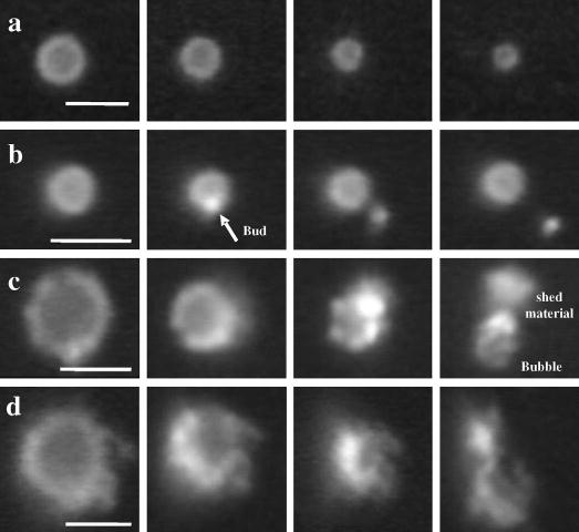
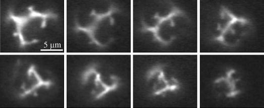
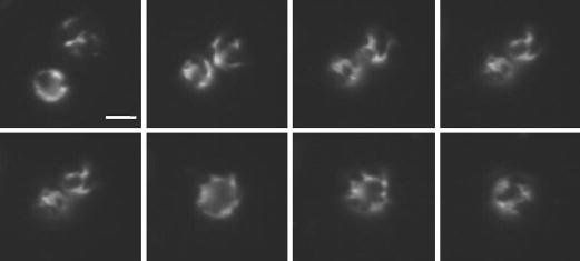
We present the first study of the effects of monolayer shell physicochemical properties on the destruction of lipid-coated microbubbles during insonification with single, one-cycle pulses at 2.25 MHz and low-duty cycles. Shell cohesiveness was changed by varying phospholipid and emulsifier composition, and shell microstructure was controlled by postproduction processing. Individual microbubbles with initial resting diameters between 1 and 10 microm were isolated and recorded during pulsing with bright-field and fluorescence video microscopy. Microbubble destruction occurred through two modes: acoustic dissolution at 400 and 600 kPa and fragmentation at 800 kPa peak negative pressure. Lipid composition significantly impacted the acoustic dissolution rate, fragmentation propensity, and mechanism of excess lipid shedding. Less cohesive shells resulted in micron-scale or smaller particles of excess lipid material that shed either spontaneously or on the next pulse. Conversely, more cohesive shells resulted in the buildup of shell-associated lipid strands and globular aggregates of several microns in size; the latter showed a significant increase in total shell surface area and lability. Lipid-coated microbubbles were observed to reach a stable size over many pulses at intermediate acoustic pressures. Observations of shell microstructure between pulses allowed interpretation of the state of the shell during oscillation. We briefly discuss the implications of these results for therapeutic and diagnostic applications involving lipid-coated microbubbles as ultrasound contrast agents and drug/gene delivery vehicles.


Figures

Fig. 1
Fig. 1
Typical pulse-diameter plot of a single microbubble (DSPC:DSPE-PEG2000) undergoing acoustic dissolution at 2.25 MHz, 400 kPa PNP, 0.5 Hz PRF. Measurements are shown as scatter, and sigmoid curve fit is shown as solid line. Sigmoid behavior exhibits the following three regimes: slow acoustic dissolution at large diameters; linear regime, with a constant acoustic dissolution rate at intermediate diameters; stable microbubble regime for which size does not change over many pulses.
Fig. 2
Fig. 2
Composition dependence of pulse-diameter plots. Data is shown for single microbubbles coated with DMPC:DMPE-PEG2000 (▪) and DBPC:DSPE-PEG2000 (•).
Fig. 3
Fig. 3
Composition and initial resting diameter dependence of acoustic dissolution parameters at 2.25 MHz, 400 kPa PNP, 0.5 Hz PRF. (a) Stable diameter and (b) acoustic dissolution rate of linear regime versus initial resting diameter. Data shown for microbubbles coated with the three phospholipid chain lengths with the lipopolymer emulsifier (i.e., DMPC:DMPE-PEG2000, DSPC:DSPE-PEG2000, and DBPC:DSPE-PEG2000).
Fig. 4
Fig. 4
Composition and initial resting diameter dependence of fragmentation propensity at 2.25 MHz, 800 kPa PNP, 0.5 Hz PRF. Fragmentation diameter (Dfrag), defined as the resting diameter immediately prior to the pulse that induces fragmentation to form two or more daughter bubbles, is shown as a function of the initial resting diameter (D0) for individual microbubbles coated with the three phospholipid chain lengths and the lipopolymer emulsifier (i.e., DMPC:DMPE-PEG2000, DSPC:DSPE-PEG2000, and DBPC:DSPE-PEG2000).
Fig. 5
Fig. 5
Sequential fluorescent video micrographs showing lipid shedding mechanisms as a function of lipid shell composition during intermittent, single-cycle pulsing at 2.25 MHz, 400 kPa PNP: (a) DMPC:DMPE-PEG2000-coated microbubbles shed excess material, probably as diffuse submicron particles, without an observable buildup of collapse lipid on the shell. (b) DSPC:DSPE-PEG2000-coated microbubbles form micron-sized buds that spontaneously shed under thermal motion or due to ultrasound on a subsequent pulse. (c) DSPC:PEG-40 stearate-coated microbubbles exhibit buds that grow during pulsing and detach less readily, significantly increasing the bubble surface area. (d) DBPC:PEG-40 stearate-coated microbubbles exhibit buds and strings that build up to form large lipid particles several microns in diameter after the gas core has been completely eliminated. Scale bars represent 5 μm.
Fig. 6
Fig. 6
Schematic representation showing the progression of possible excess lipid shedding mechanisms during acoustic dissolution with increasing cohesiveness of the phospholipid/emulsifier monolayer shell. Arrows designate a change in shell composition that leads to greater cohesiveness (e.g., increasing phospholipid acyl-chain length).
Fig. 7
Fig. 7
Effect of acoustic dissolution on lipid-shell microstructure (DSPC:DSPE-PEG2000; 2.25 MHz, 600 kPa PNP, intermittent, single-cycle pulsing). Sequential images are shown in chronological order going from left to right then top to bottom. Condensed lipid-domain shapes are conserved after several pulses, and bud formation occurs in interdomain areas as evident from patches of high fluorescence intensity. Scale bar represents 5 μm.
Fig. 8
Fig. 8
Effect of ultrasound-induced coalescence on lipid-shell microstructure (DSPC:DSPE-PEG2000; 2.25 MHz, 400 kPa PNP, 0.5 Hz PRF). Sequential images are shown in chronological order going from left to right then top to bottom. Microbubbles approach each other due to secondary radiation force then coalesce. The presence of condensed-phase domains persist on the fused microbubble. Scale bar represents 5 μm.
None
Mark Andrew Borden (M’04) received the B.S. degree in chemical engineering in 1999 from the University of Arizona, Tucson, and the Ph.D. degree in chemical engineering in 2003 from the University of California, Davis. He conducted undergraduate research on competitive protein adsorption in glass capillaries and peptide affinity to immobilized-metal-ion chromatography columns, and he designed and tested an actuator to accurately control surface tension-driven sample aspiration into glass capillaries. His doctoral research involved production and characterization of diblock copolymer bilayer vesicles and modeling and characterization of the gas transport properties, such as oxygen permeability and surface morphology of lipid-coated microbubbles. He is currently a project scientist in the Department of Biomedical Engineering and Fellow in the Professors for the Future Program at the University of California, Davis. His current research interests include investigation of the physical properties of condensed multicomponent lipid monolayers and engineering-improved ultrasound contrast agents for applications in molecular imaging and drug delivery.
None
Paul Alexander Dayton graduated from Villanova University, Villanova, PA, in 1995 with B.S. degrees in physics and comprehensive science, and a minor in chemistry. As an undergraduate, he conducted research in surface science with the Physics Department and pulse sequence programming for nuclear magnetic resonance spectrometer in the Chemistry Department. He received his M.E. degree in electrical engineering and his Ph.D. degree in biomedical engineering from the University of Virginia, Charlottesville, VA, in 1998 and 2001, respectively. His research involved the study of ultrasound contrast agents. Dr. Dayton conducted post-doctoral research at the University of California, Davis, and is now an assistant research professor at the same institution. His current research interests include targeted imaging and drug delivery.
None
Katherine Whittaker Ferrara (S’82–M’87–SM’99) received the B.S. and M.S. degrees in electrical engineering in 1982 and 1983, respectively, from the California State University, Sacramento, and the Ph.D. degree in electrical engineering and computer science in 1989 from the University of California, Davis. From 1983 to 1988, she worked for Sound Imaging, Inc., Folsom, CA, and for General Electric Medical Systems, Rancho Cordova, CA, in the areas of magnetic resonance and ultrasound imaging. From 1989–1993, she was an associate professor in the Department of Electrical Engineering at California State University, Sacramento. From 1993–1995, she was a principal member of the research staff at the Riverside Research Institute, New York, NY. From 1995–1999, she was an associate professor at the University of Virginia, Charlottesville, VA. She is currently a Professor in the Department of Biomedical Engineering, University of California, Davis, having served as the founding chair of the department from 1999 to 2004. She served as a regular member of the Diagnostic Radiology Study Section (2000–2004), is a Fellow of the Acoustical Society of America, and serves on the Editorial Board of Annual Reviews in Biomedical Engineering.
None
Charles Caskey received his B.S. degree in electrical and computer engineering from the University of Texas, Austin, where he did undergraduate research in elasticity imaging. He is currently pursuing a Ph.D. degree in biomedical engineering at the University of California, Davis. His current research interests include improving vascular permeability with ultrasound contrast agents.
None
Dustin E. Kruse (S’99–M’04) received a B.A. degree in physics from the State University of New York College at Geneseo in 1996, the M.E. degree in electrical engineering from the University of Virginia, Charlottesville, VA, in 1999, and the Ph.D. degree in biomedical engineering in 2004, also from the University of Virginia. His graduate work involved the development and application of high-frequency ultrasound to image blood flow in the microcirculation. His research interests include velocity estimation, ultrasound instrumentation, and contrast-assisted imaging. He is a member of Sigma Pi Sigma.
None
Shukui Zhao (S’02) received the B.S. degree in material science and engineering in 1998 and the M.S. degree in biomedical engineering in 2001, both from Sichuan University, Chengdu, China, and the M.S. degree in electrical engineering in 2003 from St. Cloud State University, St. Cloud, MN. He is currently pursuing a Ph.D. degree in biomedical engineering in the Department of Biomedical Engineering at the University of California, Davis. His current research interests include targeted imaging with ultrasound contrast agent and ultrasound radiation force for enhanced targeting efficiency.

References

    1. Wei K, Jayaweera AR, Firoozan S, Linka A, Skyba DM, Kaul S. “Quantification of myocardial blood flow with ultrasound-induced destruction of microbubbles administered as a constant venous infusion,”. Circulation. 1998;97:473–483. - PubMed
    1. Chomas JE, Dayton P, Allen J, Morgan K, Ferrara KW. “Mechanisms of contrast agent destruction,”. IEEE Trans Ultrason, Ferroelect, Freq Contr. 2001;48:232–248. - PubMed
    1. Lindner JR. “Microbubbles in medical imaging: Current applications and future directions,”. Nature Rev Drug Discovery. 2004;3:527–532. - PubMed
    1. Unger EC, Porter T, Culp W, Labell R, Matsunaga T, Zutshi R. “Therapeutic applications of lipid-coated microbubbles,”. Adv Drug Delivery Rev. 2004;56:1291–1314. - PubMed
    1. Shortencarier MJ, Dayton PA, Bloch SH, Schumann PA, Matsunaga TO, Ferrara KW. “A method for radiation-force localized drug delivery using gas-filled lipospheres,”. IEEE Trans Ultrason, Ferroelect, Freq Contr. 2004;51:822–831. - PubMed

Publication types

MeSH terms