. 2006 Jan;117(1):47-58.
doi: 10.1111/j.1365-2567.2005.02262.x.

Augmented induction of CD8+ cytotoxic T-cell response and antitumour resistance by T helper type 1-inducing peptide

Affiliations

Augmented induction of CD8+ cytotoxic T-cell response and antitumour resistance by T helper type 1-inducing peptide

Takeshi Kikuchi et al. Immunology. 2006 Jan.

Abstract

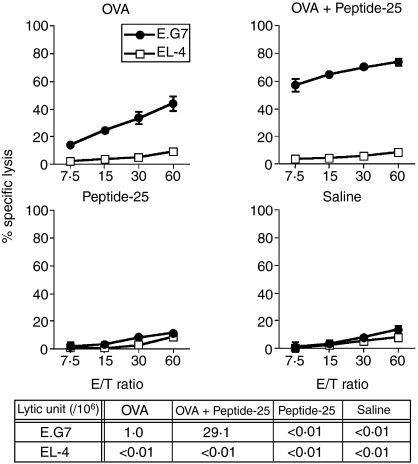
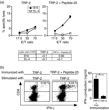
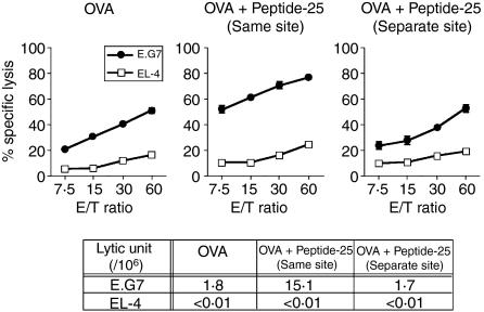
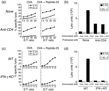
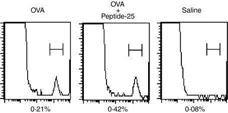
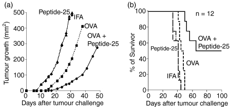
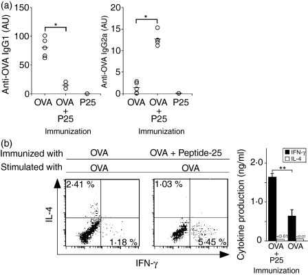
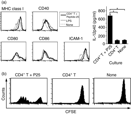
The effector CD8(+) T cells recognize major histocompatibility complex (MHC) class I binding altered self-peptides expressed in tumour cells. Although the requirement for CD4(+) T helper type 1 (Th1) cells in regulating CD8(+) T cells has been documented, their target epitopes and functional impact in antitumour responses remain unclear. We examined whether a potent immunogenic peptide of Mycobacterium tuberculosis eliciting Th1 immunity contributes to the generation of CD8(+) T cells and to protective antitumour immune responses to unrelated tumour-specific antigens. Peptide-25, a major Th epitope of Ag85B from M. tuberculosis preferentially induced CD4(+) Th1 cells in C57BL/6 mice and had an augmenting effect on Th1 generation for coimmunized unrelated antigenic peptides. Coimmunization of mice with Peptide-25 and ovalbumin (OVA) or Peptide-25 and B16 melanoma peptide [tyrosinase-related protein-2 (TRP-2)] for MHC class I led to a profound increase in CD8(+) T cells specific for OVA and TRP-2 peptides, respectively. This heightened response depended on Peptide-25-specific CD4(+) T cells and interferon-gamma-producing T cells. In tumour protection assays, immunization with Peptide-25 and OVA resulted in the enhancement of CD8(+) cytotoxic cell generation specific for OVA and the growth inhibition of EL-4 thymoma expressing OVA peptide leading to the tumour rejection. These phenomena were not achieved by immunization with OVA alone. Peptide-25-reactive Th1 cells counteractivated dendritic cells in the presence of Peptide-25 leading them to activate and present OVA peptide to CD8(+) cytotoxic T cells.


Figures

Figure 1
Figure 1
Enhanced induction of OVA-specific CD8+ CTL response in spleen by coimmunization with OVA and Peptide-25. Three groups of mice were immunized with OVA (10 μg) in IFA, OVA (10 μg) and Peptide-25 (10 μg) in IFA or Peptide-25 (10 μg) in IFA subcutaneously. Spleen cells from each group of mice 10 days after the immunization were subjected to OVA-specific CTL assay. CTL assay was conducted by incubating sensitized cells with 51Cr-labelled E.G7 or 51Cr-labelled EL-4 cells of various effector to target ratios at 37° for 4 hr. The values represent the geometric means and standard deviation of percentage specific lysis in triplicate cultures. Background lysis of the target cells was less than 10% of maximum 51Cr-release. The number of LU was calculated from the dose–response curve obtained for each group. A representative result of a series of five experiments is shown.
Figure 2
Figure 2
Enhancing effect of Peptide-25 on TRP-2-specific CD8+ CTL response by coimmunization with TRP-2. (a) Two groups of mice were immunized subcutaneously with TRP-2 (10 μg) in IFA or TRP-2 (10 μg) and Peptide-25 (P25) (10 μg) in IFA. Spleen cells from each group of mice were subjected to TRP-2-specific CTL assay 10 days after the immunization. CTL assay was conducted by incubating sensitized cells with 51Cr-labelled B16 melanoma or 51Cr-labelled EL-4 cells of various effector to target ratios at 37° for 4 hr. The values represent the geometric means and standard deviation of percentage specific lysis in triplicate cultures. Background lysis of the target cells was less than 9% of maximum 51Cr-release. The number of LU was calculated from the dose–response curve obtained with each group. A representative result of a series of three experiments is shown. (b) Ten days after immunization, spleen cells were stimulated with TRP-2 (10 μg/ml) for 2 days. Intracellular staining of IFN-γ was carried out on the recovered cells and they were examined by FACSCalibur. The percentages of IFN-γ-producing CD8+ cells are presented in the upper right region. IFN-γ production of spleen cells in the culture supernatants was quantified by ELISA. The values represent the mean and standard deviation of the triplicate cultures. *P < 0·01 by Student's t-test.
Figure 3
Figure 3
Enhanced OVA-specific CD8+ T-cell response induced by coimmunization with OVA and Peptide-25 at the same site. A group of mice was immunized with OVA (10 μg) and Peptide-25 (10 μg) in IFA at the same site subcutaneously. Another group of mice was immunized with OVA (10 μg) in IFA and Peptide-25 (10 μg) in IFA at two distant sites (separate sites). As a control, we also immunized a group of mice with OVA (10 μg) in IFA. Spleen cells from each group of mice 10 days after the immunization were subjected to OVA-specific CTL assay. CTL assay was conducted by incubating sensitized cells with 51Cr-labelled E.G7 or 51Cr-labelled EL-4 cells at various effector to target ratios at 37° for 4 hr. The values represent the geometric means and standard deviation of percentage specific lysis in triplicate cultures. Background lysis of the target cells was less than 10% of maximum 51Cr-release. The number of LU was calculated from the dose–response curve obtained with each group. A representative result of a series of three experiments is shown.
Figure 4
Figure 4
Role of CD4+ T cells and IFN-γ in the enhancing effect of Peptide-25 on CTL response. (a,b) Two groups of wild-type mice were injected with anti-CD4 mAb (GK1.5) on days −13, −12, −11, −6, −5, −4, +1, +2 and +3 relative to immunization. Each group of mice was immunized with OVA (10 μg) in IFA or a mixture of OVA (10 μg) and Peptide-25 (10 μg) in IFA on Day 0. As controls, two other groups of mice had been treated with normal rat IgG in place of GK1.5 and immunized with OVA in IFA or a mixture of OVA and Peptide-25 in IFA. Spleen cells from each group of mice were subjected to in vitro OVA-specific CTL assay 10 days after the immunization. (c,d). Either wild-type or IFN-γ–/– mice with C57BL/6 background were immunized with OVA (10 μg) in IFA or a mixture of OVA (10 μg) and Peptide-25 (P25) (10 μg) in IFA. Spleen cells from each group of mice were subjected to OVA-specific CTL assay 10 days after the immunization. CTL assay (a,c) and LU calculations (b,d) were conducted as described in Figure 1.
Figure 5
Figure 5
Frequency analysis of OVA-specific CTL. Two groups of mice were immunized with either OVA in IFA or OVA and Peptide-25 in IFA. Spleen cells from each group of mice were prepared 10 days after the immunization and stained with 4 μg of OVA peptide (SIINFEKL)-loaded H-2Kb:Ig protein and incubated for 60 min at 4°. After washing with staining buffer, cells were resuspended in 100 μl staining buffer containing appropriately diluted anti-mouse IgG1-PE and anti-CD8-FITC. After washing twice with staining buffer, cells were analysed using FACSCalibur.
Figure 6
Figure 6
Enhancement of antitumour immunity by Peptide-25. 51 (a) Suppression of E.G7 growth by augmented induction of E.G7-specific immunity. Three groups of mice were immunized with OVA (10 μg) in IFA, OVA (10 μg) and Peptide-25 (10 μg) in IFA or Peptide-25 (10 μg) in IFA subcutaneously. As a control, a group of mice was injected with IFA. All groups of mice were challenged with 5 × 10⁵ viable E.G7 cells subcutaneously 10 days after the immunization. Growth of E.G7 tumour was monitored by measuring its size periodically (2- to 3-day intervals) and expressed as mm2. (b) Survival of E.G7-bearing mice. The percentages of survivors in the respective groups shown in (a) are displayed.
Figure 7
Figure 7
Enhancement of OVA-specific Th1 response by coimmunization of C57BL/6 mice with OVA and Peptide-25. Two different groups of mice were immunized with OVA (10 μg) in IFA, OVA (10 μg) and Peptide-25 (P25) (10 μg) in IFA, or Peptide-25 (10 μg) in IFA subcutaneously. (a) Serum anti-OVA IgG1 and IgG2a were titrated by ELISA 10 days after the immunization. Each open circle represents the results of an individual mouse. The horizontal bar represents the mean value of six mice. *P < 0·01 by Student's t-test. (b) Ten days after immunization, spleen cells were stimulated in vitro with OVA (10 μg/ml) for 4 days. Intracellular staining of IL-4 and IFN-γ was carried out to the recovered cells. Cells stained were gated on live CD4+ cells and examined by FACSCalibur. The percentages of IL-4- and IFN-γ-producing CD4+ T cells are presented in the upper left and lower right regions, respectively. IL-4 and IFN-γ produced in the culture supernatants were titrated by ELISA. The values represent the mean and standard deviation of the triplicate cultures. **P < 0·05 by Student's t-test.
Figure 8
Figure 8
Activation of DCs by culturing with Peptide-25 in the presence of CD4+ T cells from P25 TCR-Tg mice. (a) Immature DCs were propagated by culturing bone marrow cells with GM-CSF (20 ng/ml) and IL-3 (20 ng/ml) for 6 days. The cells recovered (5 × 10⁵) were cultured with lipopolysaccharide (LPS; 5 μg/ml), Peptide-25 together with CD4+ T cells (5 × 10⁵) from P25 TCR-Tg mice or left untreated for 48 hr. The expression of surface markers was assessed by FACS analysis. IL-12p40 in the cultured supernatant was assessed by ELISA. (b) CFSE-labelled CD8+ T cells (5 × 10⁵) from OT-1 mice were cultured with immature DCs (5 × 10⁵) and OVA (10 μg/ml) for 4 days. The cells were cocultured with CD4+ T cells (5 × 10⁵) from P25 TCR-Tg mice and Peptide-25 (P25) (10 μg/ml) (left panel) or CD4+ T cells from P25 TCR-Tg mice (middle panel). Subsequently, cell division of the CD8+ T cells was monitored by FACSCalibur.

Similar articles

Cited by

References

    1. Boon T, Cerottini JC, Van den Eynde B, Van der Bruggen P, Van Pel A. Tumor antigens recognized by T lymphocytes. Annu Rev Immunol. 1994;12:337–65. - PubMed
    1. Wang RF. The role of MHC class II-restricted tumor antigens and CD4+ T cells in antitumor immunity. Trends Immunol. 2001;22:269–76. - PubMed
    1. Davis ID, Jefford M, Parente P, Cebon J. Rational approaches to human cancer immunotherapy. J Leukoc Biol. 2003;73:3–29. - PubMed
    1. Wagner H, Pfizenmaier K, Rollinghoff M. The role of the major histocompatibility gene complex in murine cytotoxic T cell responses. Adv Cancer Res. 1980;31:77–124. - PubMed
    1. North RJ. The murine antitumor immune response and its therapeutic manipulation. Adv Immunol. 1984;35:89–155. - PubMed

Publication types

MeSH terms