. 2006 Jan 19;49(2):215-28.
doi: 10.1016/j.neuron.2005.12.008.

Signaling mechanisms underlying Slit2-induced collapse of Xenopus retinal growth cones

Affiliations

Signaling mechanisms underlying Slit2-induced collapse of Xenopus retinal growth cones

Michael Piper et al. Neuron. .

Abstract

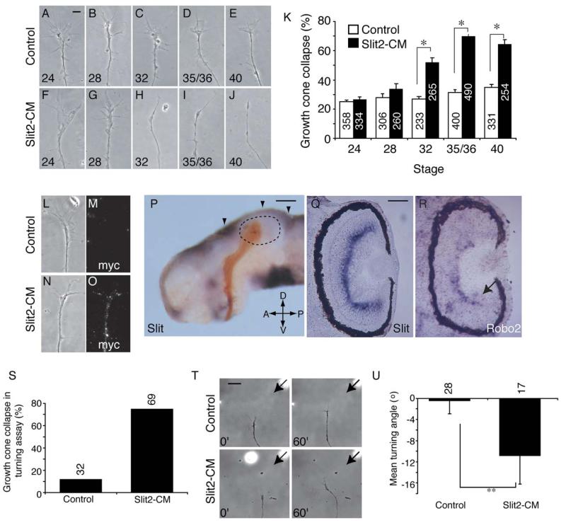
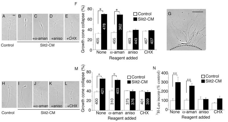
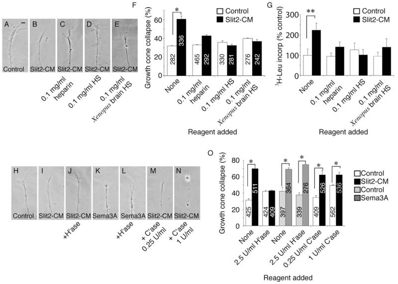
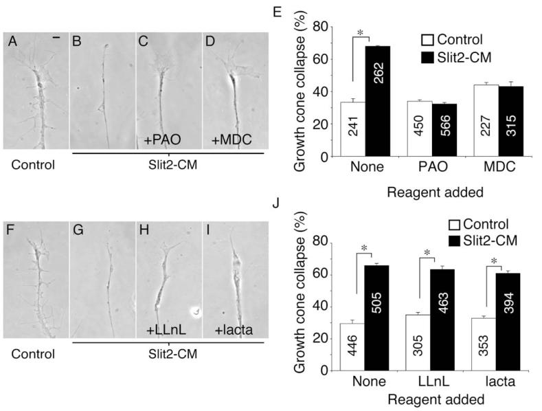
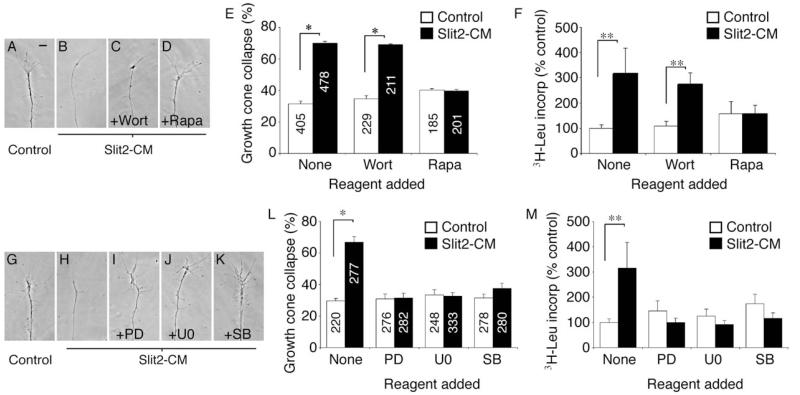
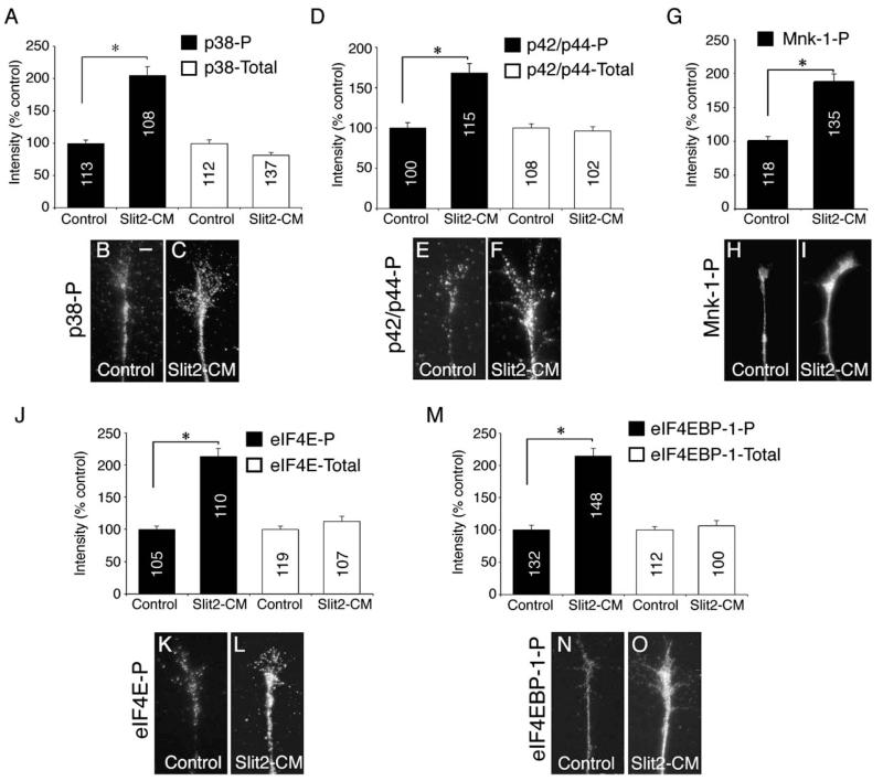
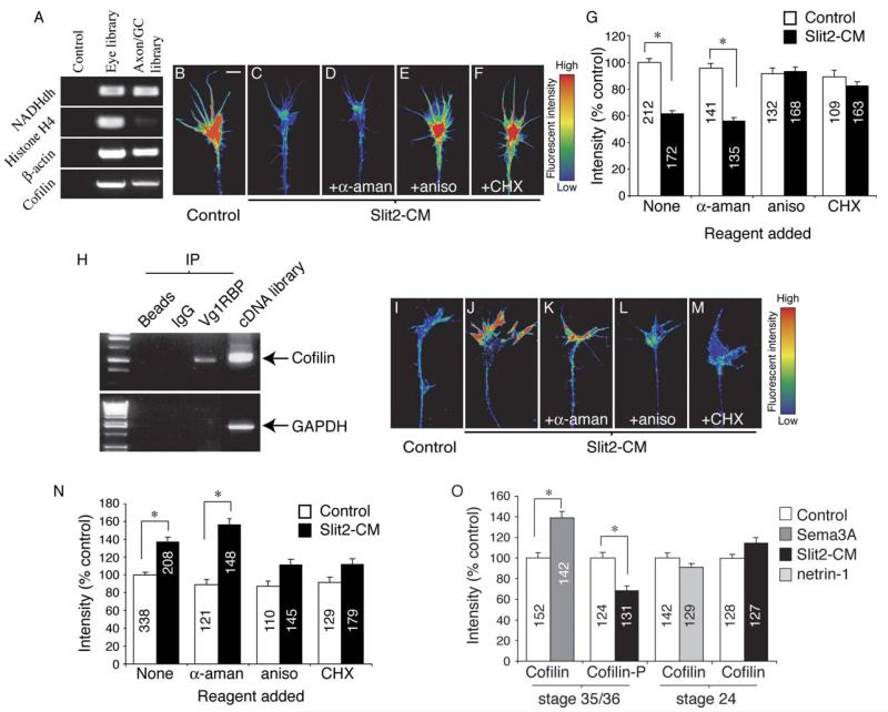
Slits mediate multiple axon guidance decisions, but the mechanisms underlying the responses of growth cones to these cues remain poorly defined. We show here that collapse induced by Slit2-conditioned medium (Slit2-CM) in Xenopus retinal growth cones requires local protein synthesis (PS) and endocytosis. Slit2-CM elicits rapid activation of translation regulators and MAP kinases in growth cones, and inhibition of MAPKs or disruption of heparan sulfate blocks Slit2-CM-induced PS and repulsion. Interestingly, Slit2-CM causes a fast PS-dependent decrease in cytoskeletal F-actin concomitant with a PS-dependent increase in the actin-depolymerizing protein cofilin. Our findings reveal an unexpected link between Slit2 and cofilin in growth cones and suggest that local translation of actin regulatory proteins contributes to repulsion.


Figures

Figure 1
Figure 1
Slit2-CM Causes Turning and Developmentally Regulated Collapse Retinal explants from stage 24 (A and F), 28 (B and G), 32 (C and H), 35/36 (D and I), and 40 (E and J) embryos were cultured for 24 hr prior to addition of Slit2-CM or control for 10 min. Slit2-CM caused growth cone collapse in those explants taken from embryos at stage 32 or later (K). After 2 min, Slit2 bound to the surface of cultured stage 35/36 growth cones was detected with an antibody against the myc epitope (L–O). An isolated brain at stage 40, with retinal axons (brown) entering the tectum (delineated with dotted lines), exhibits Slit mRNA (purple) expression at the dorsal midline and at the anterior and posterior margins of the tectum (arrowheads, [P]). Expression of Slit is also seen in the inner plexiform layer of the eye at stage 40 (Q). Developing RGCs at the periphery of the retina express Robo2 at stage 40 (arrow, [R]), and transcript is also seen in the inner nuclear layer. In turning assays, Slit2-CM caused a high level of collapse compared to the control (S). In those growth cones that did not collapse (T), Slit2-CM caused repulsive turning (U). Numbers inside bars denote growth cones tested. *p < 0.03, Mann-Whitney U test; **p < 0.05, t test. Scale bars: 10 μm (A–J), (L–O), 200 μm (P), 75 μm (Q and R), 20 μm (T).
Figure 2
Figure 2
Collapse Is Blocked by Protein Synthesis Inhibitors Slit2-CM triggers collapse of cultured stage 35/36 retinal growth cones (A and B). Collapse is not affected by the transcriptional inhibitor α-aman, but is blocked by the translational inhibitors aniso and CHX (C–F). To investigate whether this was a local effect, explants were removed prior to the assay being performed (G). Cue-induced collapse was still blocked by translational inhibitors (H–M). Slit2-CM increased the uptake of TCA-precipitated 3H leucine in isolated growth cones in comparison to the control. This effect was inhibited by aniso and CHX, but not by α-aman (N). The counts observed in the aniso- and CHX-treated samples may reflect tRNA-bound 3H leucine. Numbers inside bars denote growth cones tested. *p < 0.03, Mann-Whitney U test; **p < 0.01, Kruskal-Wallis test. Scale bars: 10 μm (A–E and H–L), 100 μm (G).
Figure 3
Figure 3
Heparan Sulfate Is Essential for Slit-Induced Protein Synthesis and Collapse In cultured stage 35/36 retinal explants, Slit2-CM-induced collapse of growth cones is inhibited by the addition of heparin, bovine HS, or HS extracted from embryonic Xenopus brains (A–F). These HSs also inhibited cue-induced PS in isolated neurites (G). Removal of HS moieties via pretreatment with heparinase I (H’ase) resulted in a failure of growth cones to collapse in response to Slit2-CM (H–J and O). Sema3A-induced collapse was unaffected by H’ase treatment (K, L, and O). Pretreatment of explants with chondroitinase ABC (C’ase) did not affect collapse of growth cones caused by Slit2-CM (M–O). Numbers inside bars denote growth cones tested. *p < 0.03, Mann-Whitney U test; **p < 0.05, Kruskal-Wallis test. Scale bar: 10 μm.
Figure 4
Figure 4
Endocytosis, but Not Degradation, Is Required for Slit-Induced Collapse The collapse of retinal growth cones after application of Slit2-CM for 10 min (A and B) was reduced to background levels when the endocytosis inhibitors PAO (C) or MDC (D) were present during the assay (E). Conversely, the proteasomal inhibitors LLnL and lacta did not block growth cone collapse induced by this cue (F–J). Numbers inside bars denote growth cones tested. *p < 0.03, Mann-Whitney U test. Scale bar: 10 μm.
Figure 5
Figure 5
Inhibition of Signaling Pathways Can Block Collapse and Protein Synthesis Specific kinase inhibitors were used to identify signaling components involved in eliciting collapse and PS in response to Slit2-CM. Wortmannin (Wort), an inhibitor of PI-3 kinase, had no effect on collapse (A–C and E) or PS (F), unlike rapamycin (Rapa), which did inhibit collapse (D and E) and PS (F). Similarly, inhibition of the MAPKs MEK1 and MEK2 with PD98059 (PD) or U0126 (U0), and the MAPK p38 with SB203580 (SB), resulted in an abrogation of collapse (G–L) and PS (M) following application of Slit2-CM. Numbers inside bars denote growth cones tested. *p < 0.03, Mann-Whitney U test; **p < 0.05, Kruskal-Wallis test. Scale bar: 10 μm.
Figure 6
Figure 6
Rapid Activation of MAPKs and Translational Regulators To assess the activation of MAPKs and translational regulators 5 min after Slit2-CM application, we used antibodies that specifically recognize the phosphorylated form of these proteins, coupled with digital quantitation of immunofluorescence. Slit2-CM caused a significant increase in phosphorylated p38 (p38-P) and phosphorylated p42/p44 (p42/p44-P) immunoreactivity when compared to controls (A–F). The immunoreactivity of total p38 and p42/p44 was not significantly altered. The level of phosphorylated Mnk-1 (Mnk-1-P) immunofluorescence within growth cones rose following application of Slit2-CM (G–I). The phosphorylated, but not total, forms of the transcriptional regulator eIF4E (J–L) and its binding protein eIF4EBP-1 (M–O) also displayed a significant increase in fluorescent signal intensity. Numbers inside bars denote growth cones tested. *p < 0.001, Kruskal-Wallis test (A, D, J, and M) or Mann-Whitney U test (G). Scale bar: 10 μm.
Figure 7
Figure 7
Slit-Induced Changes in Growth Cone F-Actin and Cofilin Are PS Dependent cDNA libraries were constructed from mRNA extracted from cultured stage 35/36 eyes or isolated stage 35/36 cultured axons and growth cones. β-actin and cofilin were identified from both samples by using PCR (A). Using digital quantitation of immunofluorescence, Slit2-CM was shown to cause an ~40% decrease in growth cone F-actin fluorescent intensity (B, C, and G). This decrease was dependent on translation, but not transcription (D–G). Cofilin mRNA was immunoprecipitated with Vg1RBP bound to protein-A beads. Beads alone or beads coupled to rabbit IgG did not immunoprecipitate cofilin mRNA. GAPDH mRNA did not immunoprecipitate with Vg1RBP. Xenopus cDNA was used as a PCR positive control (H). Slit2-CM significantly raised total growth cone cofilin immunoreactivity after 5 min. This increase was not seen when translation was inhibited by aniso or CHX (I-N). Sema3A also elicited a rise in growth cone cofilin immunoreactivity, while phosphorylated cofilin (cofilin-P) immunofluorescence decreased in response to Slit2-CM. Cultured stage 24 growth cones stimulated with netrin-1 or Slit2-CM did not demonstrate a significant change in total cofilin fluorescent intensity (O). Numbers inside bars denote growth cones tested. Growth cone images have been pseudocolored to represent fluorescent intensity: low intensity, blue; high intensity, red. *p < 0.001, Kruskal-Wallis test. Scale bar: 5 μm.

References

    1. Abe H, Obinata T, Minamide LS, Bamburg JR. Xenopus laevis actin-depolymerizing factor/cofilin: a phosphorylation-regulated protein essential for development. J. Cell Biol. 1996;132:871–885. - PMC - PubMed
    1. Agnes F, Perron M. RNA-binding proteins and neural development: a matter of targets and complexes. Neuroreport. 2004;15:2567–2570. - PubMed
    1. Aizawa H, Wakatsuki S, Ishii A, Moriyama K, Sasaki Y, Ohashi K, Sekine-Aizawa Y, Sehara-Fujisawa A, Mizuno K, Goshima Y, et al. Phosphorylation of cofilin by LIM-kinase is necessary for semaphorin 3A-induced growth cone collapse. Nat. Neurosci. 2001;4:367–373. - PubMed
    1. Alvarez J, Giuditta A, Koenig E. Protein synthesis in axons and terminals: significance for maintenance, plasticity and regulation of phenotype. With a critique of slow transport theory. Prog. Neurobiol. 2000;62:1–62. - PubMed
    1. Arber S, Barbayannis FA, Hanser H, Schneider C, Stanyon CA, Bernard O, Caroni P. Regulation of actin dynamics through phosphorylation of cofilin by LIM-kinase. Nature. 1998;393:805–809. - PubMed

Publication types

MeSH terms

LinkOut - more resources