. 2006 Jan 31;103(5):1581-6.
doi: 10.1073/pnas.0510573103. Epub 2006 Jan 19.

A multifunctional cytoprotective agent that reduces neurodegeneration after ischemia

Affiliations

A multifunctional cytoprotective agent that reduces neurodegeneration after ischemia

Zhi-Gang Jiang et al. Proc Natl Acad Sci U S A. .

Abstract

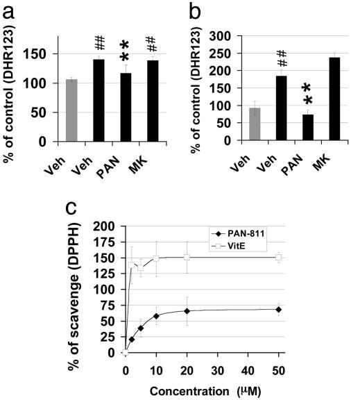
Cellular and molecular pathways underlying ischemic neurotoxicity are multifaceted and complex. Although many potentially neuroprotective agents have been investigated, the simplicity of their protective mechanisms has often resulted in insufficient clinical utility. We describe a previously uncharacterized class of potent neuroprotective compounds, represented by PAN-811, that effectively block both ischemic and hypoxic neurotoxicity. PAN-811 disrupts neurotoxic pathways by at least two modes of action. It causes a reduction of intracellular-free calcium as well as free radical scavenging resulting in a significant decrease in necrotic and apoptotic cell death. In a rat model of ischemic stroke, administration of PAN-811 i.c.v. 1 h after middle cerebral artery occlusion resulted in a 59% reduction in the volume of infarction. Human trials of PAN-811 for an unrelated indication have established a favorable safety and pharmacodynamic profile within the dose range required for neuroprotection warranting its clinical trial as a neuroprotective drug.


Figures

Fig. 1.
Fig. 1.
PAN-811 protects neurons from both ischemic and hypoxic stress. (a) Neurons were subjected to H/H for 6 h and allowed to recover for 24 h. Drug treatment, as indicated, was initiated 24 h before the stress and maintained during the period of ischemia. (b) Neurons were subjected to hypoxia for 18 h and allowed to recover for 24 h. Drug treatment, as indicated, was initiated 24 h before stressing and maintained during the periods of hypoxia and recovery. Cell viability/mitochondrial function was determined via the MTS assay. The gray and black bars indicate treatment under normoxic (control) and stressed conditions, respectively. MTS data are expressed as % protection = (compound treated - stressed)/(normoxia - stressed) × 100%. Figure symbols are ##, P < 0.01 compared with control; **, P < 0.01 compared with stressed. Veh, 1:12,500 PEG:EtOH; PAN, 2 μM PAN-811; GT, 1:200 GT; MK, 5 μM MK-801; Mem, 5 μM memantine.
Fig. 2.
Fig. 2.
Determination of the efficacy, toxicity, and treatment window for PAN-811 neuroprotection. (a and b) Efficacy of PAN-811 vis-à-vis prevention of mitochondrial dysfunction induced by H/H and hypoxia, respectively. (c and d) Efficacy of PAN-811 in suppressing H/H- and hypoxia-induced cell membrane leakage, respectively. (e and f) Neurons treated with a broad dose range of PAN-811 were quantitatively examined with the MTS assay after H/H and hypoxia, respectively. (g) Effect of administration of PAN-811 at various treatment windows on H/H-induced cell membrane leakage (Before, 24-h pretreatment before H/H; During, the 6-h period during H/H; After, for a 48-h period after H/H). (h) Posttreatment window. Two micromolar PAN-811 or 1:12,500 PEG:EtOH was administered at varying times after termination of H/H. Mitochondrial function was measured with MTS assay 24 h after termination of H/H. MTS data in a, b, e, f, and h is expressed as % protection = (treated - stressed)/(normoxia - stressed) × 100%. LDH data in (c, d, and g) are expressed as % protection = (stressed - treated)/(stressed - normoxia) × 100%. Open (or gray) and filled bars indicate vehicle (control) and stressed groups, respectively. Figure symbols are ##, P < 0.01 compared with control; **, P < 0.01 compared with stressed.
Fig. 3.
Fig. 3.
Effects of PAN-811 on the expressions of proapoptotic and antiapoptotic proteins. (a and b) Measurement of pro- and antiapoptotic proteins by Western blotting after H/H and hypoxic stresses, respectively. Band densities in the Western blots were analyzed with imagej (National Institutes of Health) and normalized against α-tubulin. Veh, 1:5,000 PEG:EtOH; PAN, 5 μM PAN-811; MK, 5 μM MK-801, Norm, normoxia, Hypo, hypoxia.
Fig. 4.
Fig. 4.
Neuroprotection by PAN-811 occurs via chelation of [Ca2+]i in H/H. (a) Fura Red dye is responsive to changes in free Ca2+ concentration. Different concentrations of CaCl2 were coincubated with 15.3 μM Fura Red at 37°C for 30 min and measured at Ex450/Em620 and Ex492/Em620 nm, respectively. The Ca2+-bound dye was determined by taking the ratio of emitted fluorescence after excitation at 450/492 nm, which increased after the binding of Ca2+. (b) Chelation of Ca2+ by PAN-811 is shown by the dose-dependent reduction in intensity of Ca2+-bound Fura Red when incubated at various concentrations of PAN-811 with 1 μM CaCl2 in a cell-free system for 30 min at 37°C. EDTA was taken as a positive control. (c and e) The [Ca2+]i levels in hypoxic and H/H stressed neurons were quantified by determination of the ratio of emitted fluorescence at 620 nm after excitation at 450/492 nm after Fura Red staining. (d and f) LDH assay for hypoxia and H/H treated neurons, respectively. In b-f, the gray and black bars indicate treatment under normoxic (control) and stressed conditions, respectively. Figure symbols are ##, P < 0.01 compared with control; **, P < 0.01 compared with stressed. Veh, 1:5,000 PEG:EtOH; PAN, 5 μM PAN-811; MK, 5 μM MK-801.
Fig. 5.
Fig. 5.
Neuroprotection by PAN-811 occurs via free radical scavenging. (a and b) Quantification of DHR123 fluorescence for hypoxia and H/H stressed neurons, respectively. PAN-811, but not MK-801, at a dose of 5 μM inhibited ROS accumulation in hypoxia- or H/H-stressed neurons. (c) Direct free radical scavenging function of PAN-811. % of scavenging = (DPPH - DPPH with compound)/(DPPH × 100%). Gray and black bars indicates treatment under normoxic (control) and stressed conditions, respectively. Figure symbols are ##, P < 0.01 compared with control; **, P < 0.01 compared with stressed. Veh, 1:5000 PEG:EtOH; PAN, 5 μM PAN-811; MK, 5 μM MK-801.
Fig. 6.
Fig. 6.
PAN-811 significantly reduces infarct volume in an animal model of stroke. PAN-811 was administered i.c.v. (50 μg per rat) 1 h after initiation of ischemia (1 h before reperfusion). (a) Rats were killed 22 h after reperfusion and consecutive coronal sections (2 mm thick) were stained with 2,3,5-triphyltetrazolium chloride. Unstained areas indicate the infarcted tissue (see arrow). (b) Infarct volumes were quantified by computer-assisted image analysis and evaluated statistically at a significance level of 5% with the Student t test (**, P < 0.005).

References

    1. Zerwic, J. J., Ennen, K. & DeVon, H. A. (2002) AAOHN J. 50, 354-359. - PubMed
    1. Folbergrova, J., Zhao, Q., Katsura, K. & Siesjo, B. K. (1995) Proc. Natl. Acad. Sci. USA 92, 5057-5061. - PMC - PubMed
    1. Liu, S., Shi, H., Liu, W., Furuichi, T., Timmins, G. S. & Liu, K. J. (2004) J. Cereb. Blood Flow Metab. 24, 343-349. - PubMed
    1. MacGregor, D. G., Avshalumov, M. V. & Rice, M. E. (2003) J. Neurochem. 85, 1402-1411. - PubMed
    1. Kristian, T., Gido, G., Kuroda, S., Schutz, A. & Siesjo, B. K. (1998) Exp. Brain Res. 120, 503-509. - PubMed

Publication types

MeSH terms