. 2006 Jan 31;103(5):1593-8.
doi: 10.1073/pnas.0508901103. Epub 2006 Jan 19.

Leptin: a potential novel antidepressant

Affiliations

Leptin: a potential novel antidepressant

Xin-Yun Lu et al. Proc Natl Acad Sci U S A. .

Abstract

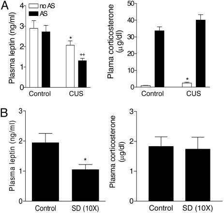
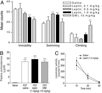
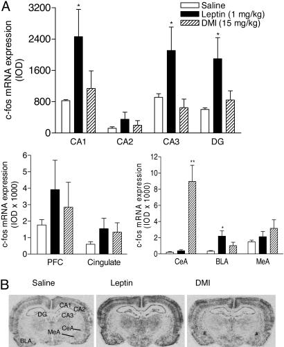
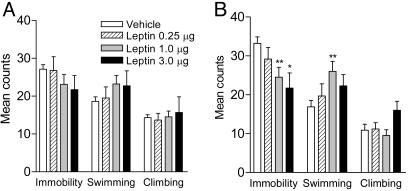
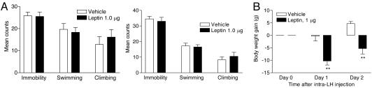
Leptin, a hormone secreted from adipose tissue, was originally discovered to regulate body weight. The localization of the leptin receptor in limbic structures suggests a potential role for leptin in emotional processes. Here, we show that rats exposed to chronic unpredictable stress and chronic social defeat exhibit low leptin levels in plasma. Systemic leptin treatment reversed the hedonic-like deficit induced by chronic unpredictable stress and improved behavioral despair dose-dependently in the forced swim test (FST), a model widely used for screening potential antidepressant efficacy. The behavioral effects of leptin in the FST were accompanied by increased neuronal activation in limbic structures, particularly in the hippocampus. Intrahippocampal infusion of leptin produced a similar antidepressant-like effect in the FST as its systemic administration. By contrast, infusion of leptin into the hypothalamus decreased body weight but had no effect on FST behavior. These findings suggest that: (i) impaired leptin production and secretion may contribute to chronic stress-induced depression-like phenotypes, (ii) the hippocampus is a brain site mediating leptin's antidepressant-like activity, and (iii) elevating leptin signaling in brain may represent a novel approach for the treatment of depressive disorders.


Figures

Fig. 1.
Fig. 1.
Plasma leptin levels after CUS or chronic social defeat. (A) Effect of CUS for 14 days on plasma leptin levels and corticosterone at 24 h after the cessation of the final stressor or after 30 min of acute restraint stress (AS) on day 15. Control, n = 13; control with acute stress, n = 9; CUS, n = 12; CUS with acute stress, n = 11. ANOVAs indicated significant effects for CUS [F(1,40) = 13.37, P < 0.001 for leptin; F(1,40) = 5.55, P < 0.05 for corticosterone] and acute stress [F(1,40) = 4.923, P < 0.05 for leptin; F(1,40), P < 0.0001 for corticosterone]. *, P < 0.05 compared with controls; ++, P < 0.01 compared with control animals subjected to acute stress. (B) Effect of chronic social defeat (SD) for 10 days on plasma leptin levels at 24 h after the last SD. Control, n = 6; SD, n = 6. Two-tailed t test, P < 0.05.
Fig. 2.
Fig. 2.
Effect of leptin treatment on CUS-induced reduction in sucrose preference. (A) Sucrose preference expressed as a ratio of the volume of sucrose intake to the volume of water intake. CUS rats display a substantially decreased sucrose preference. This effect of CUS was reversed by leptin treatment (1 mg/kg, i.p.). ANOVA revealed a significant effect on sucrose preference for time after injection [F(1,26) = 37.38, P < 0.0001] and drug treatment [F(2,13) = 7.498, P < 0.01]. **, P < 0.01 compared with controls. (B) Total amount of fluid intake. The total fluid intake was not affected by leptin treatment. Rats subjected to CUS significantly drink less than handled controls [F(1,28) = 6.525, P < 0.05]. *, P < 0.05. The empty underbar indicates measurements in the light cycle; the filled underbar shows measurements in the dark cycle.
Fig. 3.
Fig. 3.
Behavioral and neuroendocrine effects of leptin treatment. (A) FST. Subacute leptin treatments (23.5, 2.5, and 0.5 h before the test session, s.c.) caused a reduction in immobility time [F(3,31) = 7.043, P = 0.001], accompanied by an increase in swimming time [F(3,31) = 10.11, P < 0.0001] with no significant change in climbing time [F(3,31) = 2.512, P > 0.05]. Saline, n = 14; leptin 0.1 mg/kg, n = 5; leptin 0.5 mg/kg, n = 5; leptin 1.0 mg/kg, n = 12; DMI 15 mg/kg, n = 5. Post hoc analyses indicate that leptin significantly decreases immobility at 1.0 mg/kg (**, P < 0.01) and increased swimming at 0.5 and 1.0 mg/kg (**, P < 0.01), as compared with saline treatment. DMI treatment significantly decreases immobility (**, P < 0.01) and increased climbing (**, P < 0.01). (B) Plasma corticosterone levels 30 min after the FST. The FST increased corticosterone levels markedly compared with nonstressed controls [n = 5-6 per group, F(1,19) = 120.5, P < 0.0001]. This effect was not attenuated by either leptin or DMI treatments [n = 5-6 per group, F(2,12) = 0.466, P = 0.639]. ***, P < 0.001. (C) Locomotor activity. At 24 h after the first exposure to forced swim, leptin treatment (23.5, 2.5, and 0.5 h before the locomotion test, 1.0 mg/kg, s.c.) did not significantly alter locomotor activity (10-min bin) [n = 5 per group; F(1,28) = 0.035, P = 0.852].
Fig. 4.
Fig. 4.
c-fos induction in limbic areas in response to treatments with leptin or DMI after the FST. (A) Changes in mRNA levels of c-fos in the hippocampal formation, prefrontal cortex, cingulate cortex, and amygdala after the FST in rats treated with saline, leptin (1.0 mg/kg), or DMI (15 mg/kg) (23.5, 2.5, and 0.5 h before testing, s.c.). IOD, integrated optical density; CA1, CA2, CA3, fields of the hippocampus; DG, dentate gyrus; PFC, prefrontal cortex; CeA, central nucleus of amygdala; BLA, basolateral nucleus of amygdala; MeA, medial nucleus of amygdala. n = 5 per group. *, P < 0.05; **, P < 0.01 compared with saline treatment. (B) Autoradiograms showing c-fos induction in the hippocampal formation and amygdala.
Fig. 5.
Fig. 5.
Antidepressant-like effect in the FST induced by intrahippocampal injection of leptin. Artificial cerebrospinal fluid (vehicle) or rat leptin at different doses (0.25, 1.0, and 3.0 μg per side) was infused into the hippocampus. Three days later, the FST behavior recorded in the first 5 min of the training session and the 5-min test session was scored. Vehicle, n = 18; leptin 0.25 μg, n = 6; leptin 1.0 μg, n = 13; leptin 3.0 μg, n = 6. (A) Training session (first 5 min). Immobility, F(3,39) = 1.165, P = 0.3354; swimming, F(3,39) = 1.276, P = 0.2961; climbing, F(3,39) = 0.1427, P = 0.934. (B) Test session. Immobility, F(3,39) = 4.509, P < 0.01; swimming, F(3,39) = 3.472, P < 0.05; climbing, F(3,39) = 1.769, P = 0.1689. Post hoc tests, *, P < 0.05; **, P < 0.01 compared with vehicle controls.
Fig. 6.
Fig. 6.
Effect of intrahypothalamic injection of leptin on FST behaviors and body weight. Artificial cerebrospinal fluid (vehicle) or rat leptin (1 μg per side) was stereotaxically infused into the lateral hypothalamus. (A) FST behaviors recorded in the first 5 min of the training session (Left) and the 5-min test session (Right). Vehicle, n = 6; leptin 1 μg, n = 6. (B) Body weight. ANOVA with repeated measures revealed a significant effect of intrahypothalamic injection of leptin (1 μg per side) on body weight [F(1,30) = 13.98, P < 0.01]. **, P < 0.01 compared with vehicle controls.

References

    1. Wong, M. L. & Licinio, J. (2001) Nat. Rev. Neurosci. 2, 343-351. - PubMed
    1. Lenox, R. H. & Frazer, A. (2002) Mechanism of Action of Antidepressants and Mood Stabilizers (Williams & Wilkins, Philadelphia).
    1. Cassano, P. & Fava, M. (2004) Ann. Clin. Psychiatry 16, 15-25. - PubMed
    1. Banks, W. A., Kastin, A. J., Huang, W., Jaspan, J. B. & Maness, L. M. (1996) Peptides 17, 305-311. - PubMed
    1. Ahima, R. S. & Osei, S. Y. (2004) Physiol. Behav. 81, 223-241. - PubMed

Publication types

MeSH terms