. 2006 Feb 1;176(3):1561-70.
doi: 10.4049/jimmunol.176.3.1561.

TLR agonists abrogate costimulation blockade-induced prolongation of skin allografts

Affiliations

TLR agonists abrogate costimulation blockade-induced prolongation of skin allografts

Thomas B Thornley et al. J Immunol. .

Abstract

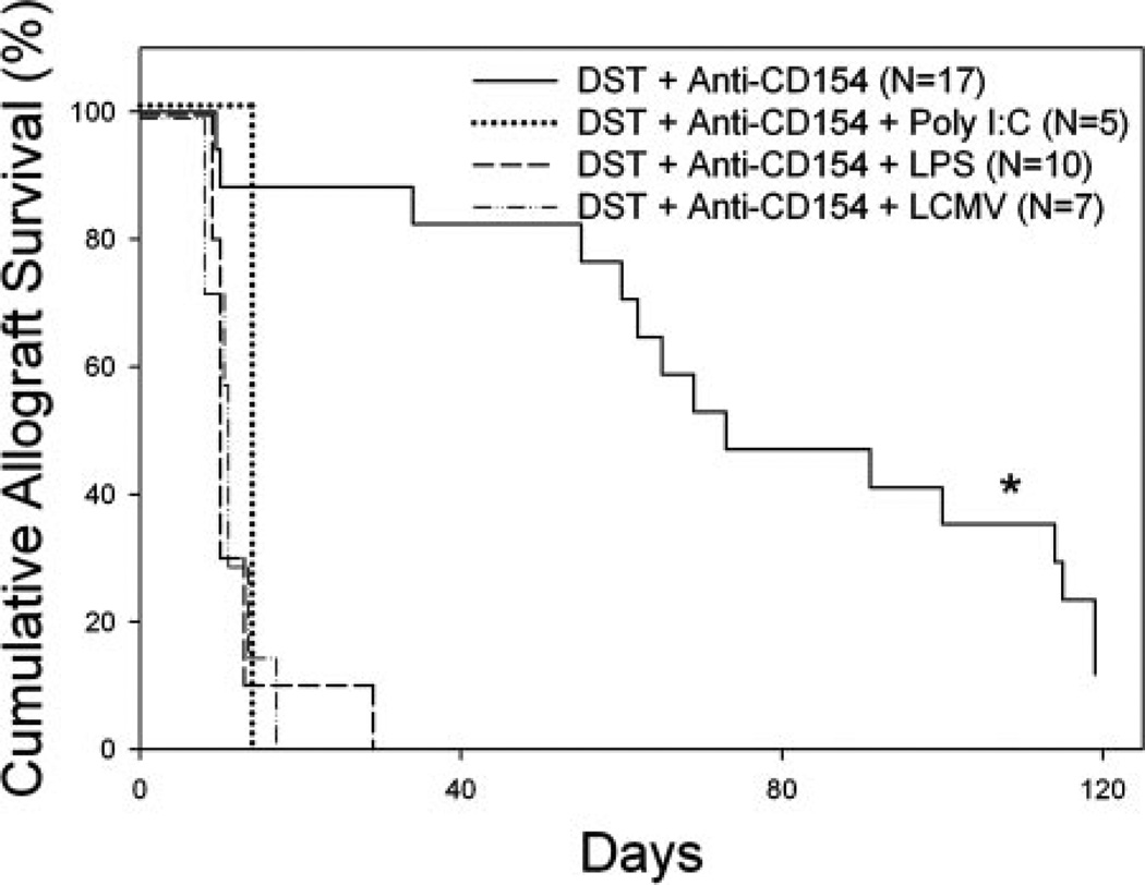
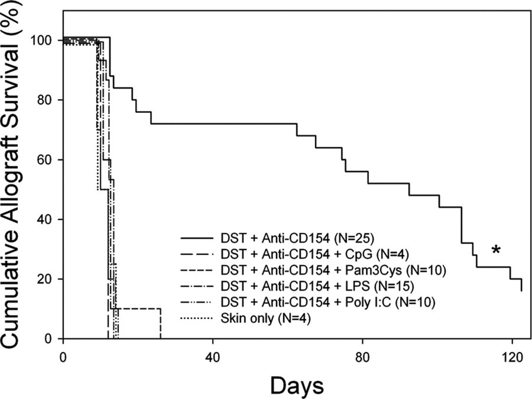
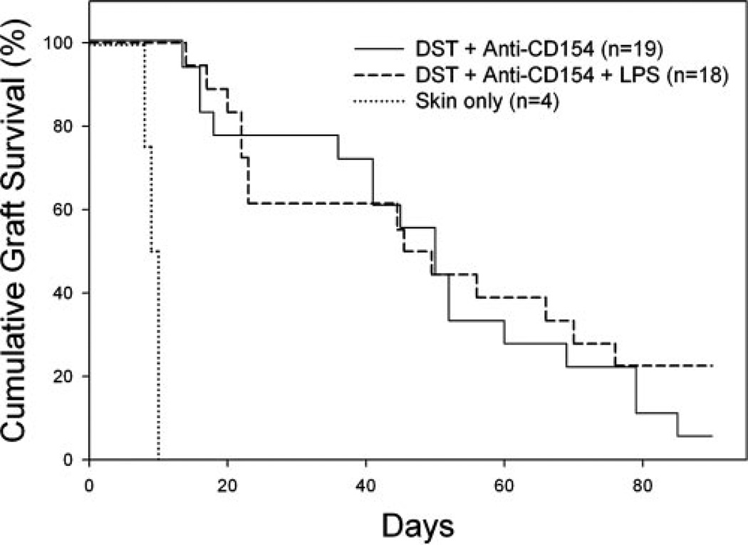
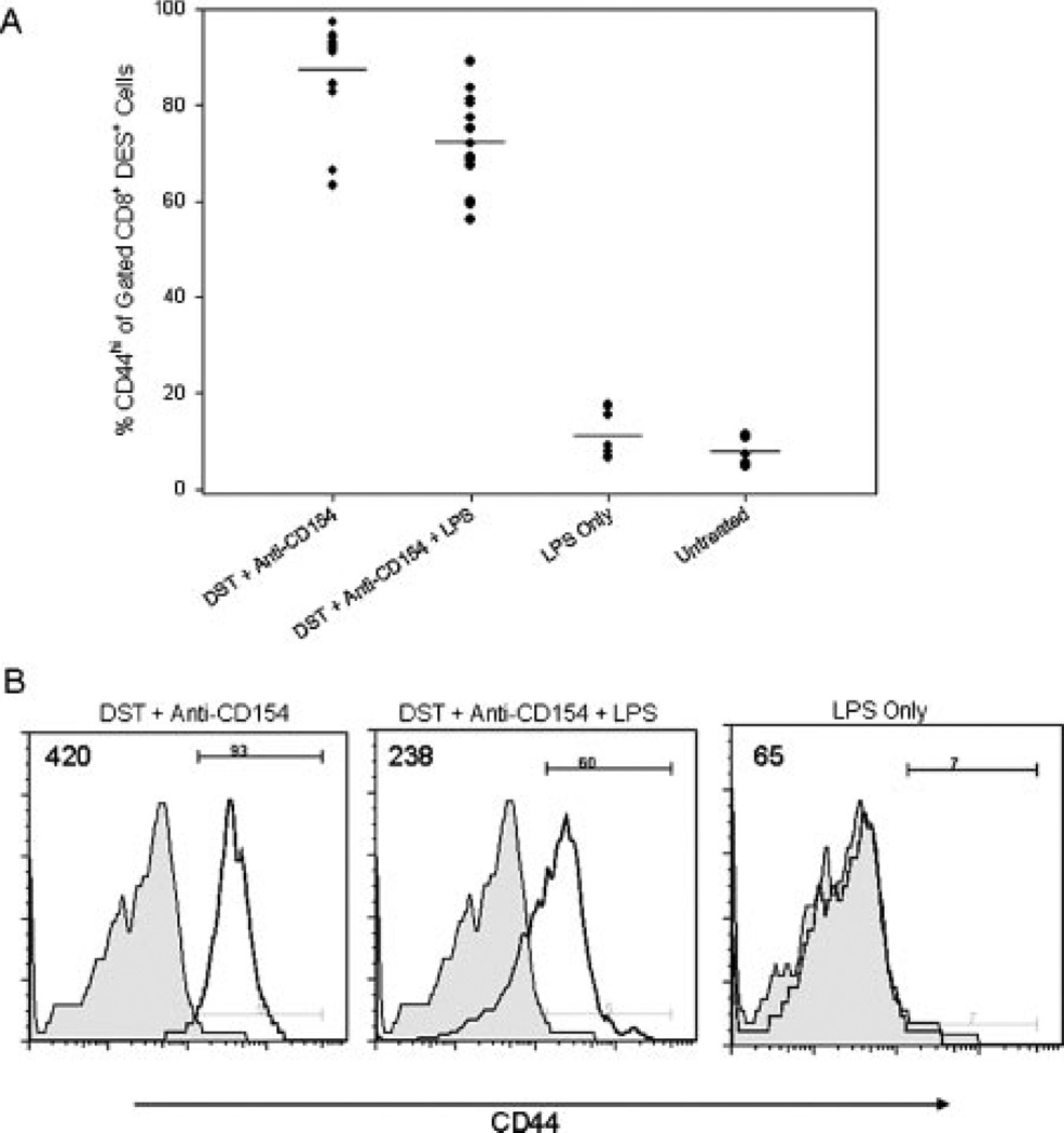
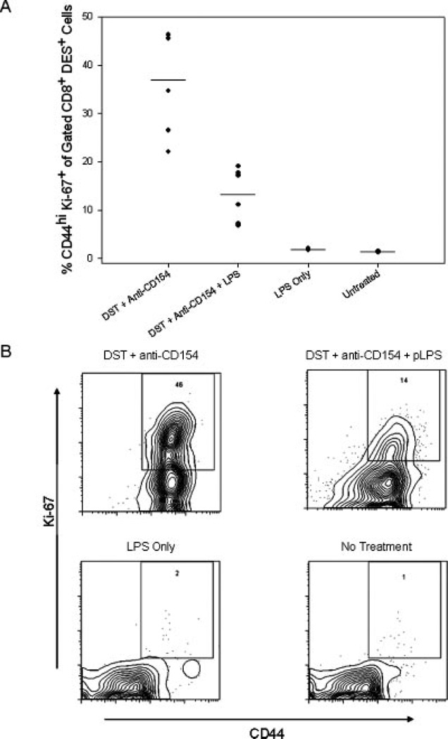
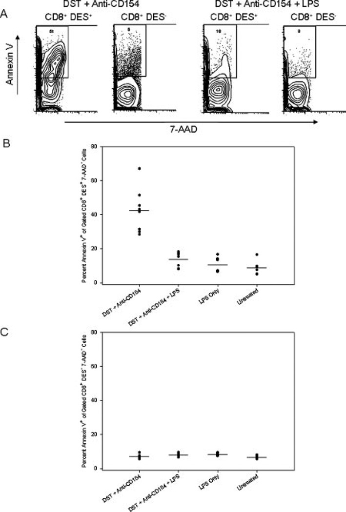
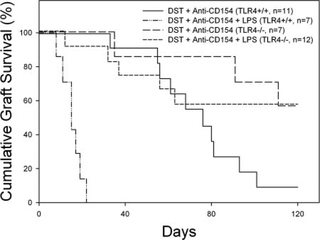
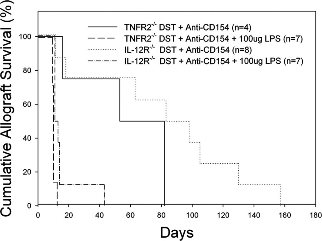
Costimulation blockade protocols are effective in prolonging allograft survival in animal models and are entering clinical trials, but how environmental perturbants affect graft survival remains largely unstudied. We used a costimulation blockade protocol consisting of a donor-specific transfusion and anti-CD154 mAb to address this question. We observed that lymphocytic choriomeningitis virus infection at the time of donor-specific transfusion and anti-CD154 mAb shortens allograft survival. Lymphocytic choriomeningitis virus 1) activates innate immunity, 2) induces allo-cross-reactive T cells, and 3) generates virus-specific responses, all of which may adversely affect allograft survival. To investigate the role of innate immunity, mice given costimulation blockade and skin allografts were coinjected with TLR2 (Pam3Cys), TLR3 (polyinosinic:polycytidylic acid), TLR4 (LPS), or TLR9 (CpG) agonists. Costimulation blockade prolonged skin allograft survival that was shortened after coinjection by TLR agonists. To investigate underlying mechanisms, we used "synchimeric" mice which circulate trace populations of anti-H2b transgenic alloreactive CD8+ T cells. In synchimeric mice treated with costimulation blockade, coadministration of all four TLR agonists prevented deletion of alloreactive CD8+ T cells and shortened skin allograft survival. These alloreactive CD8+ T cells 1) expressed the proliferation marker Ki-67, 2) up-regulated CD44, and 3) failed to undergo apoptosis. B6.TNFR2-/- and B6.IL-12R-/- mice treated with costimulation blockade plus LPS also exhibited short skin allograft survival whereas similarly treated B6.CD8alpha-/- and TLR4-/- mice exhibited prolonged allograft survival. We conclude that TLR signaling abrogates the effects of costimulation blockade by preventing alloreactive CD8+ T cell apoptosis through a mechanism not dependent on TNFR2 or IL-12R signaling.


Conflict of interest statement

Disclosures

The authors have no financial conflict of interest.

Figures

FIGURE 1
FIGURE 1
LPS and poly I:C abrogate prolonged allograft survival induced by costimulation blockade in C57BL/6 mice. C57BL/6 mice were treated with BALB/c DST and anti-CD154 mAb according to our standard protocol with or without injection of the indicated TLR agonist or infection with LCMV on day −7 relative to transplantation with a BALB/c skin allograft on day 0. *, Statistically significantly different than all others; p < 0.0001.
FIGURE 2
FIGURE 2
TLR agonists abrogate prolonged allograft survival induced by costimulation blockade in synchimeric CBA/J mice. KB5 synchimeras were treated with C57BL/6 DST and anti-CD154 mAb according to our standard protocol with or without injection of the indicated TLR agonist on day −7 relative to transplantation with a C57BL/6 skin allograft on day 0. Mice receiving the “‘skin only” treatment received no preconditioning before transplantation. *, Statistically significantly different than all others; p < 0.0001.
FIGURE 3
FIGURE 3
CD8+ cells are required for LPS to shorten allograft survival induced by costimulation blockade. B6.CD8α−/− mice were given DST and anti-CD154 mAb according to our standard protocol with or without injection of LPS on day −7 relative to skin grafting on day 0. The difference between groups receiving DST and anti-CD154 mAb or DST, anti-CD154 mAb, and LPS was not significant; p = NS.
FIGURE 4
FIGURE 4
Alloreactive CD8+ T cells have an activated phenotype 72 h after treatment with DST and anti-CD154 mAb. KB5 synchimeras were treated with DST and anti-CD154 mAb in the presence or absence of coinjection with LPS and analyzed 72 h later. Splenocytes were stained with purified DES mAb, followed by the DES detection mAb (anti-mIgG2a-FITC), anti-CD8-PerCP mAb, and anti-CD44-allophycocyanin mAb. Samples were fixed and analyzed by flow cytometry. Samples were gated on DES+CD8+ lymphocytes and analyzed for CD44 expression. A, A summary of experimental mice in four independent trials with means indicated by the solid lines (n = 7–13/group). B, Representative flow histograms showing CD44 expression on gated DES+CD8+ lymphocytes. Shaded histograms represent untreated control mice. The MFI of a representative sample from each experimental group is indicated in the upper left corner of each panel. The percentage of CD8+ T cells that was CD44high in the DST and anti-CD154 mAb-treated group was significantly higher than that observed in the DST, anti-CD154 mAb, and LPS-treated group; p < 0.001.
FIGURE 5
FIGURE 5
Alloreactive CD8+ T cells are proliferating in the spleen 72 h after treatment with DST and anti-CD154 mAb. KB5 synchimeras were treated with DST and anti-CD154 mAb in the presence or absence of injection of LPS and analyzed 72 h later. Splenocytes were stained with the clonotypic Ab DES, followed by the DES detection mAb (anti-mIgG2a-FITC), anti-CD8-PerCP mAb and anti-CD44-allophycocyanin mAb. Cells were then fixed, permeabilized, and stained for the nuclear proliferation Ag Ki-67 with an anti-Ki-67-PE mAb. Samples were gated on DES+CD8+ lymphocytes and analyzed for Ki-67 expression. A, A summary of experimental mice from two independent experiments with the mean indicated by the solid lines (n = 3–6 mice/group). B, Representative flow dot plots showing CD44 and Ki-67 expression in gated DES+CD8+ lymphocytes.
FIGURE 6
FIGURE 6
LPS protects alloreactive CD8+ T cells from apoptosis in mice treated with costimulation blockade. KB5 synchimeras were treated with DST and anti-CD154 mAb in the presence or absence of coinjection of LPS and analyzed 72 h later. Splenocytes were stained with the clonotypic Ab DES, followed by the DES detection mAb (anti-mIgG2a–FITC), anti-CD8-PerCP mAb, Annexin VPE, and the viability dye 7-AAD. A, Representative contour plot showing annexin V staining after gating on live (7AAD) transgenic (CD8+DES+) or nontransgenic (CD8+ DES) T cells. B, Percent of live (7-AAD) transgenic CD8+ T cells binding annexin V. C, Percent of live nontransgenic CD8+ T cells binding annexin V. Graphs represent combined data from two independent experiments (n = 6–9 mice/group).
FIGURE 7
FIGURE 7
TLR4 is required on host cells but is not required on donor cells. C57BL/10ScSn (TLR4+/+) and C57BL/10ScN (TLR4−/−) mice were given DST, anti-CD154 mAb, and skin allografts according to our standard protocol with or without coinjection of LPS at the time of co-stimulation blockade. Skin allograft survival in tolerized TLR4+/+ C57BL/ 10ScSn mice was significantly prolonged (MST = 76 days, n = 11) as compared with allograft survival in mice coinjected with LPS (MST = 15 days, n = 7, p < 0.0001). Skin allograft survival in tolerized TLR4−/− C57BL/10ScN mice was similar in the presence (MST >128 days, n = 7) or absence (MST >128 days, n = 12, p = NS) of LPS.
FIGURE 8
FIGURE 8
TNFR2 and IL-12R are not required for LPS to shorten allograft survival induced by costimulation blockade. B6.TNFR2−/− and B6.IL-12R−/− mice were given DST and anti-CD154 mAb according to our standard protocol with or without injection of LPS on day −7 relative to skin grafting on day 0. Skin allograft survival in tolerized B6.TNFR2−/− mice was significantly prolonged (MST = 68 days, n = 4) as compared with allograft survival in mice coinjected with LPS (MST = 11 days,p < 0.005). Skin allograft survival in tolerized B6.IL-12R−/− mice significantly prolonged (MST = 91 days, n = 8) as compared with allograft survival in mice coinjected with LPS (MST = 12 days, n = 4, p < 0.005).

References

    1. Rickels MR, Schutta MH, Markmann JF, Barker CF, Naji A, Teff KL. β-cell function following human islet transplantation for type 1 diabetes. Diabetes. 2005;54:100–106. - PubMed
    1. Street CN, Lakey JRT, Shapiro AMJ, Imes S, Rajotte RV, Ryan EA, Lyon JG, Kin T, Avila J, Tsujimura T, Korbutt GS. Islet graft assessment in the Edmonton protocol-implications for predicting long-term clinical outcome. Diabetes. 2004;53:3107–3114. - PubMed
    1. Shapiro AMJ, Lakey JRT, Ryan EA, Korbutt GS, Toth E, Warnock GL, Kneteman NM, Rajotte RV. Islet transplantation in seven patients with type 1 diabetes mellitus using a glucocorticoid-free immunosuppressive regimen. N Engl. J. Med. 2000;343:230–238. - PubMed
    1. Ryan EA, Lakey JRT, Paty BW, Imes S, Korbutt GS, Kneteman NM, Bigam D, Rajotte RV, Shapiro AMJ. Successful islet transplantation-continued insulin reserve provides long-term glycemic control. Diabetes. 2002;51:2148–2157. - PubMed
    1. Teraoka S, Kawai T, Yamaguchi Y, Fujita S, Tojinbara T, Nakajima T, Hayashi T, Nakagawa Y, Fujikawa H, Honda H, et al. Mechanism of the preventive effect of aminobenzoic acid salt on pancreatic β cell toxicity by cyclosporin. Transplant. Proc. 1990;22:863–866. - PubMed

Publication types

MeSH terms