. 2006 Jan 20;311(5759):388-91.
doi: 10.1126/science.1122142.

Chromosomes can congress to the metaphase plate before biorientation

Affiliations

Chromosomes can congress to the metaphase plate before biorientation

Tarun M Kapoor et al. Science. .

Abstract

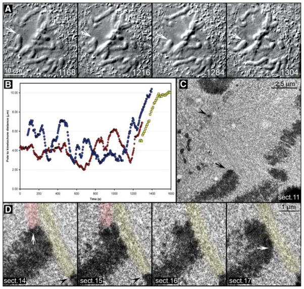
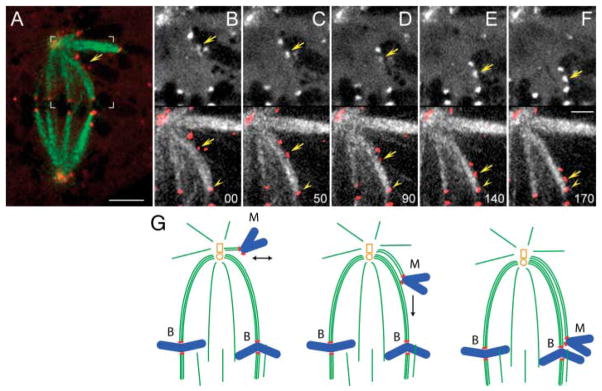
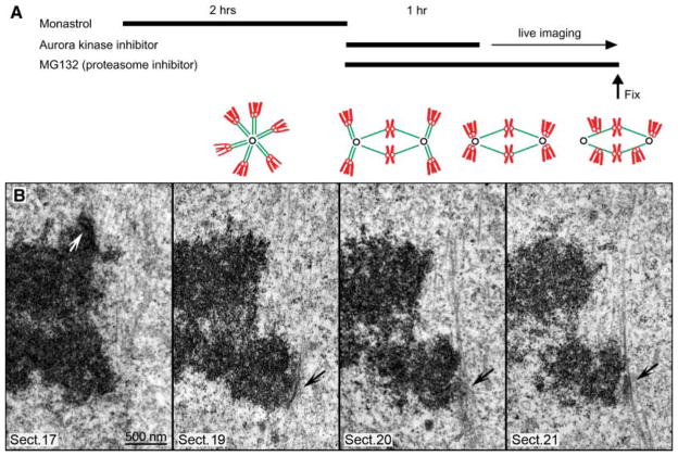
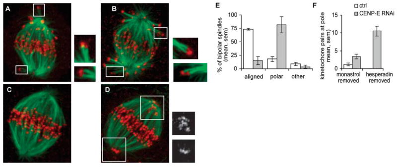
The stable propagation of genetic material during cell division depends on the congression of chromosomes to the spindle equator before the cell initiates anaphase. It is generally assumed that congression requires that chromosomes are connected to the opposite poles of the bipolar spindle ("bioriented"). In mammalian cells, we found that chromosomes can congress before becoming bioriented. By combining the use of reversible chemical inhibitors, live-cell light microscopy, and correlative electron microscopy, we found that monooriented chromosomes could glide toward the spindle equator alongside kinetochore fibers attached to other already bioriented chromosomes. This congression mechanism depended on the kinetochore-associated, plus end-directed microtubule motor CENP-E (kinesin-7).


Figures

Fig. 1
Fig. 1
Leading kinetochores are not properly attached to microtubules during a chromosome’s attempt to congress. (A) Selected frames from a DIC time-lapse recording (also see Movie S2). The cell was fixed as one chromosome (arrows) moved toward the spindle equator (1304 s). (B) Distance versus time plot confirmed that the chromosome’s movement (red curve) was typical for chromosome congression [compare with blue and yellow curves, which represent movements of chromosomes shown in fig. S1 and Fig. 2, respectively]. (C) Lower-magnification EM image of the cell showing the position of the chromosome of interest (arrow) with respect to the spindle pole (arrowhead). (D) Selected 100-nm EM section from a full series through the centromere region of the chromosome. Note the prominent bundle of microtubules (highlighted red) connecting the trailing kinetochore (white arrow in section 14) to the proximal spindle pole. These microtubules approached the kinetochore at ~90° angle and terminated within the trilaminar kinetochore plate. By contrast, the leading kinetochore (white arrow, section 17) lacked attached microtubules but was laterally associated with a mature kinetochore fiber (highlighted yellow) that was attached to the kinetochore of a bioriented chromosome (black arrowheads in sections 14 and 15) positioned on the metaphase plate.
Fig. 2
Fig. 2
Monooriented chromosomes are transported toward the spindle equator along kinetochore fibers of other chromosomes. (A) Two-color fluorescence image of a live PtK1 cell in which kinetochores were labeled with CENP-F/Alexa488 (red) and microtubules with tubulin/rhodamine (green). Area marked with white brackets is enlarged in (B to F). (B to F) selected frames from the two-color time-lapse recording. In each frame, CENP-F/Alexa488 fluorescence (kinetochores) is shown alone (top) and overlaid in red on microtubules (bottom). Arrows mark the kinetochore that moved toward the spindle equator. Note that trajectory of this kinetochore coincided with a prominent kinetochore fiber that extended from the spindle pole to a kinetochore on a bioriented chromosomes already positioned on the metaphase plate (arrowhead). Time in seconds. Scale bars: (A) 5 μm, (F) 2.5 μm. (G) Schematic illustrating the sequence of events presented in (B to F).
Fig. 3
Fig. 3
Leading kinetochores are laterally associated with kinetochore fibers of other chromosomes during chromosome congression. (A) Protocol for inducing synchronous chromosome congression. Cells were arrested in mitosis with monastrol to accumulate monopolar mitosis with high incidence of syntelic chromosomes (green, microtubules; red, chromosomes). Then, monastrol was removed and Hesperadin was added with MG132 for 1 hour. This resulted in spindle bipolarization, although many chromosomes remained syntelic. After 1 hour, Hesperadin was removed, and cells were imaged live until fixation. Removal of Hesperadin resulted in simultaneous correction of syntelic attachments. Syntelic chromosomes moved to the pole, became monooriented, and then attempted to congress. (B) Selected 100-nm EM sections from a full series through the centromere of a congressing chromosome. Note that, similarly to untreated cells (Fig. 1), chromosomes congressed with their leading kinetochores unattached (black arrows) but slide alongside mature kinetochore fibers of other chromosomes. By contrast, trailing kinetochores (white arrow) were always attached to prominent kinetochore fibers that terminated within the trilaminar plate.
Fig. 4
Fig. 4
CENP-E is required for congression of monooriented chromosomes. Twenty-four hours after transfection, with mock (control) or CENP-E siRNA, synchronous chromosome congression was induced using the chemical inhibition and reactivation approach described in Fig. 3. Cells were fixed either after 1 hour in Hesperadin (A and B) or 1 hour after removal of Hesperadin (C to F) and processed for immunostaining [tubulin, green; kinetochores (CREST), red]. In the presence of Hesperadin, syntelic attachments were observed in both control (A) and CENP-E–depleted cells (B). After removal of Hesperadin, chromosomes congressed to the metaphase plate in control cells (C), but monooriented chromosomes were observed near spindle poles in CENP-E–depleted cells (Insets in D) CREST staining. For cells fixed 1 hour after removal of Hesperadin, bipolar spindles were counted and classified as fully aligned or containing polar chromosome(s) (E) (average of four experiments). To quantify the number of chromosomes at the pole, the number of kinetochore pairs with no detectable K fiber was counted in three-dimensional confocal images [(F), averages from 19 CENP-E–depleted or 7 control cells for each condition, two experiments]. All images presented as maximal-intensity projections. (Insets in A and B) Optical sections at 2× magnification.

Comment in

References

    1. Rieder CL, Salmon ED. Trends Cell Biol. 1998;8:310. - PMC - PubMed
    1. Rajagopalan H, Lengauer C. Nature. 2004;432:338. - PubMed
    1. Roos UP. Chromosoma. 1976;54:363. - PubMed
    1. Rieder CL, Alexander SP. J Cell Biol. 1990;110:81. - PMC - PubMed
    1. Kirschner M, Mitchison T. Cell. 1986;45:329. - PubMed

Publication types

MeSH terms