. 2006 May 15;107(10):3976-82.
doi: 10.1182/blood-2005-11-4551. Epub 2006 Jan 19.

Enhancement of infectivity and persistence in vivo by HBZ, a natural antisense coded protein of HTLV-1

Affiliations

Enhancement of infectivity and persistence in vivo by HBZ, a natural antisense coded protein of HTLV-1

Joshua Arnold et al. Blood. .

Abstract

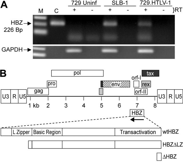
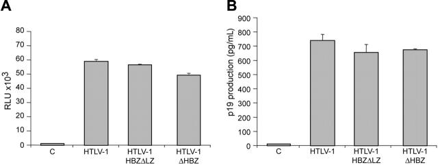
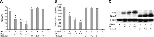
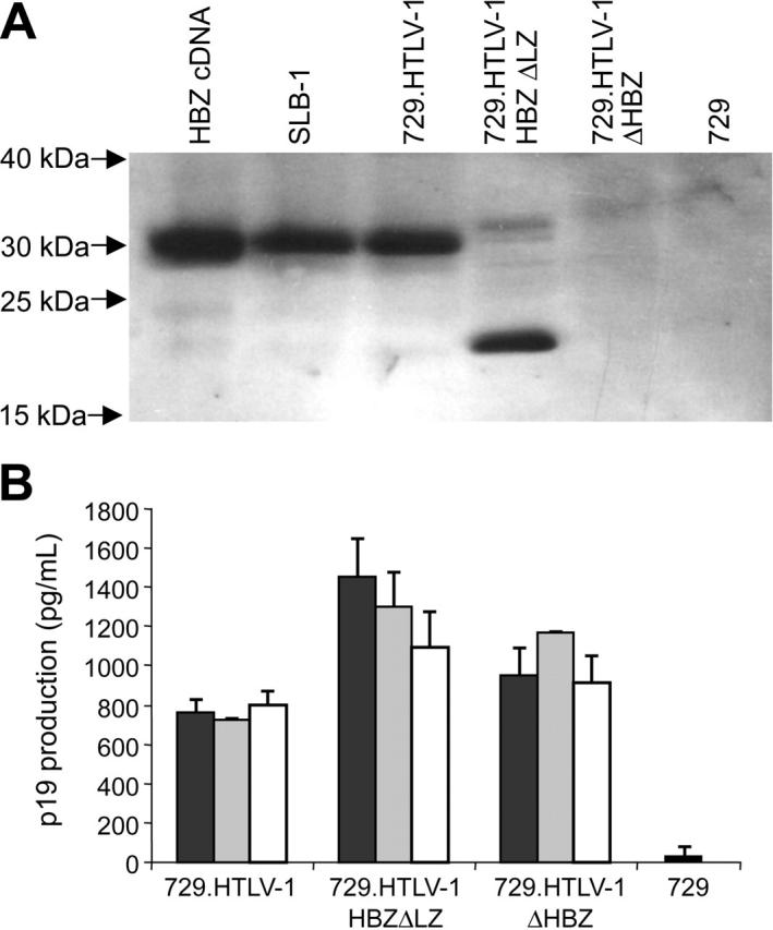
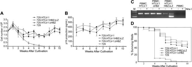
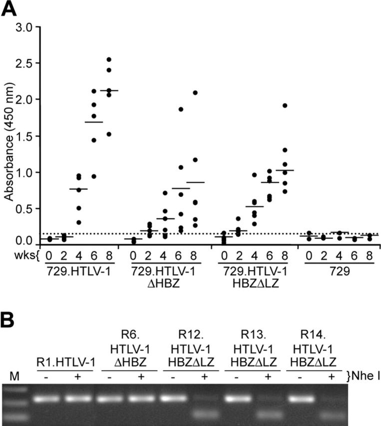
Natural antisense viral transcripts have been recognized in retroviruses, including human T-cell leukemia virus type 1 (HTLV-1), HIV-1, and feline immunodeficiency virus (FIV), and have been postulated to encode proteins important for the infection cycle and/or pathogenesis of the virus. The antisense strand of the HTLV-1 genome encodes HBZ, a novel nuclear basic region leucine zipper (b-ZIP) protein that in overexpression assays down-regulates Tax oncoprotein-induced viral transcription. Herein, we investigated the contribution of HBZ to HTLV-1-mediated immortalization of primary T lymphocytes in vitro and HTLV-1 infection in a rabbit animal model. HTLV-1 HBZ mutant viruses were generated and evaluated for viral gene expression, protein production, and immortalization capacity. Biologic properties of HBZ mutant viruses in vitro were indistinguishable from wild-type HTLV-1, providing the first direct evidence that HBZ is dispensable for viral replication and cellular immortalization. Rabbits inoculated with irradiated cells expressing HTLV-1 HBZ mutant viruses became persistently infected. However, these rabbits displayed a decreased antibody response to viral gene products and reduced proviral copies in peripheral blood mononuclear cells (PBMCs) as compared with wild-type HTLV-1-infected animals. Our findings indicated that HBZ was not required for in vitro cellular immortalization, but enhanced infectivity and persistence in inoculated rabbits. This study demonstrates that retroviruses use negative-strand-encoded proteins in the establishment of chronic viral infections.


Figures

Figure 1.
Figure 1.
Detection of the HBZ RNA transcript. (A) HBZ transcript was detected in Poly A+ RNA isolated from SLB-1 and 729.HTLV-1, but not 729 uninfected (Uninf), using standard RT-PCR. First-strand synthesis with a specific oligo designed to copy only HTLV-1 antisense RNA containing the HBZ coding sequence was performed in the presence and absence of reverse transcriptase. The 226-bp PCR product was separated on a 2% agarose gel and visualized by ethidium bromide staining. (B) Schematic representation of the complete HTLV-1 proviral genome is shown. LTRs are depicted with their U3, R, and U5 regions. The location of the viral open reading frames and the opposite-strand HBZ are indicated. The reported HBZ coding sequence has been expanded showing the transactivation domain, basic region, and leucine-zipper region, as well as the 2 HBZ truncation mutants generated for this study (HBZΔLZ and ΔHBZ).
Figure 2.
Figure 2.
Characterization of proviral clones in vitro. 293 T cells (1.5 × 10⁵) were cotransfected with 2 μg wtHTLV-1, HTLV-1ΔHBZ, HTLV-1HBZΔLZ proviral clones, or negative control DNA along with 0.1 μg LTR-1–Luc and 0.01 μg TK-Renilla. All transfections were performed in triplicate and normalized to TK-Renilla to control for transfection efficiency. Cell lysates or supernatants were harvested 48 hours after transfection. Histograms present the average values from 3 independent experiments; error bars denote (SD). (A) Measure of Tax activity presented as relative luciferase units. (B) Measure of p19 Gag in the cellular supernatants.
Figure 3.
Figure 3.
Exogenously expressed HBZ results in dose-dependent repression of Tax-mediated transcription and p19 Gag production. 293T cells (1.5 × 10⁵) were cotransfected with 1 μg wtHTLV-1 proviral clone or negative control DNA, 0.1 μg LTR-1–Luc, and 0.01 μg TK-Renilla, and varying concentrations (0.1-0.4 μg) of HBZ or HBZΔLZ expression vectors as indicated. (A) Tax function was measured as firefly luciferase activity (RLU indicates relative light units) from LTR-Luc normalized to Renilla luciferase activity. (B) Culture supernatant was collected from cells in panel A and assayed for p19 Gag production by ELISA. (C) Western blot analysis to confirm increasing concentrations of HBZ and HBZΔLZ used in panels A and B. β-actin levels were assessed as a loading control. *Statistically significant dose-dependent reduction of Tax transactivation activity or p19 Gag production. Statistical significance was determined by analysis of variance (ANOVA) followed by Tukey test. The histogram presents the average values from 3 independent experiments; error bars denote SDs.
Figure 4.
Figure 4.
HBZ and p19 Gag protein expression in stably transfected cell lines. (A) HBZ protein was detected in stable provirus expressing cell lines by Western blot using a rabbit polyclonal antibody against HBZ. HBZ was not detected in 729 control and 729.HTLV-1ΔHBZ as expected. HBZ polypeptide of the expected molecular weight was detected in SLB-1, 729.HTLV-1, and 729.HTLV-1HBZΔLZ. (B) HTLV-1 p19 Gag was quantified by ELISA from 3 independently isolated stable 729 transfectants expressing wild-type HTLV-1, HTLV-1ΔHBZ, or HTLV-1HBZΔLZ. HBZ mutant virus producers expressed statistically greater amounts of p19 Gag than wild-type virus producers, which was consistent with a repressive role of HBZ on viral transcription. p19 Gag production of the 2 mutant virus producers (HTLV-1ΔHBZ and HTLV-1HBZΔLZ) were not statistically significant. Statistical significance was determined by ANOVA followed by Tukey test. The histogram presents the average values from 3 independent experiments; error bars denote SDs.
Figure 5.
Figure 5.
HTLV-1 T-lymphocyte proliferation and immortalization assays. PBMC (2 × 10⁶) donor cells were cultured with (106) irradiated producer cells as indicated in 24-well plates. (A) Representative growth curve is presented showing cell viability at weekly intervals. The mean and standard deviation of each time point was determined from 3 random independent samples. (B) HTLV-1 gene expression was quantified by detection of Gag protein in the culture supernatant using ELISA. (C) The HTLV-1 genome fragment containing the HBZ coding region was amplified by PCR from DNA of immortalized PBMCs as indicated (HTLV-1HBZΔLZ DNA was cut by NheI). (D) Prestimulated PBMCs (104) were cocultured with 2000 irradiated 729 stable producer cells in 96-well plates. The Kaplan-Meier plot shows the percentages of proliferating wells as a function of time (weeks). Results indicated that the percentage of wells containing proliferating lymphocytes was similar between wtHTLV-1 and HBZ mutant viruses.
Figure 6.
Figure 6.
Assessment of HTLV-1 infection in rabbits. (A) Antibody response against HTLV-1 from each rabbit was measured by anti–HTLV-1 ELISA assay, using both HTLV-1 Gag and envelope proteins as antigens. Each dot represents the absorbance value of a single inoculated rabbit at 0, 2, 4, 6, and 8 weeks after inoculation within each group. Inoculum as indicated at bottom includes 729.HTLV-1 (n = 5), 729.HTLV-1ΔHBZ (n = 6), 729.HTLV-1HBZΔLZ (n = 6), or 729/media (n = 2). The horizontal line represents the average of the rabbit group at each weekly time point and the dotted line represents 3 times the standard deviation of uninfected control values. (B) The HTLV-1 genome fragment containing the HBZ coding region was amplified by PCR from DNA of PBMCs from a representative of at least 1 rabbit of each group (week 8 after inoculation). The expected HBZ mutations in rabbit PBMCs were confirmed using the diagnostic restriction enzyme NheI for HTLV-1HBZΔLZ and further confirmed by nucleotide sequencing (data not shown).

References

    1. Green PL, Chen ISY. Human T-cell leukemia virus types 1 and 2. In: Knipe DM, Howley P, Griffin D, Lamb R, Martin M, Straus S, eds. Virology. 4th ed. Philidelphia, PA: Lippincott Williams & Wilkins; 2001: 1941-1969.
    1. Bartoe JT, Albrecht B, Collins ND, et al. Functional role of pX open reading frame II of human T-lymphotropic virus type 1 in maintenance of viral loads in vivo. J Virol. 2000;74: 1094-1100. - PMC - PubMed
    1. Collins ND, Newbound GC, Albrecht B, Beard J, Ratner L, Lairmore MD. Selective ablation of human T-cell lymphotropic virus type 1 p12I reduces viral infectivity in vivo. Blood. 1998;91: 4701-4707. - PubMed
    1. Silverman LR, Phipps AJ, Montgomery A, Ratner L, Lairmore MD. Human T-cell lymphotropic virus type 1 open reading frame II-encoded p30II is required for in vivo replication: evidence of in vivo reversion. J Virol. 2004;78: 3837-3845. - PMC - PubMed
    1. Wycuff DR, Marriott SJ. The HTLV-1 Tax oncoprotein: hypertasking at the molecular level. Front Biosci. 2005;10: 620-642. - PubMed

Publication types

Substances