Comparative Study
. 2006 Jan 20:7:4.
doi: 10.1186/1471-2121-7-4.

Wnt signaling induces epithelial differentiation during cutaneous wound healing

Affiliations
Comparative Study

Wnt signaling induces epithelial differentiation during cutaneous wound healing

Carrie Fathke et al. BMC Cell Biol. .

Abstract

Background: Cutaneous wound repair in adult mammals does not regenerate the original epithelial architecture and results in altered skin function. We propose that lack of regeneration may be due to the absence of appropriate molecular signals to promote regeneration. In this study, we investigated the regulation of Wnt signaling during cutaneous wound healing and the consequence of activating either the beta-catenin-dependent or beta-catenin-independent Wnt signaling on epidermal architecture during wound repair.

Results: We determined that the expression of Wnt ligands that typically signal via the beta-catenin-independent pathway is up-regulated in the wound while the beta-catenin-dependent Wnt signaling is activated in the hair follicles adjacent to the wound edge. Ectopic activation of beta-catenin-dependent Wnt signaling with lithium chloride in the wound resulted in epithelial cysts and occasional rudimentary hair follicle structures within the epidermis. In contrast, forced expression of Wnt-5a in the deeper wound induced changes in the interfollicular epithelium mimicking regeneration, including formation of epithelia-lined cysts in the wound dermis, rudimentary hair follicles and sebaceous glands, without formation of tumors.

Conclusion: These findings suggest that adult interfollicular epithelium is capable of responding to Wnt morphogenic signals necessary for restoring epithelial tissue patterning in the skin during wound repair.


Figures

Figure 1
Figure 1
(a) Representative normal murine skin shows the presence of an orderly collagen in the dermis and hair follicles, with associated sebacecous glands. (b) After a full-thickness wound has healed, the skin forms a stratified epithelium over a fibroblast-rich dermis, lacking the epithelial appendages seen in 1a. (c) PCR analysis of normal murine skin shows the presence of Wnt-4, -5a, -7a, -10b and -11. (d) Real time PCR analysis of wounds examined at several time points shows Wnt-4 expression peaking at day 7 after wounding, whereas Wnt 5a and -11 expression levels peak 21 days after wounding, or 1 week after epithelialization has completed.
Figure 2
Figure 2
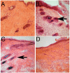
(a) β-galactosidase staining of normal skin from TOPGAL mice, which express the β-galactosidase reporter gene under the control of a promoter responsive to β-catenin-dependent signaling shows little to no β-galactosidase activity. (b) β-galactosidase staining 7 days after wounding shows activity present in the differentiated hair follicle shafts immediately adjacent to the wound edge (arrow). The actual wound is to the right of the arrow. (c) The staining for β-galactosidase was most intense 21 days post wounding, with the wound being to the right of the arrow. (d) At no time point was β-gal activity detected within the wound itself (a representative 21 day wound is shown).
Figure 3
Figure 3
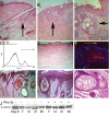
To determine whether Wnt signaling may induce epithelial patterning in wound epithelium as occurs during development, LiCl was applied continuously to the wounds of mice. (a) The adjacent normal skin of a 30 day LiCl treated mouse wound shows normal epithelial appendages and dermal architecture. (b) The same animal, but the wound has been treated with LiCl for 14 days and which shows the presence of epithelial inclusion cysts throughout the wound (arrow; day 30). Note the difference in the dermal architecture between the wounded tissue and normal skin; LiCl treatment had no observable effect on the dermal wound healing response. (c) Occasional larger and more mature appearing epithelial appendages can be seen within the wound (different animal; same time point), demonstrating the formation of sebaceous glands (arrow). Retroviral infected wounds were examined after 21 days and shows CD5 expression in 17% of the wound cells by flow cytometry (d) and (e) shows the histologic effect on the control retroviral treated wound at day 30: lacking any epithelial appendages overlying the wound. (f) Immunofluorescent detection of CD5 shows the infection has occurred in the dermal cells (red staining) adjacent to the normal skin, hair follicles in normal skin can be seen on the left and the wound is to the right of the arrow. By day 30 and persisting through day 90, numerous large epithelia-lined cysts can be seen in the wound (g & h) immediately adjacent to the mature normal skin hair follicles (normal skin is left of the line; wound to the right). (i) These cysts were also associated with sebaceous gland formation. (j) Wounds from PMXWnt-5a treated mice were examined for β-catenin stabilization and compared with control retroviral treated wounds. No significant difference in the amount of free β-catenin was seen between the two groups.

References

    1. Martin P. Wound healing--aiming for perfect skin regeneration. Science. 1997;276:75–81. doi: 10.1126/science.276.5309.75. - DOI - PubMed
    1. Tumbar T, Guasch G, Greco V, Blanpain C, Lowry WE, Rendl M, Fuchs E. Defining the epithelial stem cell niche in skin. Science. 2004;303:359–363. doi: 10.1126/science.1092436. - DOI - PMC - PubMed
    1. Blanpain C, Lowry WE, Geoghegan A, Polak L, Fuchs E. Self-renewal, multipotency, and the existence of two cell populations within an epithelial stem cell niche. Cell. 2004;118:635–648. doi: 10.1016/j.cell.2004.08.012. - DOI - PubMed
    1. Zuk PA, Zhu M, Ashjian P, De Ugarte DA, Huang JI, Mizuno H, Alfonso ZC, Fraser JK, Benhaim P, Hedrick MH. Human adipose tissue is a source of multipotent stem cells. Mol Biol Cell. 2002;13:4279–4295. doi: 10.1091/mbc.E02-02-0105. - DOI - PMC - PubMed
    1. Fathke C, Wilson L, Hutter J, Kapoor V, Smith A, Hocking A, Isik F. Contribution of bone marrow-derived cells to skin: collagen deposition and wound repair. Stem Cells. 2004;22:812–822. doi: 10.1634/stemcells.22-5-812. - DOI - PMC - PubMed

Publication types

MeSH terms

LinkOut - more resources