. 2006 Apr 10;347(2):261-76.
doi: 10.1016/j.virol.2005.11.053. Epub 2006 Jan 19.

Roles for herpes simplex virus type 1 UL34 and US3 proteins in disrupting the nuclear lamina during herpes simplex virus type 1 egress

Affiliations

Roles for herpes simplex virus type 1 UL34 and US3 proteins in disrupting the nuclear lamina during herpes simplex virus type 1 egress

Susan L Bjerke et al. Virology. .

Abstract

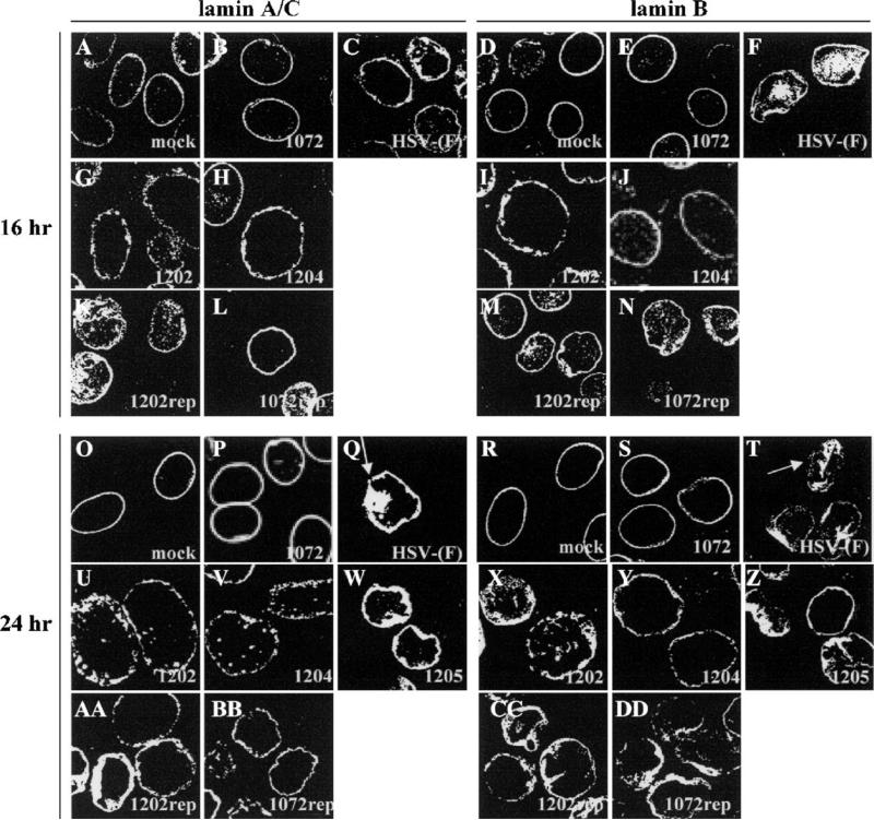
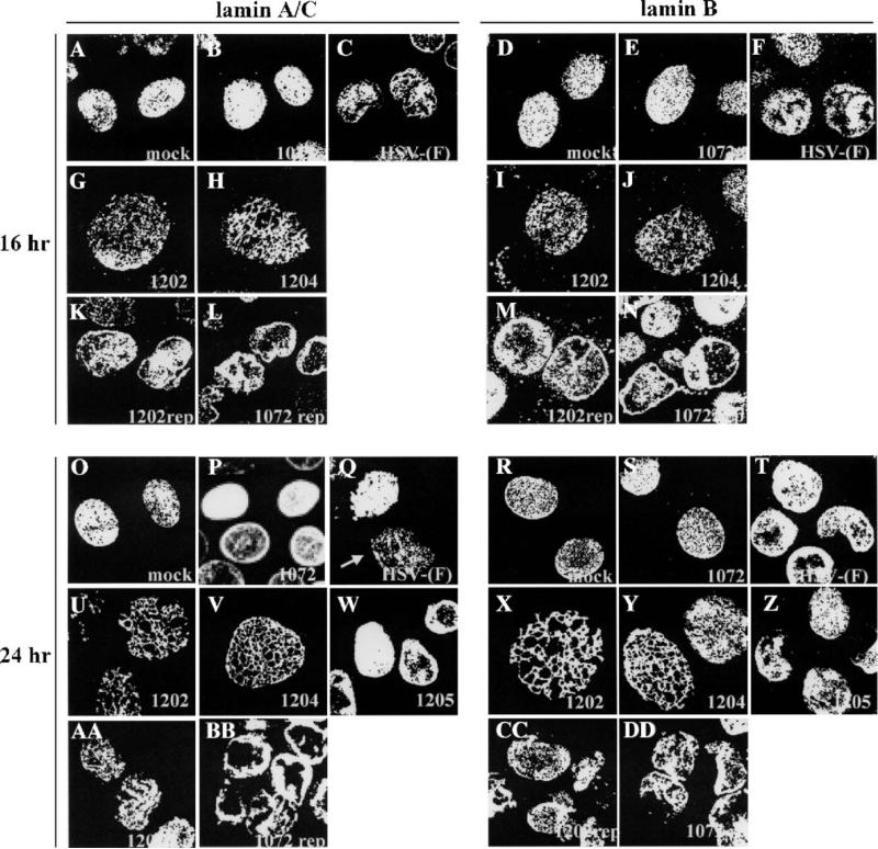
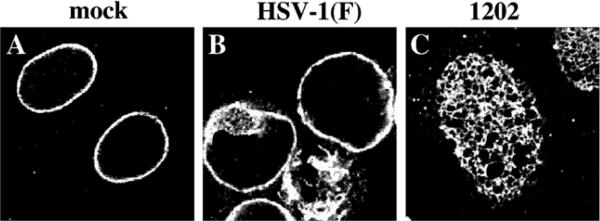
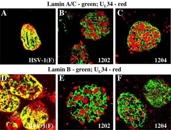
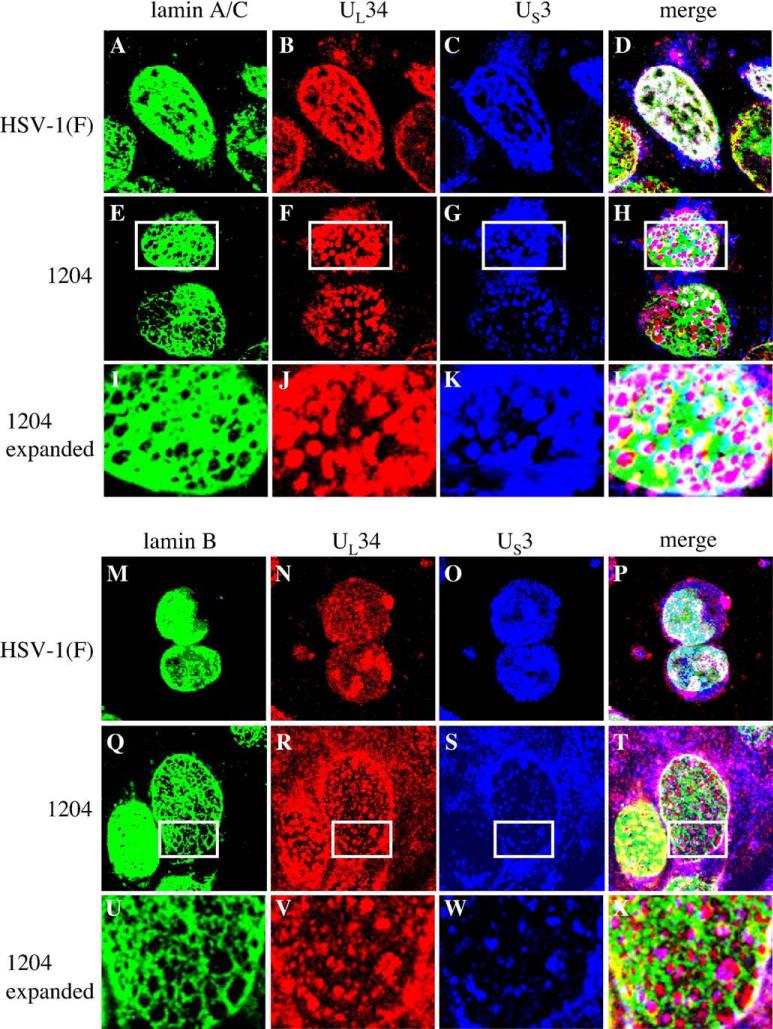
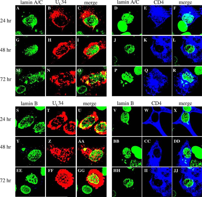
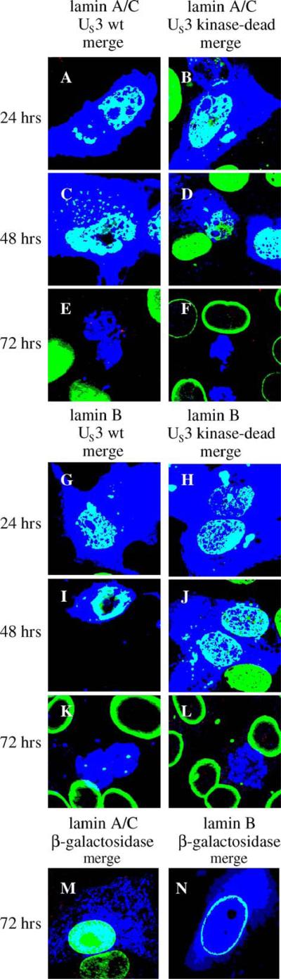
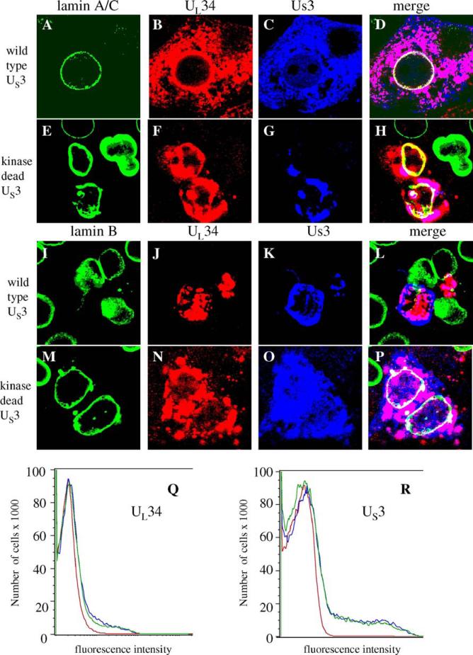
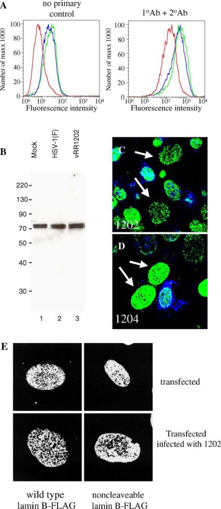
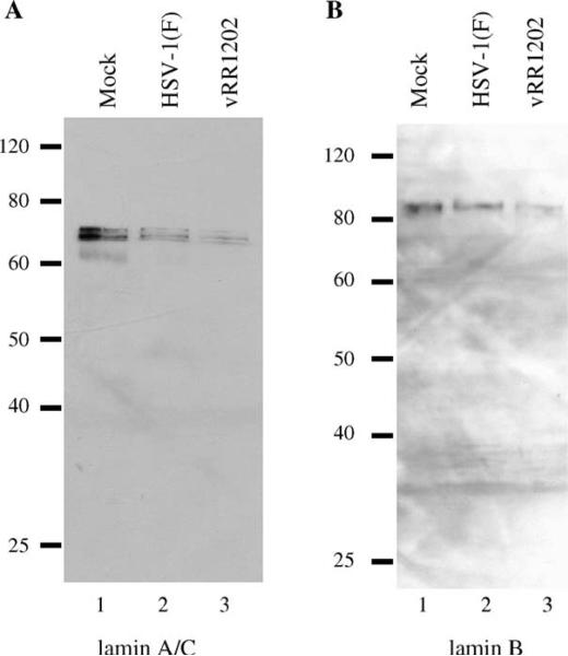
Cells infected with wild type HSV-1 showed significant lamin A/C and lamin B rearrangement, while UL34-null virus-infected cells exhibited few changes in lamin localization, indicating that UL34 is necessary for lamin disruption. During HSV infection, US3 limited the development of disruptions in the lamina, since cells infected with a US3-null virus developed large perforations in the lamin layer. US3 regulation of lamin disruption does not correlate with the induction of apoptosis. Expression of either UL34 or US3 proteins alone disrupted lamin A/C and lamin B localization. Expression of UL34 and US3 together had little effect on lamin A/C localization, suggesting a regulatory interaction between the two proteins. The data presented in this paper argue for crucial roles for both UL34 and US3 in regulating the state of the nuclear lamina during viral infection.


Figures

Fig. 1
Fig. 1
UL34- and US3-dependent disruption of the nuclear lamina of Vero cells. Shown are digital confocal images of optical sections taken near the middle of the cell nucleus showing the disruption of lamin A/C and lamin B localization in infected Vero cells. For all panels, Vero cells were mock infected or infected with the UL34-null virus vRR1072, HSV-1(F), the US3-null virus vRR1202, the US3 kinase-dead virus vRR1204, the UL34 phosphorylation mutant virus vRR1205, the UL34 repair virus vRR1072Rep, or the US3 repair virus vRR1202Rep for 16 or 24 h at an MOI of 5. The infecting virus is indicated in the lower right corner of each panel. The time of infection is indicated to the left of the figure. The primary antibody used for staining is indicated at the top of the figure. Cells stained for lamin A/C were fixed with formaldehyde; those stained for lamin B were fixed with cold methanol. Cover slips were also co-stained with a viral protein to ascertain infection.
Fig. 2
Fig. 2
UL34- and US3-dependent formation of perforations in the nuclear lamina of Vero cells. Shown are digital confocal images of optical sections taken near the top of the cell nucleus showing perforations in lamin A/C and lamin B localization in infected Vero cells. For all panels, Vero cells were mock infected or infected with the UL34-null virus vRR1072, HSV-1(F), the US3-null virus vRR1202, the US3 kinase-dead virus vRR1204, the UL34 phosphorylation mutant virus vRR1205, the UL34 repair virus vRR1072Rep, or the US3 repair virus vRR1202Rep for 16 or 24 h at an MOI of 5. The infecting virus is indicated in the lower right corner of each panel. The time of infection is indicated to the left of the figure. The primary antibody used for staining is indicated at the top of the figure. Cells stained for lamin A/C were fixed with formaldehyde; those stained for lamin B were fixed with cold methanol. Cover slips were also co-stained with a viral protein to ascertain infection.
Fig. 3
Fig. 3
Alterations of lamin staining in infected cells are not due to monoclonal antibody epitope masking. Shown are digital confocal images showing the localization of lamin A/C in infected Vero cells. Vero cells were mock infected (A), or infected with HSV-1(F) (B) or vRR1202 (C) for 24 h at an MOI of 5. Cells were fixed with formaldehyde and immunostained with chicken polyclonal anti-lamin A/C detected with AlexaFluor 548 goat anti-chicken.
Fig. 4
Fig. 4
Association of UL34 with lamin perforations in cells infected with US3 mutant viruses. Shown are digital confocal images showing the localization of UL34 and lamin A/C or lamin B in infected Vero cells. Green: lamins, red: UL34. Vero cells were infected with HSV-1(F) (A and D) vRR1202 (B and E) or vRR1204 (C and F) for 24 h at an MOI of 5. Cells were stained for UL34 (A–F) and for either lamin A/C (A–C) or lamin B (D–F).
Fig. 5
Fig. 5
Association of US3 with lamin perforations in cells infected with kinase-dead US3 mutant virus. Shown are digital confocal images of UL34, US3 and lamin A/C or lamin B localizations in HSV-1(F) or vRR1204 infected Vero cells. Green: lamins, red: UL34, blue: US3. For all panels, Vero cells were infected with the HSV-1(F) or vRR1204 for 24 h at an MOI of 5. Infecting viruses are indicated to the left of the figure. Panels I–L and U–X each show an enlarged view of the area enclosed in the white box in the panel immediately above.
Fig. 6
Fig. 6
Disruption of Lamin A/C and lamin B localization by expression of transfected UL34 or CD4 genes. Shown are digital confocal images showing the localization of UL34, CD4 and lamin A/C or lamin B in Vero cells expressing either the UL34 or CD4 protein for various times. Green: lamins, red: UL34, blue: CD4. For all panels, Vero cells were transfected with plasmids expressing either UL34 or CD4 for 24, 48, or 72 h. Time after transfection is indicated to the left of each row of panels. The protein being detected is indicated above each column of panels.
Fig. 7
Fig. 7
Disruption of lamin A/C and lamin B localization by expression of a transfected US3 gene. Shown are digital merged images showing the localization of wild type US3, mutant US3, beta-galactosidase and lamin A/C or lamin B in Vero cells transfected with plasmids expressing US3 or beta-galactosidase protein. Green: lamins, blue: US3 or beta-galactosidase. For all panels A–L, Vero cells were transfected with a plasmid expressing wild-type or kinase dead US3 for 24, 48 and 72 h. For panels M and N, only the 72 h time point is shown. Time after transfection is indicated to the left of each row of panels. The proteins being detected are indicated above each column of panels.
Fig. 8
Fig. 8
Regulation of UL34-mediated lamin disruption by co-expression of US3. For all panels, Vero cells were transfected with plasmids expressing UL34 and US3or kinase-dead US3 for 72 h. (A–Q) Digital confocal images showing the localization of UL34, US3 or kinase-dead US3 and lamin A/C or lamin B in Vero cells transfected with plasmids expressing UL34 and US3 or kinase-dead US3. All cells were transfected with UL34-expressing plasmid. The identity of the US3-expressing plasmid is indicated to the left of each row. The protein being detected is indicated above each column of panels. Green: lamins, red: UL34, blue: US3 (Q and R) Cells transfected with UL34 or US3 alone or together were analyzed by flow cytometry. (Q) UL34 staining profile for untransfected cells (red) or cells transfected with UL34 (blue) or UL34 and US3 (green). (R) US3 staining profile for untransfected cells (red) cells transfected with US3 (blue) or US3 and UL34 (green).
Fig. 9
Fig. 9
Disruptions in lamin localization are not accompanied by loss of lamin protein or cleavage of PARP. (A) Lamin A/C protein levels in Vero cells mock-infected (red), or infected with HSV-1(F) (blue) or vRR1202 (green), as analyzed by flow cytometry. The left histogram shows the results of detection with no primary antibody; the right graph shows the results of detection with both primary and secondary antibodies. (B) Lamin B protein levels in Vero cells mock-infected (lane 1), or infected with HSV-1(F) (lane 2) or vRR1202 (lane 3), as analyzed by western blot. Total cellular protein was extracted as described in Materials and methods. Coomassie staining was used to equilibrate loading (not shown). These experiments were done three times. Representative images are shown. Lane 3 is from the same blot and the same exposure as lanes 1 and 2, but was not adjacent to these lanes. (C and D) Digital confocal merged images showing the presence of cleaved PARP positive cells in Vero cells infected with vRR1202 or vR1204 at an MOI of 5 for 24 h. This experiment was done five times. A representative experiment is shown. The infecting virus is indicated on each panel. Green: lamin A/C; blue: cleaved PARP. The arrows point to cells that have perforations in the lamina, but do not stain positive for cleaved PARP. (E) Vero cells were transfected with a plasmid expressing either wild type lamin B-FLAG or non-cleavable lamin B-FLAG, then mock-infected or infected with vRR1202. The identity of the transfecting plasmid is indicated below each column. The state of infection is indicated to the right of each row.
Fig. 10
Fig. 10
Lamin disruption in infected HEp-2 cells is associated with loss of lamin protein. Shown are digital images of western blots of protein from HEp-2 cells infected for 24 h probed for lamin A/C (A) and lamin B (B). Coomassie staining was used to equilibrate loading (not shown). These experiments were done three times. Representative images are shown. The infecting virus is indicated above each lane.

References

    1. Asano S, Honda T, Goshima F, Watanabe D, Miyake Y, Sugiura Y, Nishiyama Y. US3 protein kinase of herpes simplex virus type 2 plays a role in protecting corneal epithelial cells from apoptosis in infected mice. J. Gen. Virol. 1999;80:51–56. - PubMed
    1. Asano S, Honda T, Goshima F, Nishiyama Y, Sugiura Y. US3 protein kinase of herpes simplex virus protects primary afferent neurons from virus-induced apoptosis in ICR mice. Neurosci. Lett. 2000;294:105–108. - PubMed
    1. Aubert M, O'Toole J, Blaho J. Induction and prevention of apoptosis in human HEp-2 cells by herpes simplex virus type 1. J. Virol. 1999;73:10359–10370. - PMC - PubMed
    1. Baines JD, Roizman B. The UL11 gene of herpes simplex virus 1 encodes a function that facilitates nucleocapsid envelopment and egress from cells. J. Virol. 1992;66:5168–5174. - PMC - PubMed
    1. Benetti L, Munger J, Roizman B. The herpes simplex virus 1 US3 protein kinase blocks caspase-dependent double cleavage and activation of the proapoptotic protein BAD. J. Virol. 2003;77:6567–6573. - PMC - PubMed

Publication types

MeSH terms

Substances

LinkOut - more resources