. 2006 Jan 31;103(5):1498-503.
doi: 10.1073/pnas.0510046103. Epub 2006 Jan 20.

Foamy virus vector integration sites in normal human cells

Affiliations

Foamy virus vector integration sites in normal human cells

Grant D Trobridge et al. Proc Natl Acad Sci U S A. .

Abstract

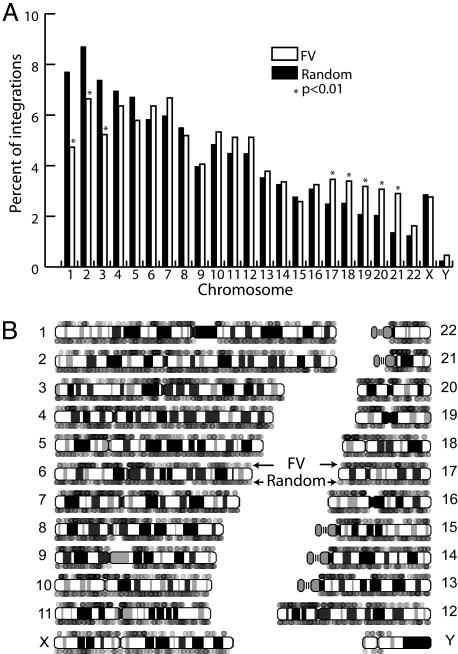
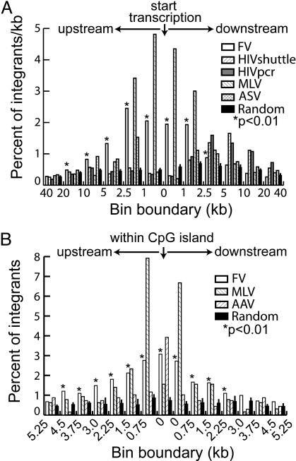
Foamy viruses (FVs) or spumaviruses are retroviruses that have been developed as vectors, but their integration patterns have not been described. We have performed a large-scale analysis of FV integration sites in unselected human fibroblasts (n = 1,008) and human CD34(+) hematopoietic cells (n = 1,821) by using a bacterial shuttle vector and a comparable analysis of lentiviral vector integration sites in CD34(+) cells (n = 1,331). FV vectors had a distinct integration profile relative to other types of retroviruses. They did not integrate preferentially within genes, despite a modest preference for integration near transcription start sites and a significant preference for CpG islands. The genomewide distribution of FV vector proviruses was nonrandom, with both clusters and gaps. Transcriptional profiling showed that gene expression had little influence on integration site selection. Our findings suggest that FV vectors may have desirable integration properties for gene therapy applications.


Figures

Fig. 1.
Fig. 1.
FV shuttle vector rescue strategy. The shuttle vector ΔΦPFmcsNO is based on the ΔΦ (deleted foamy) backbone (25) and is shown with its PGK promoter, GFP transgene, multiple cloning site, neo gene driven by a Tn5-derived bacterial promoter, p15A bacterial replication origin, and U3-deleted LTRs (ΔLTR). To recover provirus junctions, genomic DNA from infected cells was digested with multiple cloning site enzyme(s) that have compatible sticky ends (e.g., PciI and BspHI). Fragments containing the Tn5 promoter, neo gene, p15A origin, 3′ LTR, and flanking chromosomal DNA were circularized by ligation and transferred to bacteria, where they replicate and confer kanamycin resistance. Unwanted fragments containing 5′ vector sequences or bacterial plasmid DNA were destroyed by digestion with the rare-cutting endonuclease I-SceI and the methylation-sensitive enzyme DpnI, respectively. Bacterial colonies were picked and their LTR/chromosome junctions were sequenced with a primer in the LTR.
Fig. 2.
Fig. 2.
Chromosomal distribution of integration sites. (A) FV vector integration sites from both CD34+ cells and fibroblasts (n = 2,829) are plotted as a percentage of all integrants in each chromosome and compared with a set of random sites (n = 10,000). Asterisks mark chromosomes with significantly different integration frequencies (P < 0.01). (B) The same FV vector integration sites are represented as individual dots positioned above each human chromosome and compared with 2,829 random sites placed below the chromosomes. Dots are 20% opaque to display multiple overlapping integrants.
Fig. 3.
Fig. 3.
Clusters and gaps of FV vector integration sites. (A) The distances between adjacent unique FV vector integration sites were determined and binned by size, with the percent of proviruses within each bin plotted. (B) The number of FV vector integration hotspots (defined as three integrations) was determined for each sequence window size from 2.5 to 100 kb. Three size-matched sets of 2,829 random sites were also plotted with standard deviations as controls. Asterisks mark significant differences (P < 0.01).
Fig. 4.
Fig. 4.
Integration and transcription units. (A) The positions of FV (n = 2,829), HIV sites isolated by shuttle vector rescue (HIVshuttle, n = 1,331), HIV sites isolated by PCR (HIVpcr, n = 1,757), MLV (n = 644), and ASV (n = 480) integration sites were mapped relative to RefSeq gene transcription start sites, binned into different size sequence windows, and plotted as the percent of all integrations per kb. (B) The positions of FV (n = 2,829), MLV (n = 644), and AAV (n = 670) integration sites were mapped relative to those of CpG islands (GC content ≥50%, length >200 bp, ratio of observed to expected number of CpG dinucleotides >0.6). Integration sites in CpG islands and 14 0.75-kb windows flanking each island (the average length of a CpG island is 764 bp) were binned and plotted as the percent of all sites. A set of random sites (n = 10,000) were included as controls, and asterisks mark significant differences (FV vs. random; P < 0.01). See Table 1 for details on non-FV vector integration sites.
Fig. 5.
Fig. 5.
Transcriptional profiling of FV vector integration sites in CD34+ cells. RNA samples from uninfected and FV vector-infected CD34+ cells were hybridized to the human U133A plus 2.0 gene chip array (Affymetrix), and the expression levels of RefSeq genes with unique probe sets were ranked and plotted (gray dots, opacity 50%). RefSeq genes containing FV vector integrations are plotted as red circles. The average expression ranks are shown for all genes containing an FV vector integrant (n = 423; blue square), genes containing an FV vector integrant within 10 kb of the transcription start site (n = 389; green square), genes containing computer-generated random sites (n = 2,567; yellow square), and all RefSeq genes analyzed (n = 13,069; white circle).

References

    1. Hacein-Bey-Abina, S., von Kalle, C., Schmidt, M., Le Deist, F., Wulffraat, N., McIntyre, E., Radford, I., Villeval, J. L., Fraser, C. C., Cavazzana-Calvo, M. & Fischer, A. (2003) N. Engl. J. Med. 348, 255-256. - PubMed
    1. Schroder, A. R., Shinn, P., Chen, H., Berry, C., Ecker, J. R. & Bushman, F. (2002) Cell 110, 521-529. - PubMed
    1. Wu, X., Li, Y., Crise, B. & Burgess, S. M. (2003) Science 300, 1749-1751. - PubMed
    1. Narezkina, A., Taganov, K. D., Litwin, S., Stoyanova, R., Hayashi, J., Seeger, C., Skalka, A. M. & Katz, R. A. (2004) J. Virol. 78, 11656-11663. - PMC - PubMed
    1. Mitchell, R. S., Beitzel, B. F., Schroder, A. R., Shinn, P., Chen, H., Berry, C. C., Ecker, J. R. & Bushman, F. D. (2004) PLoS Biol. 2, E234. - PMC - PubMed

Publication types

MeSH terms

LinkOut - more resources