. 2006 Feb;188(3):894-901.
doi: 10.1128/JB.188.3.894-901.2006.

Topology and boundaries of the aerotaxis receptor Aer in the membrane of Escherichia coli

Affiliations

Topology and boundaries of the aerotaxis receptor Aer in the membrane of Escherichia coli

Divya N Amin et al. J Bacteriol. 2006 Feb.

Abstract

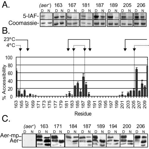
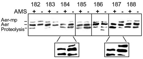
Escherichia coli chemoreceptors are type I membrane receptors that have a periplasmic sensing domain, a cytosolic signaling domain, and two transmembrane segments. The aerotaxis receptor, Aer, is different in that both its sensing and signaling regions are proposed to be cytosolic. This receptor has a 38-residue hydrophobic segment that is thought to form a membrane anchor. Most transmembrane prediction programs predict a single transmembrane-spanning segment, but such a topology is inconsistent with recent studies indicating that there is direct communication between the membrane flanking PAS and HAMP domains. We studied the overall topology and membrane boundaries of the Aer membrane anchor by a cysteine-scanning approach. The proximity of 48 cognate cysteine replacements in Aer dimers was determined in vivo by measuring the rate and extent of disulfide cross-linking after adding the oxidant copper phenanthroline, both at room temperature and to decrease lateral diffusion in the membrane, at 4 degrees C. Membrane boundaries were identified in membrane vesicles using 5-iodoacetamidofluorescein and methoxy polyethylene glycol 5000 (mPEG). To map periplasmic residues, accessible cysteines were blocked in whole cells by pretreatment with 4-acetamido-4'-maleimidylstilbene-2, 2' disulfonic acid before the cells were lysed in the presence of mPEG. The data were consistent with two membrane-spanning segments, separated by a short periplasmic loop. Although the membrane anchor contains a central proline residue that reaches the periplasm, its position was permissive to several amino acid and peptide replacements.


Figures

FIG. 1.
FIG. 1.
Hypothetical membrane anchor topology for the dimeric Aer receptor. (A) A helix-loop-helix membrane anchor would accommodate a cytosolic placement for both PAS and signaling domains. (B) An expanded view of the membrane region (highlighted in gray) for one Aer monomer, including the residues examined in this study.
FIG. 2.
FIG. 2.
Rates and extent of Aer cysteine cross-linking in intact cells after the addition of the oxidant copper phenanthroline. (A) The rate of cross-linking for representative classes of cysteine replacements, as discussed in the text. Except for Aer-A184C (□) (left) and Aer-V187C (▪) (left), the 10-min time point was in the linear range of the reaction for all cross-linking cysteine replacements, e.g., F182C (▵) and V209C (▴) (right). (B) Western blots showing (i) that the dimerization reaction for Aer-A184C was nearly complete within 2 min (2′) and (ii) the presence of a dimer band in Aer-V187C before the addition of copper phenanthroline (0′). (C) Representative Western blots comparing the extent of cross-linking at 10 min, which was the time point chosen to compare the extent of cross-linking for each cysteine replacement in the membrane anchor region. Abbreviations: M, monomer; D, dimer.
FIG. 3.
FIG. 3.
Summary of cysteine cross-linking in Aer mutants at 23°C and 4°C. The extent of Aer dimers with cross-linked cysteines in the membrane anchor demonstrates a central region of high proximity and flexibility, consistent with the PSA server secondary structure prediction. (A) Contour plot of secondary-structure probabilities (PSA server) (60, 61, 66). Rows indicate the secondary structure state; columns indicate each residue position. The probability of each structural state is depicted with contour lines in probability increments of 0.1. The β-strand prediction value was <0.1 and therefore is not included. (B) Extent of in vivo cross-linking for all 48 cysteine replacements after incubation of cells with copper phenanthroline for 10 min at 23°C. (C) Extent of in vivo cross-linking for the same cells shown in panel B, incubated with copper phenanthroline for 20 min at 4°C.
FIG. 4.
FIG. 4.
Surface accessibility of Aer cysteine replacements to the sulfhydryl-reactive reagents 5-IAF and mPEG in membrane vesicles. (A) SDS-PAGE gels showing the reactivity of strategic cysteine replacements towards 5-IAF. (Top) Negative fluorescent images of samples reacted with 5-IAF under native (N) and denaturing (D) conditions for 5 min. (Bottom) The same bands stained with Coomassie blue to estimate the total protein in each band. Note the unknown labeled protein in the aer control lane under denaturing conditions. (B) Bar graph summarizing the percent accessibility of cysteine replacements to 5-IAF during incubations for 5 min at 23°C. The results are the average of three or more independent experiments. The arrows at the top of the graph represent the accessibility boundaries for mPEG at 23°C (solid lines) and at 4°C (dotted lines). (C) Representative Western blots showing the presence (residues 163, 184, 187, and 206) or absence (residues 171, 189, 194, and 200) of a mobility shift in Aer (Aer-mp) after incubation of membrane vesicles with mPEG under native (N) conditions for 1 h. All cysteine replacements were PEGylated under denaturing (D) conditions.
FIG. 5.
FIG. 5.
Pretreatment with AMS in intact cells blocks periplasmic cysteine replacements from subsequent PEGylation. Cells were reacted with (+) and without (−) AMS for 45 min before being washed and probed with mPEG under denaturing conditions as described in the text. Aer-mPEG adducts (Aer-mp) showed a mobility shift. The enlarged lanes in the lower panel are higher exposures taken from different Western blots; these highlight replacements 184 and 187, which were reproducibly blocked by AMS.

References

    1. Bibikov, S. I., L. A. Barnes, Y. Gitin, and J. S. Parkinson. 2000. Domain organization and flavin adenine dinucleotide-binding determinants in the aerotaxis signal transducer Aer of Escherichia coli. Proc. Natl. Acad. Sci. USA 97:5830-5835. - PMC - PubMed
    1. Bibikov, S. I., R. Biran, K. E. Rudd, and J. S. Parkinson. 1997. A signal transducer for aerotaxis in Escherichia coli. J. Bacteriol. 179:4075-4079. - PMC - PubMed
    1. Boldog, T., and G. L. Hazelbauer. 2004. Accessibility of introduced cysteines in chemoreceptor transmembrane helices reveals boundaries interior to bracketing charged residues. Protein Sci. 13:1466-1475. - PMC - PubMed
    1. Butler, S. L., and J. J. Falke. 1998. Cysteine and disulfide scanning reveals two amphiphilic helices in the linker region of the aspartate chemoreceptor. Biochemistry 37:10746-10756. - PMC - PubMed
    1. Chen, C. P., A. Kernytsky, and B. Rost. 2002. Transmembrane helix predictions revisited. Protein Sci. 11:2774-2791. - PMC - PubMed

Publication types

MeSH terms

Substances

LinkOut - more resources