. 2006 Feb;26(3):898-911.
doi: 10.1128/MCB.26.3.898-911.2006.

Glycogen synthase kinase 3 and h-prune regulate cell migration by modulating focal adhesions

Affiliations

Glycogen synthase kinase 3 and h-prune regulate cell migration by modulating focal adhesions

Tsuyoshi Kobayashi et al. Mol Cell Biol. 2006 Feb.

Abstract

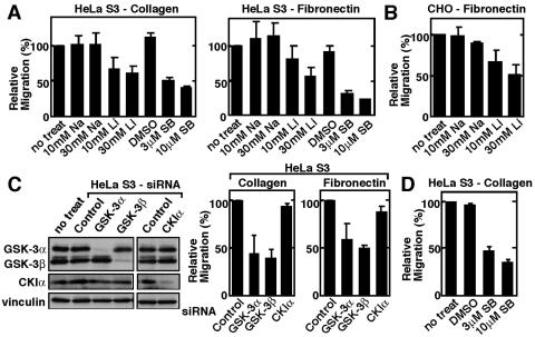
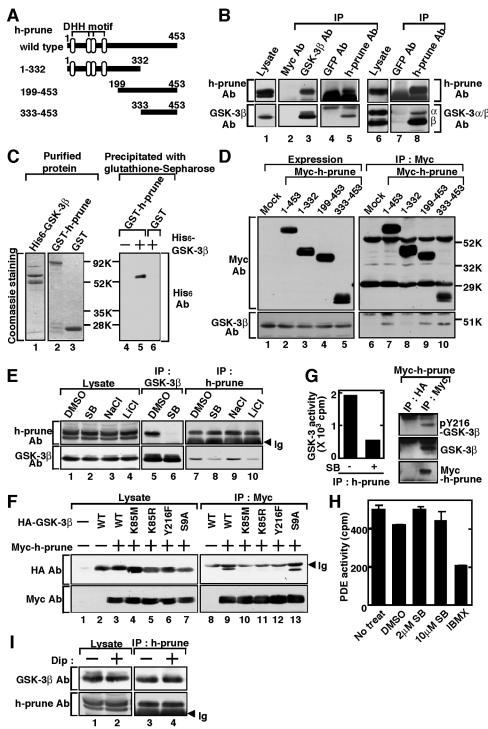
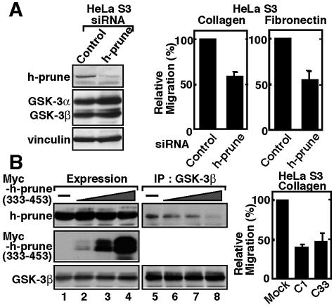
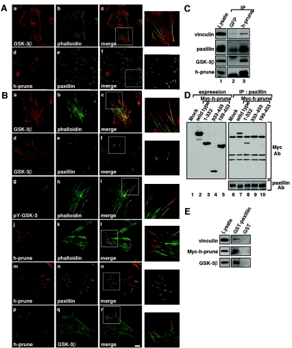
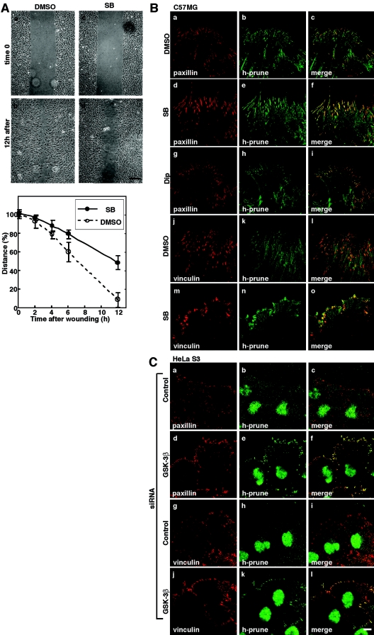
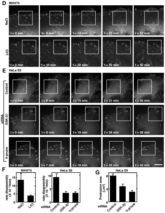
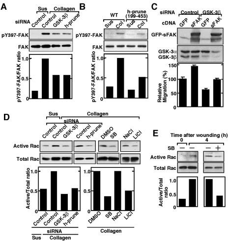
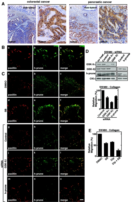
h-prune, which has been suggested to be involved in cell migration, was identified as a glycogen synthase kinase 3 (GSK-3)-binding protein. Treatment of cultured cells with GSK-3 inhibitors or small interfering RNA (siRNA) for GSK-3 and h-prune inhibited their motility. The kinase activity of GSK-3 was required for the interaction of GSK-3 with h-prune. h-prune was localized to focal adhesions, and the siRNA for GSK-3 or h-prune delayed the disassembly of paxillin. The tyrosine phosphorylation of focal adhesion kinase (FAK) and the activation of Rac were suppressed in GSK-3 or h-prune knocked-down cells. GSK-3 inhibitors suppressed the disassembly of paxillin and the activation of FAK and Rac. Furthermore, h-prune was highly expressed in colorectal and pancreatic cancers, and the positivity of the h-prune expression was correlated with tumor invasion. These results suggest that GSK-3 and h-prune cooperatively regulate the disassembly of focal adhesions to promote cell migration and that h-prune is useful as a marker for tumor aggressiveness.


Figures

FIG. 1.
FIG. 1.
Involvement of GSK-3 in cell migration. (A) HeLa S3 cells treated with the indicated inhibitors were subjected to the Transwell migration assay. (B) CHO cells treated with NaCl or LiCl were subjected to the Transwell migration assay. (C) Left panel, the lysates of HeLa S3 cells transfected with the indicated siRNAs were probed with the indicated antibodies. Right panel, HeLa S3 cells transfected with the indicated siRNAs were subjected to the Transwell migration assay. A single-strand RNA for GSK-3β was used as a control. (D) HeLa S3 cells treated with SB216763 were subjected to the random migration assay. The results shown are means ± standard errors of the means from four independent experiments. DMSO, dimethyl sulfoxide; Na, NaCl; Li, LiCl; SB, SB216763; no treat, no treatment.
FIG.2.
FIG.2.
Interaction of h-prune with GSK-3. (A) Schematic representation of the deletion mutants of h-prune used in this study. (B) The lysates of HeLa S3 cells (lanes 1 and 6) were immunoprecipitated with anti-GSK-3β or anti-h-prune antibody, and the immunoprecipitates were probed with the indicated antibodies (lanes 3, 5, and 8). The immunoprecipitates formed with anti-Myc and anti-GFP antibodies were used as controls (lanes 2, 4, and 7). GSK-3α/β Ab is an antibody that recognizes both GSK-3α (α) and GSK-3β (β). (C) Recombinant His6-GSK-3β, GST-h-prune, and GST (0.5 μg of protein) were stained with Coomassie brilliant blue (lanes 1 to 3). After 0.4 μM His6-GSK-3β was incubated with 0.5 μM GST-h-prune or GST immobilized on glutathione-Sepharose in 100 μl of reaction mixture (20 mM Tris/HCl, pH 7.5, and 1 mM dithiothreitol) for 1 h at 4°C, GST-h-prune and GST were precipitated by centrifugation, and then the precipitates were probed with the anti-His6 antibody (lanes 4 to 6). (D) The lysates of COS cells expressing the deletion mutants of Myc-h-prune were probed with anti-GSK-3β or anti-Myc antibody (lanes 1 to 5). The same lysates were immunoprecipitated with anti-Myc antibody, and the immunoprecipitates were probed with the indicated antibodies (lanes 6 to 10). (E) HeLa S3 cells treated with 10 μM SB216763 or 30 mM LiCl were lysed, and the lysates were probed with anti-GSK-3β or anti-h-prune antibody (lanes 1 to 4). The same lysates were immunoprecipitated with anti-GSK-3β (lanes 5 to 6) or anti-h-prune antibody (lanes 7 to 10), and the immunoprecipitates were probed with the indicated antibodies. The lower bands detected by anti-h-prune antibody in Fig. 2B and E are nonspecific bands. (F) The lysates of COS cells expressing HA-GSK-3β mutants and Myc-h-prune were probed with anti-HA or anti-Myc antibody (lanes 1 to 7). The same lysates were immunoprecipitated with anti-Myc antibody, and the immunoprecipitates were probed with the indicated antibodies (lanes 8 to 13). (G) Left panel, the kinase activity of GSK-3 in the immunoprecipitates from HeLa S3 cells with anti-h-prune antibody was measured in the presence or absence of SB216763 in vitro. Right panel, the lysates of HeLa S3 cells expressing Myc-h-prune were immunoprecipitated with anti-Myc or anti-HA antibody, and the immunoprecipitates were probed with anti-GSK-3β antibody and the phospho-specific antibody to GSK-3β Tyr216 (pY216-GSK-3β). (H) The PDE activity of h-prune in HeLa S3 cells was measured after treatment with SB216763 in intact cells or with IBMX in vitro. (I) After HeLa S3 cells were treated with 10 μM dipyridamole for 4 h, h-prune was immunoprecipitated from the lysates and the immunoprecipitates were probed with anti-GSK-3β and anti-h-prune antibodies. HA, hemagglutinin; IP, immunoprecipitation; Ab, antibody; SB, SB216763; Dip, dipyridamole; Ig, immunoglobulin; DMSO, dimethyl sulfoxide; no treat, no treatment; Mock, control.
FIG. 3.
FIG. 3.
Involvement of h-prune in cell migration. (A) Left panel, the lysates of HeLa S3 cells transfected with control siRNA or siRNA for h-prune were probed with the indicated antibodies. Right panel, HeLa S3 cells transfected with control siRNA or siRNA for h-prune were subjected to the Transwell migration assay. (B) Left panel, the lysates of HeLa S3 cells expressing Myc-h-prune(333-453) were probed with anti-h-prune or anti-GSK-3β antibody (lanes 1 to 4). The same lysates were immunoprecipitated with anti-GSK-3β antibody (lanes 5 to 8). Right panel, two different clones (C1 and C33) of HeLa S3 cells stably expressing Myc-h-prune(199-453) were subjected to the Transwell migration assay. Cells transfected with vectors alone were used as a control (Mock). The results shown are means ± standard errors of the means from three independent experiments. IP, immunoprecipitation.
FIG. 4.
FIG. 4.
Localization of h-prune and GSK-3 to focal adhesions. (A) HeLa S3 cells were stained with anti-GSK-3β (a), anti-h-prune (d), or antipaxillin (e) antibody or phalloidin-fluorescein isothiocyanate (FITC) (b). Merged images are shown in panels c and f. The regions in white boxes are shown magnified. (B) C57MG cells were stained with anti-GSK-3β (a and d), anti-pY216-GSK-3β (g), anti-h-prune (j, m, and p), or antipaxillin (e and n) antibody or phalloidin-FITC (b, h, and k). To show the localization of GSK-3 and h-prune simultaneously, anti-GSK-3 antibody was labeled with a Zenon labeling kit (Molecular Probes) (q). Merged images are shown in panels c, f, i, l, o, and r. The regions in white boxes are shown magnified. Scale bar, 10 μm. (C) The lysates of C57MG cells (lane 1) were immunoprecipitated with anti-h-prune antibody, and the immunoprecipitates were probed with the indicated antibodies (lane 3). The immunoprecipitates obtained with anti-GFP antibody were used as a control (lane 2). (D) The lysates of COS cells expressing deletion mutants of h-prune were probed with anti-Myc antibody (lanes 1 to 5). The same lysates were immunoprecipitated with antipaxillin antibody, and the immunoprecipitates were probed with the indicated antibodies (lanes 6 to 10). The results shown are representative of three independent experiments. (E) HeLa S3 cells expressing Myc-h-prune were lysed, and the lysates were incubated with 0.1 μM GST-paxillin or GST immobilized on glutathione-Sepharose. After GST-paxillin or GST was precipitated by centrifugation, the precipitates were probed with the indicated antibodies. IP, immunoprecipitation; Ab, antibody; Mock, control.
FIG. 5.
FIG. 5.
Involvement of GSK-3 and h-prune in dynamics of focal adhesions. (A) Upper panel, monolayers of C57MG cells on collagen-coated coverslips were treated with 10 μM SB216763 for 4 h. After wounding, wounded monolayers were allowed to heal for 12 h. Scale bar, 0.2 mm. Lower panel, the length of the wounds was measured and expressed as a percentage of the initial distance at time zero. Open circles, dimethyl sulfoxide (DMSO) treatment; filled circles, SB216763 (SB) treatment. The results shown are means ± standard errors of the means from three independent experiments. (B) C57MG cells treated with 10 μM SB216763 (d to f, m to o) or 10 μM dipyridamole (Dip) (g to i) were wounded. Six hours after wounding, the cells were stained with antipaxillin (a, d, and g), antivinculin (j and m), or anti-h-prune (b, e, h, k, and n) antibody. Merged images are shown in panels c, f, i, l, and o. (C) HeLa S3 cells transfected with the siRNA for GSK-3β (d to f and j to l) were wounded. Twelve hours after wounding, the cells were stained with antipaxillin (a and d), antivinculin (g and j), or anti-h-prune (b, e, h, and k) antibody. Merged images are shown in panels c, f, i, and l. Scale bar, 10 μm. The results shown are representative of three independent experiments. (D and E) Dynamics of GFP-paxillin in migrating NIH 3T3 cells treated with NaCl or LiCl (D) and those in HeLa S3 cells treated with the indicated siRNAs (E) were visualized by time-lapse fluorescence microscopy. For each sequence, “t = 0 min” is the frame in which the adhesions in the white box were clearly observed. Scale bar, 5 μm. (F) Rate constants for disassembly of GFP-paxillin in Fig. 5D and E were calculated. Quantifications of GFP-paxillin disassembly show means ± standard errors of the means. (G) The lamellipodium protrusion area was quantified in HeLa S3 cells transfected with siRNA for GSK-3β or h-prune.
FIG. 5.
FIG. 5.
Involvement of GSK-3 and h-prune in dynamics of focal adhesions. (A) Upper panel, monolayers of C57MG cells on collagen-coated coverslips were treated with 10 μM SB216763 for 4 h. After wounding, wounded monolayers were allowed to heal for 12 h. Scale bar, 0.2 mm. Lower panel, the length of the wounds was measured and expressed as a percentage of the initial distance at time zero. Open circles, dimethyl sulfoxide (DMSO) treatment; filled circles, SB216763 (SB) treatment. The results shown are means ± standard errors of the means from three independent experiments. (B) C57MG cells treated with 10 μM SB216763 (d to f, m to o) or 10 μM dipyridamole (Dip) (g to i) were wounded. Six hours after wounding, the cells were stained with antipaxillin (a, d, and g), antivinculin (j and m), or anti-h-prune (b, e, h, k, and n) antibody. Merged images are shown in panels c, f, i, l, and o. (C) HeLa S3 cells transfected with the siRNA for GSK-3β (d to f and j to l) were wounded. Twelve hours after wounding, the cells were stained with antipaxillin (a and d), antivinculin (g and j), or anti-h-prune (b, e, h, and k) antibody. Merged images are shown in panels c, f, i, and l. Scale bar, 10 μm. The results shown are representative of three independent experiments. (D and E) Dynamics of GFP-paxillin in migrating NIH 3T3 cells treated with NaCl or LiCl (D) and those in HeLa S3 cells treated with the indicated siRNAs (E) were visualized by time-lapse fluorescence microscopy. For each sequence, “t = 0 min” is the frame in which the adhesions in the white box were clearly observed. Scale bar, 5 μm. (F) Rate constants for disassembly of GFP-paxillin in Fig. 5D and E were calculated. Quantifications of GFP-paxillin disassembly show means ± standard errors of the means. (G) The lamellipodium protrusion area was quantified in HeLa S3 cells transfected with siRNA for GSK-3β or h-prune.
FIG. 6.
FIG. 6.
Involvement of GSK-3 and h-prune in the activation of FAK and Rac. (A and B) FAK activation. HeLa S3 cells transfected with the indicated siRNAs (A) or expressing Myc-h-prune(199-453) (B) were suspended in serum-free medium and were kept in suspension (Sus) or replated onto collagen-coated dishes (Col). Top panel, the cells were lysed at 1 h after plating, and the lysates were probed with the phospho-specific antibody to FAK pTyr397 (pY397 FAK) or anti-FAK antibody. Bottom panel, the FAK activity was measured as the ratio of phosphorylated FAK (active FAK) to total FAK. (C) GFP or GFP-FAKK578E/K581E (sFAK) was expressed in NIH 3T3 cells transfected with siRNA for GSK-3β, and the cells were subjected to the Transwell migration assay. Migrated GFP-labeled cells were normalized with transfection efficiency. Top panel, the protein levels of GFP-FAKK578E/K581E and GSK-3β were shown by anti-FAK and GSK-3 antibodies. Bottom panel, migration ability of the cells used in this assay. (D) Rac activation. HeLa S3 cells were transfected with the indicated siRNAs or treated with the indicated GSK-3 inhibitors. The cells were replated onto collagen-coated dishes, and the lysates were incubated with GST-CRIB immobilized on glutathione-Sepharose. Top panel, the total lysates and precipitates were probed with anti-Rac-1 antibody. Bottom panel, the Rac activity was measured as the ratio of the amount of CRIB-bound Rac (active Rac) to that of Rac in total cell lysates (total Rac). (E) Multiple wounds were made several times in HeLa S3 cells treated with 10 μM SB216763 (SB). The Rac activity was measured at 4 h after wounding. DMSO, dimethyl sulfoxide.
FIG. 7.
FIG. 7.
Correlation of h-prune expression with tumor aggressiveness. (A) Immunohistochemical analyses of h-prune in human colorectal cancer (a and b) and human pancreatic cancer (c and d). (a) Magnification, ×13; (c) magnification, ×33. Expression levels of h-prune in the nontumor and tumor regions were compared. (b and d) Magnification, ×135. The tumor regions were enlarged. (B) SW480 cells were stained with antipaxillin (a) or anti-h-prune (b) antibody. The merged image is shown in panel c. (C) SW480 cells treated with 10 μM SB216763 (d to f) or transfected with the siRNA for GSK-3β (j to l) or h-prune (m to o) were wounded. Twelve hours after wounding, the cells were stained with antipaxillin (a, d, g, j, and m) or anti-h-prune (b, e, h, k, and n) antibody. Merged images are shown in panels c, f, i, l, and o. Scale bar, 10 μm. (D) Upper panel, the lysates of SW480 cells transfected with the indicated siRNAs were probed with the indicated antibodies. Lower panel, SW480 cells transfected with the indicated siRNAs were subjected to the Transwell migration assay. (E) SW480 cells treated with 10 μM SB216763 and/or 10 μM dipyridamole were subjected to the Transwell migration assay on collagen. SB, SB216763; Dip, dipyridamole; DMSO, dimethyl sulfoxide. The results shown are means ± standard errors of the means from three independent experiments.

References

    1. Andoh, T., Y. Hirata, and A. Kikuchi. 2000. Yeast glycogen synthase kinase 3 is involved in protein degradation in cooperation with Bul1, Bul2, and Rsp5. Mol. Cell. Biol. 20:6712-6720. - PMC - PubMed
    1. Benard, V., and G. M. Bokoch. 2002. Assay of Cdc42, Rac, and Rho GTPase activation by affinity methods. Methods Enzymol. 345:349-359. - PubMed
    1. Bhat, R. V., J. Shanley, M. P. Correll, W. E. Fieles, R. A. Keith, C. W. Scott, and C.-M. Lee. 2000. Regulation and localization of tyrosine216 phosphorylation of glycogen synthase kinase-3β in cellular and animal models of neuronal degeneration. Proc. Natl. Acad. Sci. USA 97:11074-11079. - PMC - PubMed
    1. Biggs, J., N. Tripoulas, E. Hersperger, C. Dearolf, and A. Shearn. 1988. Analysis of the lethal interaction between the prune and Killer of prune mutations of Drosophila. Genes Dev. 2:1333-1343. - PubMed
    1. Cohen, P., and S. Frame. 2001. The renaissance of GSK3. Nat. Rev. Mol. Cell Biol. 2:769-776. - PubMed

Publication types

MeSH terms