. 2006 Feb;26(3):1063-76.
doi: 10.1128/MCB.26.3.1063-1076.2006.

Protection against fatty liver but normal adipogenesis in mice lacking adipose differentiation-related protein

Affiliations

Protection against fatty liver but normal adipogenesis in mice lacking adipose differentiation-related protein

Benny Hung-Junn Chang et al. Mol Cell Biol. 2006 Feb.

Abstract

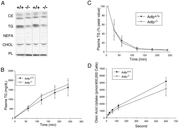
Adipose differentiation-related protein (ADFP; also known as ADRP or adipophilin), is a lipid droplet (LD) protein found in most cells and tissues. ADFP expression is strongly induced in cells with increased lipid load. We have inactivated the Adfp gene in mice to better understand its role in lipid accumulation. The Adfp-deficient mice have unaltered adipose differentiation or lipolysis in vitro or in vivo. Importantly, they display a 60% reduction in hepatic triglyceride (TG) and are resistant to diet-induced fatty liver. To determine the mechanism for the reduced hepatic TG content, we measured hepatic lipogenesis, very-low-density lipoprotein (VLDL) secretion, and lipid uptake and utilization, all of which parameters were shown to be similar between mutant and wild-type mice. The finding of similar VLDL output in the presence of a reduction in total TG in the Adfp-deficient liver is explained by the retention of TG in the microsomes where VLDL is assembled. Given that lipid droplets are thought to form from the outer leaflet of the microsomal membrane, the reduction of TG in the cytosol with concomitant accumulation of TG in the microsome of Adfp-/- cells suggests that ADFP may facilitate the formation of new LDs. In the absence of ADFP, impairment of LD formation is associated with the accumulation of microsomal TG but a reduction in TG in other subcellular compartments.


Figures

FIG. 1.
FIG. 1.
ADFP gene targeting. (A) ADFP partial genomic structure, gene targeting construct, and expected homologous recombinant allele. Restriction enzyme sites: A, AccI; B, BamHI; E, EcoRI; X, XbaI; and Xh, XhoI. (B) Southern blot genotyping of the wild type (+/+) and heterozygous (+/−), and homozygous (−/−) knockouts. (C) Northern blot analysis of RNA from four different tissues (L, liver; M, muscle; (B) brain; and W, white adipose tissue) in wild-type and homozygous knockouts. (D) Western blot of total protein extracted from wild-type and homozygous knockout livers.
FIG. 2.
FIG. 2.
Adipocyte differentiation of primary MEFs and in vitro lipolysis. Primary fibroblasts were isolated from embryos (embryonic day, 13.5) of Adfp+/+ and Adfp−/− mice for induction of adipocyte differentiation, as described in Materials and Methods. (A) Northern blot analysis of expression of various adipocyte differentiation marker genes using RNA isolated from day 0 (D0) to day 8 (D8) of differentiation. (B) Cellular triglyceride content of fully differentiated cells isolated at day 8. (C and D) Basal [ISO(−)]- and isoprotenerol [ISO(+)]-induced in vitro lipolysis measured by NEFA (C) and glycerol (D) in the media from fully differentiated MEF-derived adipocytes. (E and F) In vivo lipolysis measured using Adfp+/+ and Adfp−/− mice (three mice per group) as plasma NEFA (E) or glycerol (F) concentrations under basal (CL−) or CL-316243 (β3-adrenergic receptor agonist; CL+)-stimulated conditions.
FIG. 3.
FIG. 3.
PAT domain protein gene expression in Adfp+/+ (+/+) and Adfp−/− (−/−) mice. (A) Northern blot analysis. A total of 20 μg of total RNA isolated from WAT, BAT, muscle, and liver was separated on a 1% agarose gel; the RNAs were transferred to a nylon membrane and probed with cDNA of perilipin (PlinA), Tip47, S3-12, and Adfp. (B) Western blot analysis. A total of 20 μg of total protein extracts isolated from WAT, BAT, muscle, and liver was separated by polyacrylamide gel electrophoresis (PAGE) using a 4 to 15% gradient gel; the proteins were transferred to a nylon membrane and detected using antisera raised against perilipin and Tip47 and enhanced chemiluminescence kits.
FIG. 4.
FIG. 4.
Liver triglyceride fatty acid composition. Liver triglyceride fatty acids from Adfp+/+ and Adfp−/− mice (four mice per group) were analyzed by gas chromatography after methyl esterification, as described in Materials and Methods. Major species of fatty acids are presented (A), with classes grouped (B) as follows: Sat, saturated; Mono, monunsaturated; Poly, polyunsaturated; n-6, n-6 polyunsaturated; and n-3, n-3 polyunsaturated fatty acids. The relative percentage of each individual (C) or grouped (D) fatty acids out of the total fatty acids is also presented. *, significant difference (P < 0.01) between Adfp+/+ and Adfp−/− mice.
FIG. 5.
FIG. 5.
Hepatic lipid analysis and lipid metabolism. (A) Thin-layer chromatography analysis of lipids isolated from Adfp+/+ (+/+) and Adfp−/− (−/−) mouse livers. Lipids were extracted using 0.2 g of liver from an individual mouse, dried under nitrogen, reconstituted in chloroform, and loaded on to a TLC plate. Lipid standards were used to identify each lipid band. Lipid fractions were visualized using iodine vapor. (B) VLDL secretion after inhibition of lipoprotein lipase by Triton WR1339 treatment. Eight-week-old mice (male; n = 5) were fasted for 4 h, and plasma samples were taken as baseline (time zero). Plasma TG was measured at time zero and every hour for 4 h after Triton WR1339 treatment. (D) Intralipid clearance. Eight-week-old mice (n = 6) were infused with intralipid through the tail vein. Plasma TG levels are expressed as a percentage of the peak value measured 5 min after intralipid injection. (D) Oleic acid uptake in primary hepatocytes. Hepatocytes were isolated from male mice (n = 4). Individually isolated cells were separated into tubes for fatty acid uptake assay (Materials and Methods) in the presence of [3H]oleic acid; the reaction was stopped by the addition of phloretin solution at different time points. Cells were washed and pelleted by centrifugation, and radioactivity was determined by scintillation counting.
FIG. 6.
FIG. 6.
Hepatic β-oxidation parameters. (A) β-Oxidation in primary hepatocytes. Primary hepatocytes were isolated from Adfp+/+ and Adfp−/− mice (three mice per group). Viable cells were counted, and 5 × 10⁵ viable cells were isolated from each mouse and used for the assay. (B) Plasma β-hydroxybutyrate concentrations in Adfp+/+ and Adfp−/− mice (four mice per group) after 12-h fasting.
FIG. 7.
FIG. 7.
(A) In vivo hepatic fatty acid synthesis using the [3H]H2O labeling method (39, 40). Male Adfp+/+ and Adfp−/− mice (four mice per group) were synchronized to 3-h (8 to 11 a.m.) feeding each day for 2 weeks before experiments. Livers were removed 1 h after [3H]H2O injection. Total lipids were extracted, saponified, and separated by TLC; fatty acid fractions were scraped into scintillation vials to determine radioactivity. (B) Liver FAS activity of liver extracts from Adfp+/+ and Adfp−/− mice (four mice per group). (C) Representative Western blot of liver FAS and ACCI from Adfp+/+ and Adfp−/− mice. (D) Liver microsomal DGAT activity assay (Materials and Methods) of microsome preparations from Adfp+/+ and Adfp−/− mice (four mice per group). The reaction products from the DGAT assay were separated by TLC, the TG fraction was scrapped, and radioactivity was determined by scintillation counting.
FIG. 8.
FIG. 8.
TLC analysis of cytosolic and microsomal lipid distribution. A total of 0.4 g of liver from Adfp+/+ and Adfp−/− mice (four mice per group) was homogenized and separated into nuclear, microsomal, and cytosolic compartments. Lipids were extracted from each compartment and analyzed by TLC. Two lipid standards were loaded in the center lanes as a control. (B) Western blot analysis of subcellular fractions. A total of 20 μg of proteins from each subcellular fraction (prepared as described for panel A) was analyzed by PAGE, transferred to a nylon membrane, and immunoblotted with anti-MTP (microsomal marker), anti-GAPDH (cytosolic marker), and antihistone (nuclear marker). T, total liver protein extract; C, cytosolic fraction; N, nuclear fraction; M, microsomal fraction. +/+, Adfp+/+ mice; −/−, Adfp−/− mice. (C) Intracellular lipid partitioning in Adfp−/− and Adfp+/+ mice. Lipid (TG, NEFA, and PL) abundance was analyzed by TLC, quantified by densitometry, and expressed according to each lipid's subcellular compartment.
FIG. 9.
FIG. 9.
Liver histology and LD number and size distribution in HFD-fed mice. Adfp+/+ and Adfp−/− mice (8 weeks old; four mice per group) were fed with HFD for 4 weeks. (A and B) Photomicroscopy of liver section from HFD-fed Adfp+/+ (A) and Adfp−/− (B) mice. (C and D) Average number and size distribution of LDs in liver sections of HFD-fed Adfp+/+ (C) and Adfp−/− (D) mice.

References

    1. Bildirici, I., C. R. Roh, W. T. Schaiff, B. M. Lewkowski, D. M. Nelson, and Y. Sadovsky. 2003. The lipid droplet-associated protein adipophilin is expressed in human trophoblasts and is regulated by peroxisomal proliferator-activated receptor-gamma/retinoid X receptor. J. Clin. Endocrinol. Metab. 88:6056-6062. - PubMed
    1. Bligh, E. G., and W. J. Dyer. 1959. A rapid method of total lipid extraction and purification. Can. J. Biochem. Physiol. 37:911-917. - PubMed
    1. Brasaemle, D. L., T. Barber, N. E. Wolins, G. Serrero, E. J. Blanchette-Mackie, and C. Londos. 1997. Adipose differentiation-related protein is an ubiquitously expressed lipid storage droplet-associated protein. J. Lipid Res. 38:2249-2263. - PubMed
    1. Brasaemle, D. L., G. Dolios, L. Shapiro, and R. Wang. 2004. Proteomic analysis of proteins associated with lipid droplets of basal and lipolytically stimulated 3T3-L1 adipocytes. J. Biol. Chem. 279:46835-46842. - PubMed
    1. Buechler, C., M. Ritter, C. Q. Duong, E. Orso, M. Kapinsky, and G. Schmitz. 2001. Adipophilin is a sensitive marker for lipid loading in human blood monocytes. Biochim. Biophys. Acta 1532:97-104. - PubMed

Publication types

MeSH terms