. 2006 Feb;26(3):1109-23.
doi: 10.1128/MCB.26.3.1109-1123.2006.

Respiratory failure due to differentiation arrest and expansion of alveolar cells following lung-specific loss of the transcription factor C/EBPalpha in mice

Affiliations

Respiratory failure due to differentiation arrest and expansion of alveolar cells following lung-specific loss of the transcription factor C/EBPalpha in mice

Daniela S Bassères et al. Mol Cell Biol. 2006 Feb.

Abstract

The leucine zipper family transcription factor CCAAT enhancer binding protein alpha (C/EBPalpha) inhibits proliferation and promotes differentiation in various cell types. In this study, we show, using a lung-specific conditional mouse model of C/EBPalpha deletion, that loss of C/EBPalpha in the respiratory epithelium leads to respiratory failure at birth due to an arrest in the type II alveolar cell differentiation program. This differentiation arrest results in the lack of type I alveolar cells and differentiated surfactant-secreting type II alveolar cells. In addition to showing a block in type II cell differentiation, the neonatal lungs display increased numbers of proliferating cells and decreased numbers of apoptotic cells, leading to epithelial expansion and loss of airspace. Consistent with the phenotype observed, genes associated with alveolar maturation, survival, and proliferation were differentially expressed. Taken together, these results identify C/EBPalpha as a master regulator of airway epithelial maturation and suggest that the loss of C/EBPalpha could also be an important event in the multistep process of lung tumorigenesis. Furthermore, this study indicates that exploring the C/EBPalpha pathway might have therapeutic benefits for patients with respiratory distress syndromes.


Figures

FIG. 1.
FIG. 1.
Generation of mice with lung-specific conditional deletion of the transcription factor C/EBPα. (A) Generation of conditional C/EBPα mice. The targeted allele (C/EBPαloxP) used to generate C/EBPα-conditional mice is compared to the wild-type (wt) C/EBPα allele gene locus and with the excised allele (C/EBPαΔ). The position of the probe used for Southern blot analysis is indicated, as well as the expected sizes of BamHI restriction fragments. The positions of primers used for genotyping of the C/EBPαloxP allele are indicated by arrows. B, BamHI restriction site. (B) Generation of transgenic mice with lung-specific Cre expression. The constructs used to generate transgenic mice for doxycycline-regulatable Cre expression in the lung are schematized. The SPC-rtTA transgene consists of 3.7 kb of the human SPC promoter, the 1-kb rTtA coding sequence, and a 0.45-kb simian virus 40 (SV40) polyadenylation signal (65). The Tet(O)7-CMV-Cre transgene consists of seven copies of the tet operator, a cytomegalovirus minimal promoter, the Cre recombinase coding sequence, and the MT-1 polyadenylation sequence (46). In the presence of doxycycline (Dox), rtTA binds to the tet operator and activates Cre recombinase expression (“tet-on” system). (C) The C/EBPα gene is excised in the lungs of C/EBPαΔ/Δ mice. Excision of the C/EBPα gene was evaluated by Southern blot analysis of genomic DNA from C/EBPαΔ/Δ mice, C/EBPα4TG mice, and control littermates as indicated. The sizes of the targeted (10.9-kb) and excised (4.7-kb) alleles are indicated. ΔrtTA, mice lacking the SPC-rtTA transgene; ΔCre, mice lacking the Tet(O)7-CMV-Cre transgene. (D) C/EBPα expression is decreased in the lungs of C/EBPαΔ/Δ mice (mC/EBPα). C/EBPα expression in the lungs of 6 C/EBPαΔ/Δ mice and six control littermates was analyzed by quantitative real-time PCR analysis. “Control” indicates littermates that do not have at least one of the transgenic alleles and/or both targeted alleles. The mean expression level and standard deviation are presented as a percentage of 18S RNA expression. The asterisk indicates a significant difference (P value < 0.05) based on a two-tailed t test for samples of unequal variance. The P value for each sample is indicated.
FIG. 2.
FIG. 2.
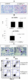
Liver architecture, C/EBPα expression in the liver and granulocytic differentiation are unaffected in C/EBPαΔ/Δ mice. (A) C/EBPαΔ/Δ mice have a normal liver architecture. Representative histological sections of liver stained with hematoxylin and eosin from C/EBPαΔ/Δ mice and control littermates (littermates that do not have at least one of the transgenic alleles and/or both targeted alleles). (B) C/EBPα RNA levels are unaffected in the livers of C/EBPαΔ/Δ mice. Quantitative real-time PCR analysis was performed using fetal liver RNA from C/EBPαΔ/Δ mice or C/EBPα−/− mice compared to that of their respective control littermates. C/EBPα expression is presented as a percentage of 18S RNA expression. This figure shows mean results plus standard deviations from six C/EBPαΔ/Δ mice and six control littermates (upper panel) or four C/EBPα−/− mice and five control littermates (two C/EBPα+/+ and three C/EBPα+/− mice) (lower panel). The asterisk indicates a significant difference (P value < 0.05) based on a two-tailed t test for samples of unequal variance. The P value for each sample is indicated. (C) C/EBPαΔ/Δ mice have granulocytes in the periphery. Representative peripheral blood smears stained with hematoxylin from C/EBPαΔ/Δ mice and control littermates. Granulocytes are indicated by arrows. (D) C/EBPαΔ/Δ mice have normal granulocytic differentiation. Fetal liver cells from C/EBPαΔ/Δ mice or C/EBPα−/− mice compared to those of their respective control littermates were stained with the granulocytic markers Gr-1/Mac-1 and analyzed by flow cytometry analysis. The percentage of double-positive cells in each sample is indicated. mC/EBPα, mouse C/EBPα; WT, control wild-type littermate; KO, C/EBPα-knocked out sample.
FIG. 3.
FIG. 3.
C/EBPαΔ/Δ mice show abnormal lung histopathology and decreased levels of C/EBPα protein in the lungs. (A) C/EBPαΔ/Δ mice have abnormal lung histopathology. Representative histological lung sections stained with hematoxylin-eosin of C/EBPαΔ/Δ mice and control littermates showing thickening of the alveolar walls (thin arrows), a lack of type I alveolar cells (arrowheads), and the presence of epithelial cuboidal cells with a clear cytosol (thick arrows). “Control” indicates littermates that do not have at least one of the transgenic alleles and/or both targeted alleles. The magnification used to photograph the sections is indicated. (B) C/EBPα protein expression is decreased in C/EBPαΔ/Δ mice. Immunohistochemistry for C/EBPα shows a loss of staining in the lungs of C/EBPαΔ/Δ neonates compared to that in control littermates (brown color staining). “Control” indicates littermates that do not have at least one of the transgenic alleles and/or both targeted alleles. The magnification used to photograph the sections is indicated.
FIG. 4.
FIG. 4.
C/EBPαΔ/Δ mouse alveolar epithelial cells are immature type II cells. (A) C/EBPαΔ/Δ mouse alveolar cells have a type II cell origin. Immunohistochemistry for SPC shows strong staining of C/EBPαΔ/Δ mouse epithelial cells (brown color). (B) C/EBPαΔ/Δ mouse alveolar cells lack lamellar bodies. Electron microscopy analysis of the lungs from C/EBPαΔ/Δ mice shows cuboidal alveolar cells with round nuclei and glycogen inclusions (open arrowheads). The epithelium lacks type I cells and the alveolar cells lack lamellar bodies. Type I cells with flattened nuclei and long cytoplasmic extensions can be found in the control littermates. These cells contain numerous lamellar bodies (full arrowheads). The magnification used to photograph the sections is indicated. (C) C/EBPαΔ/Δ mouse alveolar cells are glycogen rich. Periodic acid-Shiff staining shows an increased glycogen content in the alveolar cells of C/EBPαΔ/Δ mice compared to that in control littermates (pink color). (D) Immunohistochemistry for SPB confirms that C/EBPαΔ/Δ mice show decreased SPB expression (brown color) compared to control littermates (littermates that do not have at least one of the transgenic alleles and/or both targeted alleles). (E) C/EBPαΔ/Δ mice have decreased expression of genes important for respiratory function at birth. Quantitative real-time PCR analysis of SPA, SPD, and HNF3β was performed using total lung RNA from six C/EBPαΔ/Δ mice compared to that of six control littermates (littermates that do not have at least one of the transgenic alleles and/or both targeted alleles). The mean expression level and standard deviation are presented as a percentage of 18S RNA expression. The asterisk indicates a significant difference (P value < 0.05) based on a two-tailed t test for samples of unequal variance. The P value for each sample is indicated.
FIG. 5.
FIG. 5.
C/EBPαΔ/Δ mouse alveolar cells are hyperproliferative. The proliferative status of the alveolar cells of C/EBPαΔ/Δ mice was determined by immunohistochemistry for KI-67 (brown color). C/EBPαΔ/Δ mice display an increased number of Ki-67-positive cells compared to those of control littermates (littermates that do not have at least one of the transgenic alleles and/or both targeted alleles).
FIG. 6.
FIG. 6.
C/EBPαΔ/Δ mouse alveolar cells have decreased apoptosis. The ability of C/EBPαΔ/Δ mouse alveolar cells to undergo apoptosis was determined by the TUNEL assay. C/EBPαΔ/Δ mice display markedly decreased numbers of TUNEL-positive cells (brown color) compared to those in control littermates (littermates that do not have at least one of the transgenic alleles and/or both targeted alleles).
FIG. 7.
FIG. 7.
Expression profiling identifies many differentially expressed genes in the lungs of C/EBPαΔ/Δ mice. The expression histograms of the top 50 up-regulated (A) and down-regulated (B) genes are shown (for the full gene set [n = 124], see Tables S1 and S2 in the supplemental material). Genes are ranked by FDR-corrected P values (see Materials and Methods and reference 1). Duplicated samples and genes of unknown function were removed from the list. “Control” indicates littermates that do not have at least one of the transgenic alleles and/or both targeted alleles.
FIG. 8.
FIG. 8.
Confirmation of array targets by real-time PCR. Ppp1r3c and Gli-1 expression levels in C/EBPαΔ/Δ mice were compared to the levels in control littermates by quantitative real-time PCR analysis. “Control” indicates littermates that do not have at least one of the transgenic alleles and/or both targeted alleles. Expression levels are presented as a percentage of 18S RNA expression. This figure shows mean results plus standard deviations from five C/EBPαΔ/Δ mice and five control littermates. The asterisk indicates a significant difference (P value < 0.05) based on a two-tailed t test for samples of unequal variance. The P value for each sample is indicated. m, mouse.
FIG. 9.
FIG. 9.
Loss of C/EBPα leads to the formation of hypercellular areas in the lungs of adult C/EBPα4TG mice. (A) Histological lung sections stained with hematoxylin and eosin of a C/EBPα4TG mouse showing a normal area (left) and a hypercellular area (right). The magnification used to photograph the sections is indicated. (B) Lung sections were stained with a C/EBPα antibody. The pathologically normal area shows C/EBPα staining in bronchial epithelial cells and type II epithelial cells as expected (brown color). The hypercellular area does not express C/EBPα. The magnification used to photograph the sections is indicated.

Similar articles

Cited by

References

    1. Benjamini, Y., and Y. Hochberg. 1995. Controlling the false discovery rate—a practical and powerful approach to multiple testing. J. R. Stat. Soc. Ser. B 57:289-300.
    1. Bennett, C. N., S. E. Ross, K. A. Longo, L. Bajnok, N. Hemati, K. W. Johnson, S. D. Harrison, and O. A. MacDougald. 2002. Regulation of Wnt signaling during adipogenesis. J. Biol. Chem. 277:30998-31004. - PubMed
    1. Bigelow, R. L., N. S. Chari, A. B. Unden, K. B. Spurgers, S. Lee, D. R. Roop, R. Toftgard, and T. J. McDonnell. 2004. Transcriptional regulation of bcl-2 mediated by the sonic hedgehog signaling pathway through gli-1. J. Biol. Chem. 279:1197-1205. - PubMed
    1. Bourbon, J. R., M. Rieutort, M. J. Engle, and P. M. Farrell. 1982. Utilization of glycogen for phospholipid synthesis in fetal rat lung. Biochim. Biophys. Acta 712:382-389. - PubMed
    1. Breathnach, O. S., B. Freidlin, B. Conley, M. R. Green, D. H. Johnson, D. R. Gandara, M. O'Connell, F. A. Shepherd, and B. E. Johnson. 2001. Twenty-two years of phase III trials for patients with advanced non-small-cell lung cancer: sobering results. J. Clin. Oncol. 19:1734-1742. - PubMed

Publication types

MeSH terms

Substances

LinkOut - more resources