. 2006 Feb;74(2):830-8.
doi: 10.1128/IAI.74.2.830-838.2006.

Nontypeable Haemophilus influenzae adheres to intercellular adhesion molecule 1 (ICAM-1) on respiratory epithelial cells and upregulates ICAM-1 expression

Affiliations

Nontypeable Haemophilus influenzae adheres to intercellular adhesion molecule 1 (ICAM-1) on respiratory epithelial cells and upregulates ICAM-1 expression

Vasanthi Avadhanula et al. Infect Immun. 2006 Feb.

Abstract

Nontypeable Haemophilus influenzae (NTHI) is an important respiratory pathogen. NTHI initiates infection by adhering to the airway epithelium. Here, we report that NTHI interacts with intracellular adhesion molecule 1 (ICAM-1) expressed by respiratory epithelial cells. A fourfold-higher number of NTHI bacteria adhered to Chinese hamster ovary (CHO) cells transfected with human ICAM-1 (CHO-ICAM-1) than to control CHO cells (P < or = 0.005). Blocking cell surface ICAM-1 with specific antibody reduced the adhesion of NTHI to A549 respiratory epithelial cells by 37% (P = 0.001) and to CHO-ICAM-1 cells by 69% (P = 0.005). Preincubating the bacteria with recombinant ICAM-1 reduced adhesion by 69% (P = 0.003). The adherence to CHO-ICAM-1 cells of NTHI strains deficient in the adhesins P5, P2, HMW1/2, and Hap or expressing a truncated lipooligosaccharide was compared to that of parental strains. Only strain 1128f-, which lacks the outer membrane protein (OMP) P5-homologous adhesin (P5 fimbriae), adhered less well than its parental strain. The numbers of NTHI cells adhering to CHO-ICAM-1 cells were reduced by 67% (P = 0.009) following preincubation with anti-P5 antisera. Furthermore, recombinant ICAM bound to an OMP preparation from strain 1128f+, which expresses P5, but not to that from its P5-deficient mutant, confirming a specific interaction between ICAM-1 and P5 fimbriae. Incubation of respiratory epithelial cells with NTHI increased ICAM-1 expression fourfold (P=0.001). Adhesion of NTHI to the respiratory epithelium, therefore, upregulates the expression of its own receptor. Blocking interactions between NTHI P5 fimbriae and ICAM-1 may reduce respiratory colonization by NTHI and limit the frequency and severity of NTHI infection.


Figures

FIG. 1.
FIG. 1.
Adhesion of NTHI to A549 respiratory epithelial cells is blocked by anti-ICAM-1 antibodies. A549 cells were incubated with anti-ICAM-1 Ab or an isotype control (15 μg/ml) for 1 h, infected with NTHI for 1 h, and washed to remove loosely adherent bacteria. Adherent bacteria were quantified by plating serial dilutions of A549 cell lysates on agar. Preincubation of A549 cells with anti-ICAM-1 Ab (black bar) significantly reduced the adhesion of NTHI compared to preincubation with an isotype control Ab (white bar) (*, P = 0.001). Adhesion is expressed as the number of bacteria/cell. The data represent the mean ± standard error of the mean of four separate experiments.
FIG.2.
FIG.2.
Adhesion of NTHI and S. pneumoniae to CHO and CHO-ICAM-1 cells. CHO and CHO-ICAM-1 cells were incubated with H. influenzae strain 778, 781, or Rd or S. pneumoniae for 1 h and washed to remove loosely adherent bacteria. Adherent bacteria were quantified by plating serial dilutions of cell lysates on agar plates. Adhesion is expressed as numbers of bacteria/cell. (A) More NTHI cells adhered to CHO-ICAM-1 cells (black bars) than to control CHO cells (white bars) (*, P ≤ 0.005), whereas there was no significant difference in the adhesion of S. pneumoniae to CHO or CHO-ICAM-1 cells. The data represent the mean ± standard error of the mean of four separate experiments. (B) CHO-ICAM-1 cells were incubated with FITC-labeled NTHI or S. pneumoniae for 1 h, stained with anti-ICAM-1 antibody, and then washed and incubated with Alexa 594-conjugated anti-IgG secondary antibody and the double-stranded DNA specific fluorochrome To-PRO3. (Top) FITC-labeled NTHI (green) binds primarily to cells expressing ICAM-1 (red). Cell nuclei are counterstained blue. (Bottom) Few FITC-labeled S. pneumoniae cells (green) adhere to CHO-ICAM-1 cells.
FIG. 3.
FIG. 3.
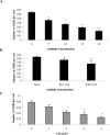
Blocking with anti-ICAM-1 antibodies and rICAM-1 reduces NTHI adhesion to CHO-ICAM-1 cells. CHO-ICAM-1 cells were incubated with anti-ICAM-1 MAb at the indicated concentrations (in μg/ml). The cells were then infected with NTHI, and the numbers of bacteria adhering to the cells were compared to the numbers of bacteria binding to untreated cells. (A) Treatment with anti-ICAM-1 MAb (LB2) resulted in a dose-dependent inhibition of NTHI adhesion. The data represent the mean ±  standard error of five independent experiments (*, P = 0.005). Preincubation of CHO-ICAM-1 cells with an isotype control antibody did not alter NTHI adherence (data not shown). (B) Adhesion of NTHI was also determined after incubating CHO-ICAM-1 cells with MAbs R1/1 and 8.4A6, which bind ICAM-1 D1 and D2, respectively. Both R1/1 and 8.4A6 at concentrations of 10 μg/ml reduced the numbers of adherent NTHI cells (*, P = 0.03 for R1/1 and P = 0.01 for 8.4A6). In contrast, preincubation of CHO-ICAM-1 cells with isotype control antibodies did not alter NTHI adherence (data not shown). The data represent the mean ± standard error of four independent experiments. (C) NTHI was preincubated with rICAM-1 at the indicated concentrations and then used to infect CHO-ICAM-1 cells. Preincubation of bacteria with rICAM-1 resulted in a dose-dependent inhibition of NTHI adhesion to ICAM-1 cells (*, P = 0.003). The data represent the mean ± standard error of five independent experiments.
FIG. 4.
FIG. 4.
An isogenic mutant strain of NTHI lacking P5 adheres poorly to CHO-ICAM-1 cells compared to its P5-sufficient parental strain. CHO-ICAM-1 cells were incubated with NTHI strains and their isogenic mutants lacking HMW1 and HMW2 (strain 12, HMW); HMW1, HMW2, and Hap proteins (strain N187, HMW, and N187, Hap); P2 fimbriae (strain Rd, P2); and P5 fimbriae (strain 1128f) and expressing LOS (strain 2019) or truncated LOS (strain 2019, pgmB::erm), and adherent bacteria were quantified. Only the P5 fimbria-deficient strain 1128f had significant reduction in adhesion to CHO-ICAM-1 cells compared to its parental strain (*, P = 0.003). The data represent the mean ± standard error of five independent experiments.
FIG. 5.
FIG. 5.
Incubation with anti-P5 antibodies inhibits adhesion of NTHI 778 to ICAM-1 cells. NTHI 778 was preincubated with increasing concentrations of rabbit anti-P5 antiserum (black bars) or control preimmune serum (white bars) at the indicated concentrations and then incubated with CHO-ICAM-1 cells. Preincubation of bacteria with anti-P5 Ab resulted in a dose-dependent inhibition of NTHI adhesion to CHO-ICAM-1 cells. The data represent the mean ± standard error of four independent experiments (*, P = 0.009).
FIG. 6.
FIG. 6.
rICAM binds to outer membrane protein P5. Whole OMPs of NTHI 1128f+ (lane 1) and 1128f (lane 2); peptides 1 to 4 (lanes 3 to 6), corresponding to the four predicted surface-exposed regions of P5; and rICAM-1 (positive control) (lane 7) were spotted on a nitrocellulose membrane and incubated with rICAM-1. Anti-ICAM-1 Ab was used to detect bound ICAM-1. rICAM-1 binds to OMP preparations from P5-sufficient strain 1128f+, but not P5-deficient strain 1128f, and to a peptide corresponding to loop 4 of P5 fimbriae.
FIG. 7.
FIG. 7.
NTHI infection increases ICAM-1 expression on A549 cells. A549 cells were infected by NTHI 778 at an MOI of 1, 10, or 100 for 6 h, and ICAM-1 expression was quantified by fluorescence-activated cell sorting. The histograms illustrate the fluorescence intensity (ICAM-1 expression; horizontal axis) of uninfected control cells (dotted line) compared to that of cells infected with increasing numbers of NTHI. The expression level of A549 cell ICAM-1 increased in an inoculum-dependent manner by threefold at an MOI of 10 (gray line) (P = 0.01) and by fourfold at an MOI of 100 (black line) (P = 0.001). The data are representative of three independent experiments.

References

    1. Barenkamp, S. J., and E. Leininger. 1992. Cloning, expression, and DNA sequence analysis of genes encoding nontypeable Haemophilus influenzae high-molecular-weight surface-exposed proteins related to filamentous hemagglutinin of Bordetella pertussis. Infect. Immun. 60:1302-1313. - PMC - PubMed
    1. Bella, J., P. R. Kolatkar, C. W. Marlor, J. M. Greve, and M. G. Rossmann. 1998. The structure of the two amino-terminal domains of human ICAM-1 suggests how it functions as a rhinovirus receptor and as an LFA-1 integrin ligand. Proc. Natl. Acad. Sci. USA 95:4140-4145. - PMC - PubMed
    1. Berendt, A. R., A. McDowall, A. G. Craig, P. A. Bates, M. J. Sternberg, K. Marsh, C. I. Newbold, and N. Hogg. 1992. The binding site on ICAM-1 for Plasmodium falciparum-infected erythrocytes overlaps, but is distinct from, the LFA-1-binding site. Cell 68:71-81. - PubMed
    1. Bochner, B. S., F. W. Luscinskas, M. A. Gimbrone, Jr., W. Newman, S. A. Sterbinsky, C. P. Derse-Anthony, D. Klunk, and R. P. Schleimer. 1991. Adhesion of human basophils, eosinophils, and neutrophils to interleukin 1-activated human vascular endothelial cells: contributions of endothelial cell adhesion molecules. J. Exp. Med. 173:1553-1557. - PMC - PubMed
    1. Clemans, D. L., R. J. Bauer, J. A. Hanson, M. V. Hobbs, J. W. St Geme III, C. F. Marrs, and J. R. Gilsdorf. 2000. Induction of proinflammatory cytokines from human respiratory epithelial cells after stimulation by nontypeable Haemophilus influenzae. Infect. Immun. 68:4430-4440. - PMC - PubMed

Publication types

Substances

LinkOut - more resources