. 2006 Feb;74(2):1323-38.
doi: 10.1128/IAI.74.2.1323-1338.2006.

Intracellular gene expression profile of Listeria monocytogenes

Affiliations

Intracellular gene expression profile of Listeria monocytogenes

Som Subhra Chatterjee et al. Infect Immun. 2006 Feb.

Abstract

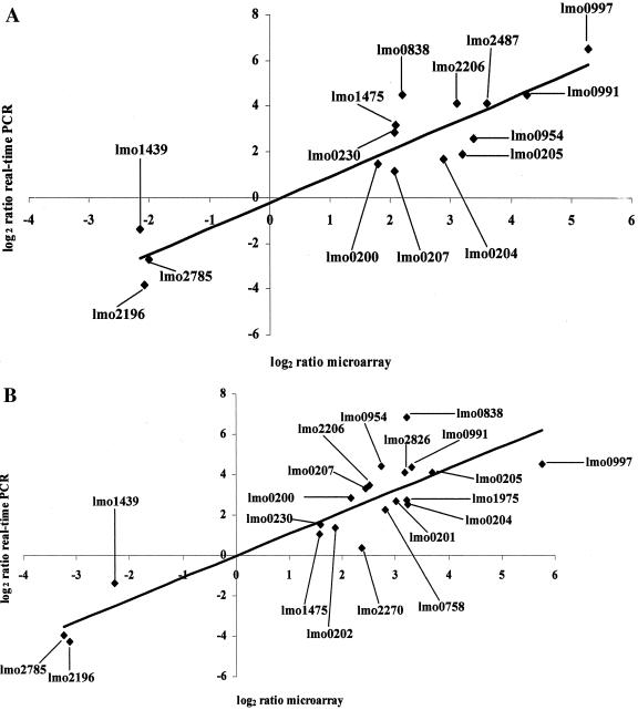
Listeria monocytogenes is a gram-positive, food-borne microorganism responsible for invasive infections with a high overall mortality. L. monocytogenes is among the very few microorganisms that can induce uptake into the host cell and subsequently enter the host cell cytosol by breaching the vacuolar membrane. We infected the murine macrophage cell line P388D1 with L. monocytogenes strain EGD-e and examined the gene expression profile of L. monocytogenes inside the vacuolar and cytosolic environments of the host cell by using whole-genome microarray and mutant analyses. We found that approximately 17% of the total genome was mobilized to enable adaptation for intracellular growth. Intracellularly expressed genes showed responses typical of glucose limitation within bacteria, with a decrease in the amount of mRNA encoding enzymes in the central metabolism and a temporal induction of genes involved in alternative-carbon-source utilization pathways and their regulation. Adaptive intracellular gene expression involved genes that are associated with virulence, the general stress response, cell division, and changes in cell wall structure and included many genes with unknown functions. A total of 41 genes were species specific, being absent from the genome of the nonpathogenic Listeria innocua CLIP 11262 strain. We also detected 25 genes that were strain specific, i.e., absent from the genome of the previously sequenced L. monocytogenes F2365 serotype 4b strain, suggesting heterogeneity in the gene pool required for intracellular survival of L. monocytogenes in host cells. Overall, our study provides crucial insights into the strategy of intracellular survival and measures taken by L. monocytogenes to escape the host cell responses.


Figures

FIG. 1.
FIG. 1.
Survival of L. monocytogenes EGD-e and the isogenic double-deletion Δhly ΔplcA mutant intacellularly. (A) P388D1 murine macrophage cells were infected with the wild-type strain EGD-e and the isogenic double-deletion Δhly ΔplcA mutant at MOIs of 10 and 500, respectively, in a 6-well tissue culture plate. One, 4, and 8 h postinfection, eukaryotic cells were lysed with cold water and plated onto agar plates for CFU counts. The data shown here are representative of results from three independent experiments. (B) Immunofluorescence microscopic image of P388D1 cells infected with wild-type EGD-e 8 h postinfection. (C to E) Confocal microscopic images of P388D1 cells infected with the Δhly ΔplcA mutant 1 h postinfection. (C) Δhly ΔplcA mutant bacteria expressing a constitutive green fluorescent protein. (D) P388D1 cells stained with LysoTracker red. (E) Merge of the diagrams in panels C and D.
FIG. 2.
FIG. 2.
Validation of microarray data with quantitative real-time PCR analysis. (A) Validation of the intravacuolarly expressed genes. (B) Validation of intracytosolically expressed genes at 4 h postinfection. (C) Validation of intracytosolically expressed genes at 8 h postinfection.
FIG. 2.
FIG. 2.
Validation of microarray data with quantitative real-time PCR analysis. (A) Validation of the intravacuolarly expressed genes. (B) Validation of intracytosolically expressed genes at 4 h postinfection. (C) Validation of intracytosolically expressed genes at 8 h postinfection.
FIG.3.
FIG.3.
Characterization of bacterial deletion mutants. (A and B) Intracellular survival of L. monocytogenes EGD-e compared to that of the ΔprfA, ΔorfX (lmo0206), ΔorfZ (lmo0207), ΔctsR (lmo0229), ΔsigB, ΔtnrA (lmo1298), ΔohrR (lmo2200), and ΔprsA (lmo2219) isogenic deletion mutants. P388D1 murine macrophage cells were infected with the wild-type strain EGD-e and its isogenic deletion mutants at MOIs of 10 in 24-well plates, and bacterial CFU were counted on agar plated following lysis of the P388D1 cells at 1, 4, and 8 h postinfection. (C) A bacterial plaque formation assay was performed with L929 murine fibroblast cells. The cells were infected with L. monocytogenes EGD-e and its isogenic deletion mutants for 2 h at 37°C in a humidified incubator and subsequently incubated for 30 min in the presence of 10 μg of gentamicin/ml. The plates were then agarose overlaid and were observed for plaques after an additional incubation at 37°C 3 days postinfection. The diameters of the different plaques obtained were determined by processing the images for Adobe Photoshop version 5.5 and are derived from measuring 25 plaques for each strain tested. The mean plaque size of the wild-type strain was defined as 100%. The mean size (%) with standard deviation is given for each strain in the experiment. n.d. (not detected) indicates that strains produced no visible plaques under the conditions used. The data shown here are representative of results of three independent experiments.
FIG.3.
FIG.3.
Characterization of bacterial deletion mutants. (A and B) Intracellular survival of L. monocytogenes EGD-e compared to that of the ΔprfA, ΔorfX (lmo0206), ΔorfZ (lmo0207), ΔctsR (lmo0229), ΔsigB, ΔtnrA (lmo1298), ΔohrR (lmo2200), and ΔprsA (lmo2219) isogenic deletion mutants. P388D1 murine macrophage cells were infected with the wild-type strain EGD-e and its isogenic deletion mutants at MOIs of 10 in 24-well plates, and bacterial CFU were counted on agar plated following lysis of the P388D1 cells at 1, 4, and 8 h postinfection. (C) A bacterial plaque formation assay was performed with L929 murine fibroblast cells. The cells were infected with L. monocytogenes EGD-e and its isogenic deletion mutants for 2 h at 37°C in a humidified incubator and subsequently incubated for 30 min in the presence of 10 μg of gentamicin/ml. The plates were then agarose overlaid and were observed for plaques after an additional incubation at 37°C 3 days postinfection. The diameters of the different plaques obtained were determined by processing the images for Adobe Photoshop version 5.5 and are derived from measuring 25 plaques for each strain tested. The mean plaque size of the wild-type strain was defined as 100%. The mean size (%) with standard deviation is given for each strain in the experiment. n.d. (not detected) indicates that strains produced no visible plaques under the conditions used. The data shown here are representative of results of three independent experiments.

References

    1. Abachin, E., C. Poyart, E. Pellegrini, E. Milohanic, F. Fiedler, P. Berche, and P. Trieu-Cuot. 2002. Formation of d-alanyl-lipoteichoic acid is required for adhesion and virulence of Listeria monocytogenes. Mol. Microbiol. 43:1-14. - PubMed
    1. Arous, S., C. Buchrieser, P. Folio, P. Glaser, A. Namane, M. Hebraud, and Y. Hechard. 2004. Global analysis of gene expression in an rpoN mutant of Listeria monocytogenes. Microbiology 150:1581-1590. - PubMed
    1. Asanuma, N., and T. Hino. 2003. Molecular characterization of HPr and related enzymes, and regulation of HPr phosphorylation in the ruminal bacterium Streptococcus bovis. Arch. Microbiol. 179:205-213. - PubMed
    1. Autret, N., C. Raynaud, I. Dubail, P. Berche, and A. Charbit. 2003. Identification of the agr locus of Listeria monocytogenes: role in bacterial virulence. Infect. Immun. 71:4463-4471. - PMC - PubMed
    1. Begley, M., R. D. Sleator, C. G. Gahan, and C. Hill. 2005. Contribution of three bile-associated loci bsh, pva, and btlB, to gastrointestinal persistence and bile tolerance of Listeria monocytogenes. Infect. Immun. 73:894-904. - PMC - PubMed

Publication types

MeSH terms

LinkOut - more resources