. 2006 Feb;9(2):180-7.
doi: 10.1038/nn1631. Epub 2006 Jan 22.

The Runx1/AML1 transcription factor selectively regulates development and survival of TrkA nociceptive sensory neurons

Affiliations

The Runx1/AML1 transcription factor selectively regulates development and survival of TrkA nociceptive sensory neurons

Frédéric Marmigère et al. Nat Neurosci. 2006 Feb.

Abstract

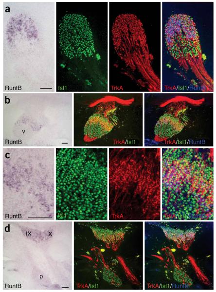
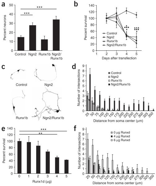
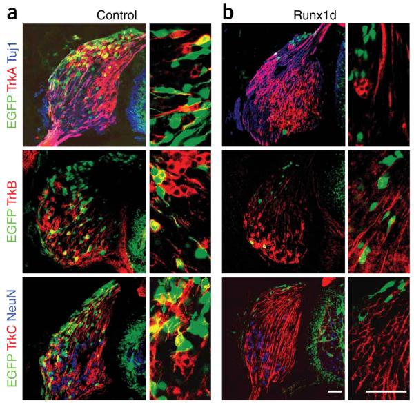
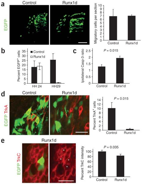
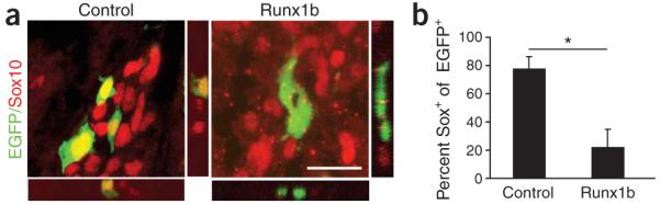
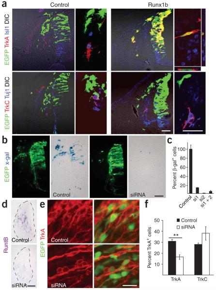
Neural crest cells (NCCs) can adopt different neuronal fates. In NCCs, neurogenin-2 promotes sensory specification but does not specify different subclasses of sensory neurons. Understanding the gene cascades that direct Trk gene activation may reveal mechanisms generating sensory diversity, because different Trks are expressed in different sensory neuron subpopulations. Here we show in chick and mouse that the Runt transcription factor Runx1 promotes axonal growth, is selectively expressed in neural crest-derived TrkA(+) sensory neurons and mediates TrkA transactivation in migratory NCCs. Inhibition of Runt activity depletes TrkA expression and leads to neuronal death. Moreover, Runx1 overexpression is incompatible with multipotency in the migratory neural crest but does not induce expression of pan-neuronal genes. Instead, Runx1-induced neuronal differentiation depends on an existing neurogenin2 proneural gene program. Our data show that Runx1 directs, in a context-dependent manner, key aspects of the establishment of the TrkA(+) nociceptive subclass of neurons.


Figures

Figure 1
Figure 1
RuntB is expressed in the TrkA+ subpopulation of neural crest-derived trunk and cranial sensory neurons. In situ hybridization for RuntB and immunohistochemical stainings for TrkA (red) and Isl1 (green) are shown (a) in the same sagittal sections of DRG, (b,c) in the trigeminal ganglion (V) and (d) in the superior (IX), jugular (X) and petrosal (p) ganglia at stage HH29 of the chick embryo. Merged images of RuntB In situ hybridization (blue pseudocolor) and immunohistochemical stainings for TrkA and Isl1 show an overlapping expression of RuntB and TrkA in trigeminal, superior, jugular and dorsal root ganglia but not in the petrosal ganglion. Scale bars, 100 μm.
Figure 2
Figure 2
Runx1b promotes survival and axonal growth of Ngn2-fated bNCSCs. (a) Percentage of neurons 3 d after differentiation with Ngn2, Runx1b or both. Ngn2 exerted a neurogenic effect, whereas Runx1 did not. (b) Camera lucida drawings of representative neurons observed 3 d after differentiation in control and with Ngn2, Runx1 and Ngn2/Runx1. Note that cotransfection with Ngn2 and Runx1 led to a marked increase in neurite growth. (c) Percentage survival of neurons at 3, 4 and 5 d after differentiation of bNCSCs. Neurons in cultures receiving only GFP plasmid (control), or after Ngn2 or Runx1b transfection, died massively between 3 and 5 days, while Ngn2/Runx1 coexpressing neurons survived. (d) Quantification by Sholl analysis of neurite length and number of branching of neurons 3 d after differentiation with Ngn2, Runx1 and Ngn2/Runx1. Cotransfection with Ngn2 and Runx1 significantly increased both neurite length and branching at all distances. (e,f) Runx1 transactivation domain mediates its survival and differentiation effects. (e) Percent survival of neurons 3 d after differentiation with Ngn2/Runx1 and increasing concentrations of Runx1d (a competitive form of Runx1b blocking the activity of the full protein) compared to survival after transfection with Ngn2/Runx1 only. Runx1d reversed the survival effect of Runx1b on Ngn2-fated bNCSCs in a dose-dependent manner. (f) Quantification of neurite length and number of branches by Sholl analysis of neurons 3 d after differentiation with Ngn2/Runx1b and increasing concentrations of Runx1d. Runx1d reversed the growth effect of Runx1b on Ngn2-fated bNCSCs. *P ≤ 0.05, **P ≤ 0.01, ***P ≤ 0.001, Student’s t-test. Error bars = s.e.m.
Figure 3
Figure 3
Blocking Runt activity is incompatible with establishment of neural crest-derived sensory neurons. Micrographs of stage HH29 DRG of (a) control and (b) Runx1d-overexpressing chick embryos stained for TrkA, TrkB or TrkC in red, the early neuronal marker Tuj1 (βIII tubulin) or NeuN in blue and corresponding high-magnification images where only the green and red channels are shown. Note in panel a that EGFP was present in TrkA+, TrkB+ and TrkC+ neurons, whereas animals overexpressing Runx1d (in panel b) showed a marked loss of EGFP-expressing cells. The remaining cells, mostly residing in the very dorsal part of the DRG, did not express TrkA, TrkB or TrkC. Scale bars, 50 μm.
Figure 4
Figure 4
Runx1d-induced apoptosis is preceded by a loss of Trk expression. (a) Runx1d does not affect NCC migration. Control and Runx1d-overexpressing chick embryos at HH19 and quantitative measurements of the number of migratory NCCs per section with and without Runx1d. (b) Percentage of EGFP+ cells relative to the total number of DRG neurons in control and Runx1d-overexpressing embryos at stages HH24 and HH29. Note that the loss of neurons occurs between stages HH24 and HH29. (c) Loss of neurons is preceded by an increase in active caspase-3. The number of active caspase-3+ cells in Runx1d-overexpressing and contralateral control DRG was counted at HH24 (the ratio of ipsi- to contralateral is presented). (d) Death of neurons is preceded by a nearly complete loss of TrkA expression at stage HH24. Micrographs showing TrkA expression in control and Runx1d-overexpressing DRG neurons and quantitative measurements of the percentage of cells expressing TrkA in control and Runx1d-overexpressing embryos. Note that the Runx1d-overexpressing EGFP+ neurons lack TrkA expression. (e) Death of neurons is also preceded by a reduction of TrkC levels at HH24. Micrographs showing TrkC expression in control and Runx1d-overexpressing DRG neurons at stage HH24 and quantitative data on the mean intensity of TrkC expression in control and Runx1d-expressing DRG neurons. Note loss of TrkC expression levels in the two Runx1d-transfected cells (green stars) as compared to adjacent untransfected control cells (yellow stars). Scale bars, 50 μm (a) and 20 μm (d,e). ***P ≤ 0.001, one-tailed t-test. Error bars = s.e.m.
Figure 5
Figure 5
Runx1b-expressing cells lose Sox10 expression. (a) Images showing control and Runx1b overexpression in chick migratory NCCs at HH19 stained for the HMG transcription factor Sox10. (b) Quantitative data from measurements of colocalization of EGFP and Sox10 showing the loss of Sox10 expression in cells expressing Runx1b. Scale bar (a), 20 μm. *P ≤ 0.05, one-tailed t-test. Error bars = s.e.m.
Figure 6
Figure 6
Runx1 selectively controls TrkA expression. (a) Micrographs show stage-HH19 control and Runx1b-overexpressing neural tube and migratory NCCs stained for TrkA and the sensory neuronal marker Isl1, as well as for TrkC and the early neuronal marker Tuj1 (βIII-tubulin). Note that Runx1b induced premature and ectopic expression of TrkA but not TrkC. Confocal Z-stacks showed that TrkA is turned on in the Runx1b-overexpressing (green) cells. (b) RuntB siRNA efficiently silences β-galactosidase translated from a LacZ construct tagged in the 3′ untranslated region with the siRNA-targeted RuntB sequence compared to control without siRNA. Micrographs show β-galactosidase (blue) and EGFP (green) expression in control and siRNA-treated chick embryos at stage HH24. (c) Percent of β-galactosidase-expressing PC12 cells 24 h after transfection with siRNA1, siRNA2 and a combination of both together with the LacZ construct tagged with the RuntB siRNA-targeted sequence. Note that siRNA induced a significant knockdown of β-galactosidase both in vitro and in vivo. (d) Expression of endogenous RuntB mRNA detected by In situ hybridization in control and siRNA-treated chick DRG at stage HH29. Note that siRNA significantly reduced endogenous RuntB mRNA expression at this stage (n = 4 animals/construct). (e,f) Reducing endogenous RuntB expression with siRNAs selectively downregulates TrkA but not TrkC expression in chick DRG at stage HH29 as shown in micrographs (e) and the corresponding quantification (f). Scale bars, 100 μm (b,d) and 20 μm (a,e). Two-tailed t-test **P ≤ 0.01. Error bars represent s.e.m.

Comment in

  • Specifying nociceptors.
    Markus A. Markus A. Nat Neurosci. 2006 Feb;9(2):162. doi: 10.1038/nn0206-162. Nat Neurosci. 2006. PMID: 16439982 No abstract available.

References

    1. Carr PA, Nagy JI. Emerging relationships between cytochemical properties and sensory modality transmission in primary sensory neurons. Brain Res. Bull. 1993;30:209–219. - PubMed
    1. Cheung M, et al. The transcriptional control of trunk neural crest induction, survival, and delamination. Dev. Cell. 2005;8:179–192. - PubMed
    1. Kim J, Lo L, Dormand E, Anderson DJ. SOX10 maintains multipotency and inhibits neuronal differentiation of neural crest stem cells. Neuron. 2003;38:17–31. - PubMed
    1. Serbedzija GN, Fraser SE, Bronner-Fraser M. Pathways of trunk neural crest cell migration in the mouse embryo as revealed by vital dye labelling. Development. 1990;108:605–612. - PubMed
    1. Kasemeier-Kulesa JC, Kulesa PM, Lefcort F. Imaging neural crest cell dynamics during formation of dorsal root ganglia and sympathetic ganglia. Development. 2005;132:235–245. - PubMed

Publication types

Substances