. 2006 Feb;13(2):147-52.
doi: 10.1038/nsmb1046. Epub 2006 Jan 22.

Allosteric signaling of ATP hydrolysis in GroEL-GroES complexes

Affiliations

Allosteric signaling of ATP hydrolysis in GroEL-GroES complexes

Neil A Ranson et al. Nat Struct Mol Biol. 2006 Feb.

Abstract

The double-ring chaperonin GroEL and its lid-like cochaperonin GroES form asymmetric complexes that, in the ATP-bound state, mediate productive folding in a hydrophilic, GroES-encapsulated chamber, the so-called cis cavity. Upon ATP hydrolysis within the cis ring, the asymmetric complex becomes able to accept non-native polypeptides and ATP in the open, trans ring. Here we have examined the structural basis for this allosteric switch in activity by cryo-EM and single-particle image processing. ATP hydrolysis does not change the conformation of the cis ring, but its effects are transmitted through an inter-ring contact and cause domain rotations in the mobile trans ring. These rigid-body movements in the trans ring lead to disruption of its intra-ring contacts, expansion of the entire ring and opening of both the nucleotide pocket and the substrate-binding domains, admitting ATP and new substrate protein.


Figures
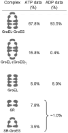
Figure 1
Figure 1
Distribution of protein complexes found in the ATP and ADP samples. Figures given are as a percentage of all side views imaged.
Figure 2
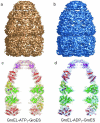
Figure 2. Solution structures of GroEL-ATP7-GroES and GroEL-ADP7-GroES
(a) Surface representation of the side view of the GroEL-ATP7-GroES complex. (b) surface representation of the side view of the GroEL-ADP7-GroES complex. (c,d) Central sections through the cryo-EM maps are shown as a semi-transparent surface in either gold (c, ATP) or blue (d, ADP), with the atomic coordinates for the GroEL equatorial (green: residues 3-136 & 410-524), intermediate (yellow: residues 137-191 & 374-409), apical (red: residues 192-373, except for 353-361 at the tip of the mobile helical hairpin in the trans ring), and GroES (magenta) fitted in. The 7-fold axis of the GroEL-GroES oligomer is vertical and in the image plane for all figures. The resolution of the ATP and ADP-bound maps are 7.7 Å and 8.7 Å respectively, determined by Fourier shell correlation (FSC) at a cut-off of 0.5. FSC curves for both reconstructions are shown in supplementary figure 1. Figures 2-6 (and supplementary figures 2-5) were created with Pymol (www.pymol.org).
Figure 3
Figure 3. Quality of EM maps and atomic structure fitting
(a) A section through the atomic coordinates for GroES fitted into the EM-derived electron density of GroEL-ATP7-GroES. The hole in the β-barrel that forms the body of the 10 kDa. subunit is clearly resolved. (b) A section through a single GroEL subunit in the cis (GroES-bound) ring with ADP.AlF3 in the nucleotide binding site shown in CPK-color space-filling mode. Although small regions corresponding to loops are not well resolved, the length and orientation of all secondary structure elements fit the EM density extremely well, regardless of their radius from the 7-fold axis (which is vertical and in the plane of the figure). (c) A section through the equatorial domains of the cis ring, perpendicular to the 7-fold axis. The helical segments of the equatorial domain are clearly resolved. In all panels, the EM-derived electron density for the GroEL-ATP7-GroES complex is contoured as a semi-transparent gold surface.
Figure 4
Figure 4. Disruption of intra-ring contacts between the equatorial domains of the trans ring
(a) The interface between neighbouring equatorial domains in the trans ring of the ATP-bound complex. The EM-derived electron density is shown as a gold mesh, with the adjacent equatorial domains in blue and magenta. In the ATP complex (and all crystal structures of GroEL complexes) two β-strands from each subunit form a 4-stranded β-sheet that is a major contact holding the ring of equatorial domains together (highlighted by a black rectangle). (b). The corresponding view of the ADP-bound complex (with EM density shown as a blue mesh and equatorial domains in green and orange) shows that a small (~3°) rotation of the equatorial domain results in the two strands from the orange subunit moving upward (in this view) and the two strands from the green subunit moving downward, pulling apart the β-sheet contact. (c & d) Structure of the rear half of the ring of equatorial domains from the ATP(c) and ADP-bound (d) complexes. The 7-fold axis is vertical and in the image plane for c & d, and these views are reached by tipping a & b forward by ~30°. (e) A comparison of the intra-strand (main chain) distances in the ATP (blue/magenta) and ADP (green/gold) complexes showing that the strand separation increases by 4-5 Å upon ATP hydrolysis.
Figure 5
Figure 5. ATP hydrolysis in cis causes radial expansion of the trans ring
(a & b) The inter-ring interface and trans ring of the ATP and ADP bound complexes. The cis equatorial (Eq) domain, together with the trans equatorial, intermediate and apical (Ap) domains are shown as a gray cartoon for both structures. The structural elements involved in communicating nucleotide binding are colored as follows: relay helix D (running from the nucleotide binding site to the Ala109 inter-ring contact) and substrate binding residues are magenta for ATP and blue for ADP. Glu461 at the second inter-ring contact is gold for ATP and orange for ADP. Finally, the β-strand contact in the trans equatorial ring is purple for ATP and green for ADP. (c) An overlay of ATP and ADP bound structures. ATP binding causes an offset between the relay helices across the ring interface. After ATP hydrolysis, the relay helices become realigned. The trans equatorial domains pivot about the Glu461 inter-ring contact, disrupting the β-strand intra-ring contact. The radial expansion is propagated throughout the trans ring, resulting in an opening of the substrate binding cavity.
Figure 6
Figure 6. Movements of key structural elements in the GroEL ATPase cycle
(a) The structures important in the allosteric mechanism are shown superimposed on the shape of an axial section through unliganded GroEL. The hydrophobic substrate-binding sites (gray space filling) line the end cavities in the high affinity state for non-native substrates, and apposed helix D's (gold) form a close contact via Ala109 between the rings. The intra-ring β-strand contacts are shown (red/blue) in the lower ring. (b) GroEL-ATP7 (EMD1047; Ref. 7). ATP binding to the upper ring causes the whole complex to elongate, with twisting of the apical domains so that the substrate binding sites are partly rotated out of the end cavity. The inter-ring interface is much weaker. (c) GroEL-ATP7-GroES. Tilt of the equatorial domains in cis causes an offset of helix D, thus moving apart the Ala109 residues across the inter-ring contact. The large rotation of the cis apical domains moves the substrate binding sites away from the cavity lining. The trans ring becomes more closed. (d) GroEL-ADP7-GroES. Rotation of the trans equatorial domains brings the helix D contact back into register but disrupts the intra-ring β-strand contact. The whole trans ring expands radially, creating the new substrate acceptor state. (e) ATP7-GroEL-ADP7-GroES. At the lower resolution to which this state was captured (EMD1046; Ref. 7), the conformation appears the same as the GroEL-ATP7-GroES state. The trans ring contracts and the intra-ring contact is re-established.

References

    1. Sigler PB, et al. Structure and function in GroEL-mediated protein folding. Annu. Rev. Biochem. 1999;67:581–608. - PubMed
    1. Saibil HR, Ranson NA. The chaperonin folding machine. Trends Biochem. Sci. 2002;27:627–632. - PubMed
    1. Hartl FU, Hayer-Hartl FU. Molecular chaperones in the cytosol: from nascent chain to folded protein. Science. 2002;295:1852–1858. - PubMed
    1. Fenton WA, Horwich AL. Chaperone-mediated protein folding: fate of substrate polypeptide. Q. Rev. Biophys. 2003;36:229–256. - PubMed
    1. Yifrach O, Horovitz A. Two lines of allosteric communication in the oligomeric chaperonin GroEL are revealed by the single mutation Arg196->Ala. J. Mol. Biol. 1994;243:397–401. - PubMed

Publication types

MeSH terms