Review
. 2005 Dec;5(12):1964-82.
doi: 10.1166/jnn.2005.446.

RNA nanotechnology: engineering, assembly and applications in detection, gene delivery and therapy

Affiliations
Review

RNA nanotechnology: engineering, assembly and applications in detection, gene delivery and therapy

Peixuan Guo. J Nanosci Nanotechnol. 2005 Dec.

Abstract

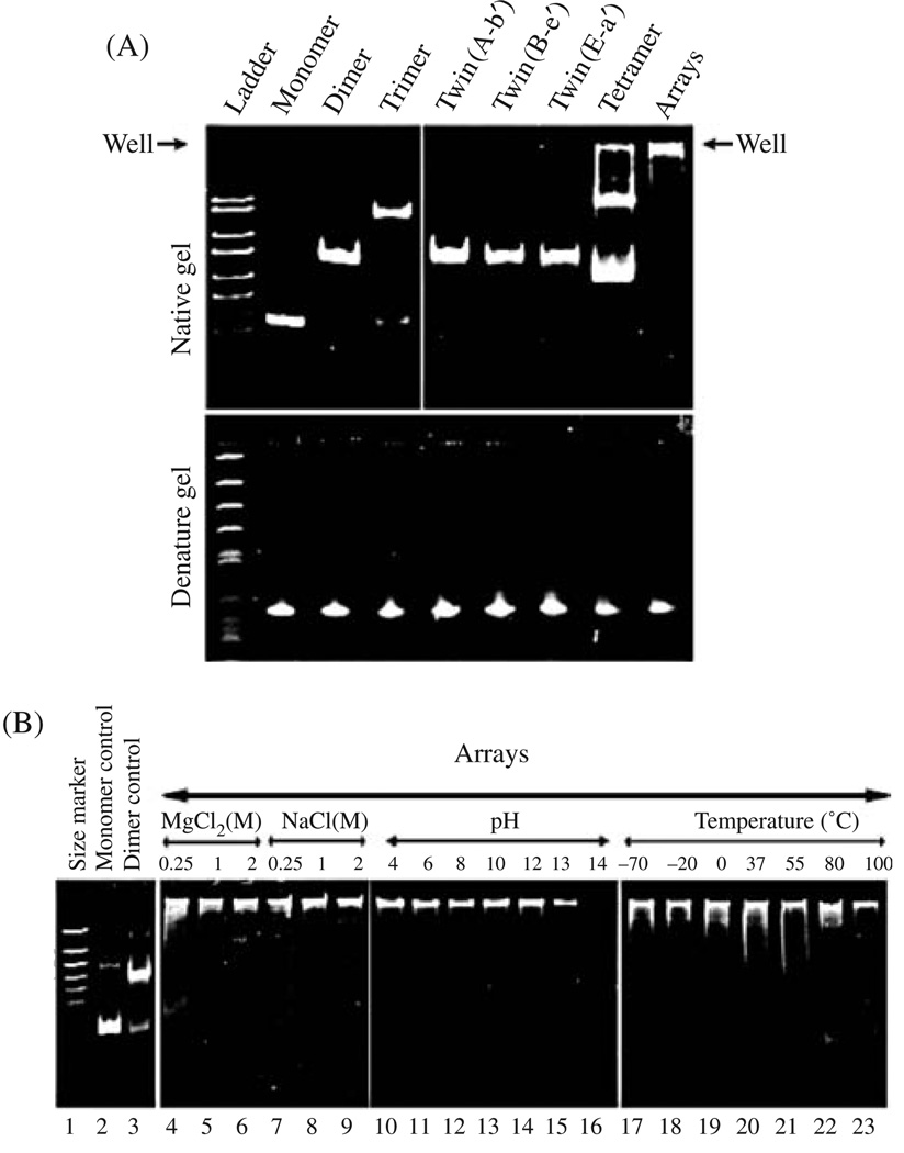
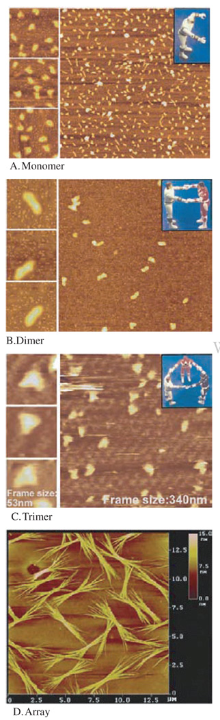
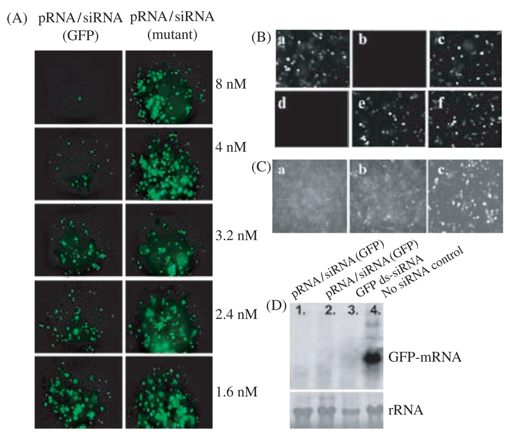
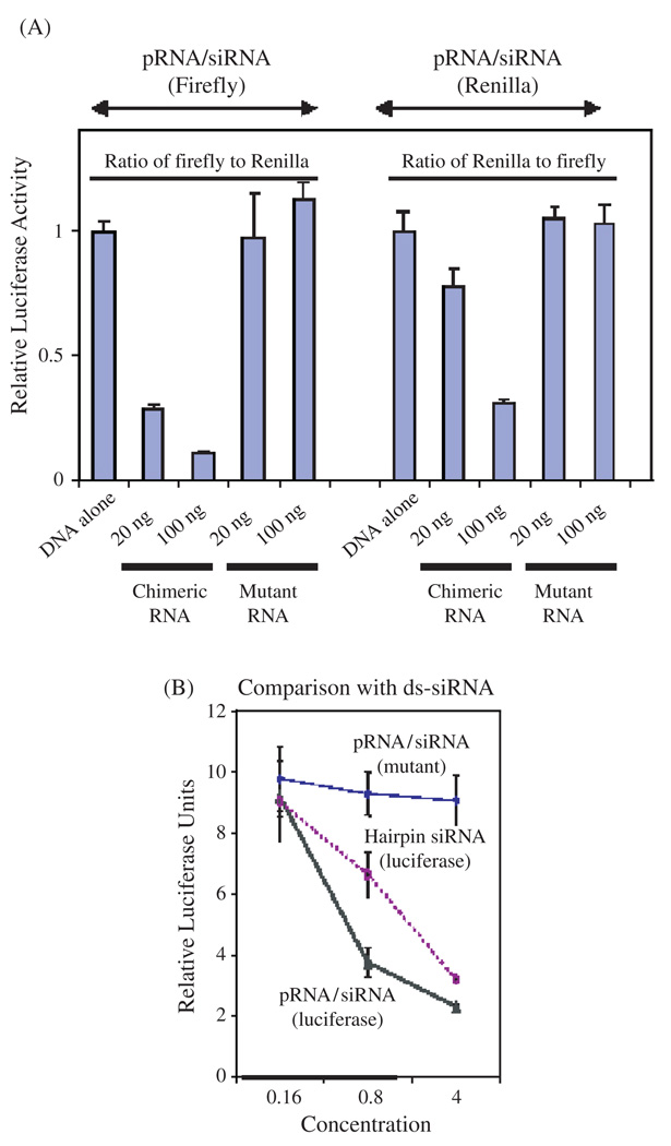
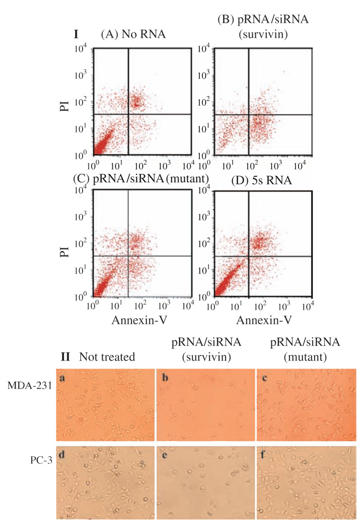
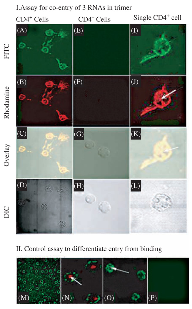
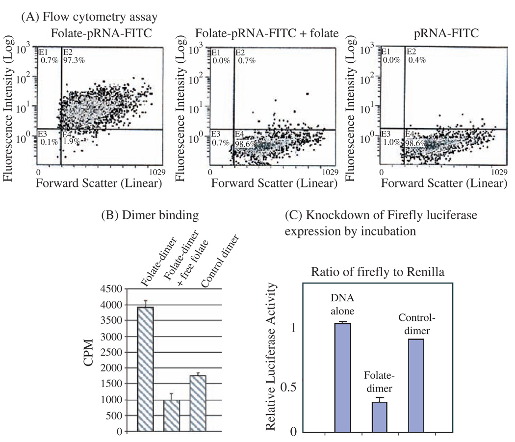
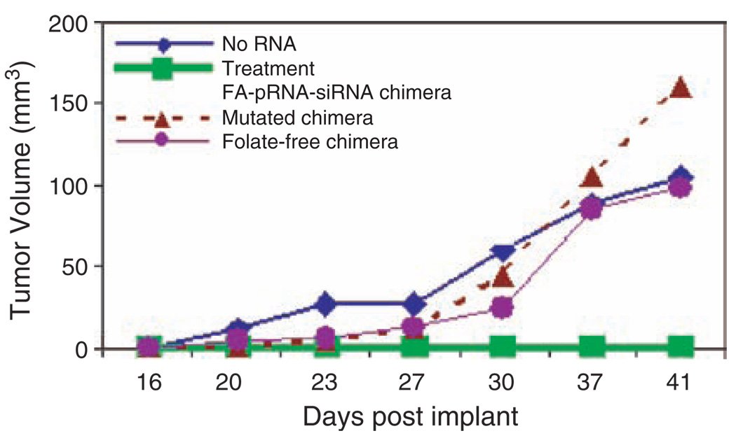
Biological macromolecules including DNA, RNA, and proteins, have intrinsic features that make them potential building blocks for the bottom-up fabrication of nanodevices. RNA is unique in nanoscale fabrication due to its amazing diversity of function and structure. RNA molecules can be designed and manipulated with a level of simplicity characteristic of DNA while possessing versatility in structure and function similar to that of proteins. RNA molecules typically contain a large variety of single stranded loops suitable for inter- and intra-molecular interaction. These loops can serve as mounting dovetails obviating the need for external linking dowels in fabrication and assembly. The self-assembly of nanoparticles from RNA involves cooperative interaction of individual RNA molecules that spontaneously assemble in a predefined manner to form a larger two- or three-dimensional structure. Within the realm of self-assembly there are two main categories, namely template and non-template. Template assembly involves interaction of RNA molecules under the influence of specific external sequence, forces, or spatial constraints such as RNA transcription, hybridization, replication, annealing, molding, or replicas. In contrast, non-template assembly involves formation of a larger structure by individual components without the influence of external forces. Examples of non-template assembly are ligation, chemical conjugation, covalent linkage, and loop/loop interaction of RNA, especially the formation of RNA multimeric complexes. The best characterized RNA multiplier and the first to be described in RNA nanotechnological application is the motor pRNA of bacteriophage phi29 which form dimers, trimers, and hexamers, via hand-in-hand interaction. phi29 pRNA can be redesigned to form a variety of structures and shapes including twins, tetramers, rods, triangles, and 3D arrays several microns in size via interaction of programmed helical regions and loops. 3D RNA array formation requires a defined nucleotide number for twisting and a palindromic sequence. Such arrays are unusually stable and resistant to a wide range of temperatures, salt concentrations, and pH. Both the therapeutic siRNA or ribozyme and a receptor-binding RNA aptamer or other ligands have been engineered into individual pRNAs. Individual chimeric RNA building blocks harboring siRNA or other therapeutic molecules have been fabricated subsequently into a trimer through hand-in-hand interaction of the engineered right and left interlocking RNA loops. The incubation of these particles containing the receptor-binding aptamer or other ligands results in the binding and co-entry of trivalent therapeutic particles into cells. Such particles were subsequently shown to modulate the apoptosis of cancer cells in both cell cultures and animal trials. The use of such antigen-free 20-40 nm particles holds promise for the repeated long-term treatment of chronic diseases. Other potentially useful RNA molecules that form multimers include HIV RNA that contain kissing loop to form dimers, tecto-RNA that forms a "jigsaw puzzle," and the Drosophila bicoid mRNA that forms multimers via "hand-by-arm" interactions. Applications of RNA molecules involving replication, molding, embossing, and other related techniques, have recently been described that allow the utilization of a variety of materials to enhance diversity and resolution of nanomaterials. It should eventually be possible to adapt RNA to facilitate construction of ordered, patterned, or pre-programmed arrays or superstructures. Given the potential for 3D fabrication, the chance to produce reversible self-assembly, and the ability of self-repair, editing and replication, RNA self-assembly will play an increasingly significant role in integrated biological nanofabrication. A random 100-nucleotide RNA library may exist in 1.6 x 10(60) varieties with multifarious structure to serve as a vital system for efficient fabrication, with a complexity and diversity far exceeding that of any current nanoscale system. This review covers the basic concepts of RNA structure and function, certain methods for the study of RNA structure, the approaches for engineering or fabricating RNA into nanoparticles or arrays, and special features of RNA molecules that form multimers. The most recent development in exploration of RNA nanoparticles for pathogen detection, drug/gene delivery, and therapeutic application is also introduced in this review.


Figures

Fig. 1
Fig. 1
Sequence and structural elucidation of phi29 motor pRNA and related assemblages. (A) Primary and secondary structure of wild-type pRNA I-i′. The binding domain (shaded area) and the DNA translocation domain (the helical region) are marked with bold lines. The four bases in the right and left loops, which are responsible for inter-RNA interactions, are boxed. (B) Three-dimensional structure of wild-type pRNA I-i′ displayed as ribbon. (C) Diagrams depicting the pRNA monomer A-b′ with unpaired right/left loops. (D) pRNA dimers (A-b′)(B-a′). (E) pRNA trimers (A-b′)(B-e′)(E-a′). (F) pRNA monomer with unpaired right/left loops A-b′ and a 6-nucleotide palindromic sequence. (G) pRNA twin A-b′. (H) Size and three-dimensional computer model of phi29 pRNA trimer. Reprinted with permission from [43], D. Shu et al., Nano Lett. 4, 1717 (2004). © 2004, American Chemical Society.
Fig. 2
Fig. 2
Polyacrylamide gel showing monomers, dimers, trimers, twins, tetramers, and arrays. (A) Native and denatured gel. (B) Test of the stability of pRNA dimers under different conditions. Reprinted with permission from [43], D. Shu et al., Nano Lett. 4, 1717 (2004). © 2004, American Chemical Society.
Fig. 3
Fig. 3
Atomic force microscopy (AFM) showing pRNA monomers (A), dimers (B), trimers (C), and arrays (D) of pRNA. The three insets at the left of each panel contain images with higher magnification, as indicated by the size of the frame. The pRNA monomers folded into a checkmark shape, dimers displayed a rod shape, trimer exhibited triangle shape, and arrays displayed as bundles. Formation of dimers requires Mg2+, while the sample on mica was briefly rinsed with water before freezing for cryo-AFM, which resulted in some dissociation of dimers or trimers even when the pRNA was already adsorbed to the activated mica surface. The color within each image reflects the thickness and height of the molecule. The brighter or whiter, the color, the thicker or taller the molecule; the darker the image, the thinner the molecule. Reprinted with permission from [43], D. Shu et al., Nano Lett. 4, 1717 (2004). © 2004, American Chemical Society.
Fig. 4
Fig. 4
A mixture of two complementary twins, A-b′ and B-a′, assembled into two distinct supramolecular structures. (A) Two complementary twins were able to form a stable tetramer (double-twins) by assembling into a circular structure. (B) Concatemers of alternating twins formed when a twin interacted with two rather than one complementary twin. Reprinted with permission from [43], D. Shu et al., Nano Lett. 4, 1717 (2004). © 2004, American Chemical Society.
Fig. 5
Fig. 5
Model of HIV-1 RNA and TectoRNA structure. (A) HIV-1 RNA secondary structure models. Model of the structural rearrangements in the TAR hairpin. TAR dimerization induced by the nucleocapsid protein gives rise to a kissing-loop complex and an extended duplex. The following sequences are marked: the central palindrome (red), extended base-pairing (blue), and secondary parallel helix (green; gray dotted lines in extended duplex). Adapted with permission from [216], E. S. Andersen et al., J. Biol. Chem. 279, 22243 (2004). © 2004, American Society for Biochemistry and Molecular Biology. (B) TectoRNAs employing two loop-receptor motifs and their modes of assembly. (a) Two modes of assembly used in this study. (b) Schematic of RNA assembly unit showing elements varied for this study. The tetraloop (L) is shown in red, the tetraloop receptor (R) in green, the linker (or hinge) in blue and the insert [comprising a helix and a second linker (or hinge)] in magenta (refer also to the 2D and 3D models in Fig. 2 and Fig. 3). Adapted with permission from [175], L. Jaeger et al., Nucleic Acids Res. 29, 455 (2001). © 2001, Oxford University Press.
Fig. 6
Fig. 6
Functional assay of chimeric pRNA/siRNA(GFP) by transfection. (A), (B), and (C) are fluorescence microscopy images to show the silencing of GFP gene by transfection. (A) Dose-dependent silencing of GFP gene by chimeric pRNA/siRNA(GFP) (the left column). A mutant pRNA/siRNA on the right column, served as negative control. (A) GFP expression of cells transfected with various RNA. (a) No RNA; (b) Synthesized double-stranded siRNA(GFP); (c) Double-stranded siRNA(LacZ) control; (d) pRNA/siRNA(GFP); (e) pRNA/siRNA(mutant); (f) pRNA vector alone (C) Comparison of the performance of chimeric pRNA/siRNA(GFP) (a) and the conventional double-stranded -siRNA(GFP) (b) at the same molar concentration. Panel (c) is the control with no siRNA treatment. (D) Northern blot to examine the effect of chimeric pRNA/siRNA(GFP) on GFP mRNA level after transfection. Lanes 1 and 2 show the effects of two different constructs of pRNA/siRNA(GFP). Lane 3 is the double-stranded siRNA and lane 4 is cells without RNA treatment. rRNA was used as loading control. Reprinted with permission from [46], S. Guo et al., Human Gene Therapy 16, 1097 (2005). © 2005, Mary Ann Liebert, Inc.
Fig. 7
Fig. 7
Functional assay of chimeric pRNA/siRNA targeting luciferase by transfection. (A) Dual reporter luciferase assay showing the specific knockdown of firefly luciferase or renilla luciferase expression by pRNA/siRNA(Firefly) or pRNA/siRNA(Renilla), respectively in a dose dependent manner. (B) Comparison of the activities of the conventional hairpin siRNA(Luciferase) and pRNA/siRNA(Luciferase). pRNA/ siRNA(mutant) with mutations in siRNA sequences was included as a nonspecific control. Reprinted with permission from [46], S. Guo et al., Human Gene Therapy 16, 1097 (2005). © 2005, Mary Ann Liebert, Inc.
Fig. 8
Fig. 8
Apoptosis and cell death induced by transfection of chimeric pRNA harboring siRNA targeting survivin. (I) Breast cancer MCF-7 cells were transfected with pRNA/siRNA(Survivin) and apoptosis was monitored using PI/annexin V double-labeling followed by flow cytometry. Cells at the lower right quadrant represent apoptotic cells. (II) Breast cancer cells MDA-231 and prostate cancer cells PC-3 were transfected with 20 pmol of pRNA/siRNA(Survivin) in 24-well plates and images were taken in 24 hours after transfection. The mutant pRNA/siRNA was transfected in parallel as a negative control. Reprinted with permission from [46], S. Guo et al., Human Gene Therapy 16, 1097 (2005). © 2005, Mary A nn Liebert, Inc.
Fig. 9
Fig. 9
Confocal microscopy showing the specific and simultaneous delivery of three components to CD4 over-expressing cells. (I) Assay for the binding of pRNA trimer containing pRNA(A-b′)/aptamer(CD4), pRNA(B-e′)-FITC, and pRNA(E-a′)-Rhodamine to CD4 over-expressing T cells (A–D of left column, and I–L of right column) and CD4 negative cells T cells (E–H of middle column). A, E, and I were imaged with an FITC filter; whereas B, F, and J were viewed with a Rhodamine filter; C, G, and K are overlays; and D, H, and L are DIC images. The right column represents a close-up view of CD4 over-expressing cells. Arrows point to the complexes that had entered the cell. (II) Section of confocal microscopy images to differentiate between binding (M) and cell entry (arrows in N and O) as well as negative control (P). Binding of FITC-labeled pRNA trimer containing CD4-binding aptamer to lymphocytes was shown as a circle and entry was shown as a green spot inside cell (arrow in O). The red color in N is a positive entry control of transferrin labeled with Texas red. Reprinted with permission from [47], A. Khaled et al., Nano Lett. 5, 1797 (2005). © 2005, American Chemical Society.
Fig. 10
Fig. 10
Specific delivery of chimeric pRNA/siRNA by folate-pRNA. (A) Flow cytometry analyses of the binding of folate-pRNA-FITC to KB cells. Cells were incubated with folate-pRNA labeled with FITC (left panel). Cells in the middle panel were pre-incubated with free folate, which served as a blocking agent to compete with folate-pRNA for binding to the receptor. Binding was also tested using folate-free pRNA labeled with FITC (right panel) as a negative control. The percentages of FITC-positive cells are shown in the upper right quadrants. (B) Specific binding of folate-pRNA dimer to KB cells. After incubation of cells with the [3H]-folate-pRNA dimer in the presence (center column) or absence (left column) of free folate, cells were isolated and subjected to scintillation counting. The right column is the [3H]-dimer without folate labeling as a negative control. (C) In the knockdown assay by incubation, folate-chimeric dimer complex containing pRNA(B-a′)/folate and pRNA(A-b′)/siRNA(firefly) was incubated with KB cells for 3 hours to allow the binding and entry of RNA. The luciferase level was measured in the next day by the dual reporter system. The control dimer was identical to the folate dimer except for its lack of folate labeling. Reprinted with permission from [46], S. Guo et al., Human Gene Therapy 16, 1097 (2005). © 2005, Mary Ann Liebert, Inc.
Fig. 11
Fig. 11
Animal trials for cancer therapy using the fabricated RNA nanoparticles. (A) Injection without the pRNA/siRNA chimera (No RNA); (B) Treatment with RNA chimera containing folate-pRNA and siRNA(Survivin); (C) Treatment with RNA chimera containing folate-pRNA and siRNA(Survivin) with mutations in the siRNA sequence; (D) Treatment with pRNA-siRNA chimera that does not contain a folate at its 5′ end. Reprinted with permission from [47], A. Khaled et al., Nano Lett. 5, 1797 (2005). © 2005, American Chemical Society.
Fig. 12
Fig. 12
The potential use of pRNA hexamers as polyvalent gene delivery vectors. Six copies of pRNA have been found to form a hexameric ring to drive the DNA-packaging motor of bacterial virus phi29. There would therefore be six positions available to carry foreign moieties for targeting, therapy, and detection, as shown in the figure. Reprinted with permission from [46], S. Guo et al., Human Gene Therapy 16, 1097 (2005). © 2005, Mary Ann Liebert, Inc.

References

    1. Niemeyer CM. Trends Biotechnol. 2002;20:395. - PubMed
    1. Schmidt OG, Eberl K. Nature. 2001;410:168. - PubMed
    1. Baneyx G, Baugh L, Vogel V. Proc. Natl. Acad. Sci. USA. 2002;99:5139. - PMC - PubMed
    1. Hyman P, Valluzzi R, Goldberg E. Proc. Natl. Acad. Sci. USA. 2002;99:8488. - PMC - PubMed
    1. Goldberger J, He R, Zhang Y, Lee S, Yan H, Choi HJ, Yang P. Nature. 2003;422:599. - PubMed

Publication types

LinkOut - more resources