

Optical coherence tomography to identify intramucosal carcinoma and high-grade dysplasia in Barrett's esophagus
- PMID: 16431303
- PMCID: PMC2703582
- DOI: 10.1053/S1542-3565(05)00746-9
Optical coherence tomography to identify intramucosal carcinoma and high-grade dysplasia in Barrett's esophagus
Abstract
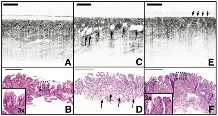
Background & aims: Optical coherence tomography (OCT) is an optical technique that produces high-resolution images of the esophagus during endoscopy. OCT can distinguish specialized intestinal metaplasia (SIM) from squamous mucosa, but image criteria for differentiating intramucosal carcinoma (IMC) and high-grade dysplasia (HGD) from low-grade dysplasia (LGD), indeterminate-grade dysplasia (IGD), and SIM without dysplasia have not been validated. The purpose of this study was to establish OCT image characteristics of IMC and HGD in Barrett's esophagus.
Methods: Biopsy-correlated OCT images were acquired from patients with Barrett's esophagus undergoing endoscopic surveillance. Two pathologists rendered consensus diagnoses of the biopsy specimens. A blinded investigator reviewed the biopsy-correlated OCT images and scored each for surface maturation and gland architecture. For each image the scores were summed to determine an OCT "dysplasia index."
Results: A total of 177 biopsy-correlated images were analyzed. The corresponding histopathology diagnosis was IMC/HGD in 49 cases, LGD in 15, IGD in 8, SIM in 100, and gastric mucosa in 5. A significant relationship was found between a histopathologic diagnosis of IMC/HGD and scores for each image feature (dysplasia index [Spearman correlation coefficient, r = 0.50, P < .0001], surface maturation [r = 0.48, P < .0001], and gland architecture [r = 0.41, P < .0001]). When a dysplasia index threshold of >or=2 was used, the sensitivity and specificity for diagnosing IMC/HGD were 83% and 75%, respectively.
Conclusions: An OCT image scoring system based on histopathologic characteristics has the potential to identify IMC and HGD in Barrett's esophagus.
Figures
Comment in
-
Detecting dysplasia with optical coherence tomography.Clin Gastroenterol Hepatol. 2006 Jan;4(1):36-7. doi: 10.1016/j.cgh.2005.10.005. Clin Gastroenterol Hepatol. 2006. PMID: 16431302 No abstract available.
References
-
- Blot WJ, Devesa SS, Kneller RW, et al. Rising incidence of adenocarcinoma of the esophagus and gastric cardia. JAMA. 1991;265:1287–1289. - PubMed
-
- Bytzer P, Christensen PB, Damkier P, et al. Adenocarcinoma of the esophagus and Barrett's esophagus: a population-based study. Am J Gastroenterol. 1999;94:86–91. - PubMed
-
- Devesa SS, Blot WJ, Fraumeni JF., Jr Changing patterns in the incidence of esophageal and gastric carcinoma in the United States. Cancer. 1998;83:2049–2053. - PubMed
-
- Lagergren J, Bergstrom R, Lindgren A, et al. Symptomatic gastroesophageal reflux as a risk factor for esophageal adenocarcinoma. N Engl J Med. 1999;340:825–831. - PubMed
-
- Sharma P, McQuaid K, Dent J, et al. A critical review of the diagnosis and management of Barrett's esophagus: the AGA Chicago Workshop. Gastroenterology. 2004;127:310–330. - PubMed
Publication types
MeSH terms
Grants and funding
LinkOut - more resources
Full Text Sources
Other Literature Sources
Medical
