. 2006 Mar 17;281(11):7429-36.
doi: 10.1074/jbc.M512138200. Epub 2006 Jan 23.

Rapid degradation of Bim by the ubiquitin-proteasome pathway mediates short-term ischemic tolerance in cultured neurons

Affiliations

Rapid degradation of Bim by the ubiquitin-proteasome pathway mediates short-term ischemic tolerance in cultured neurons

Robert Meller et al. J Biol Chem. .

Abstract

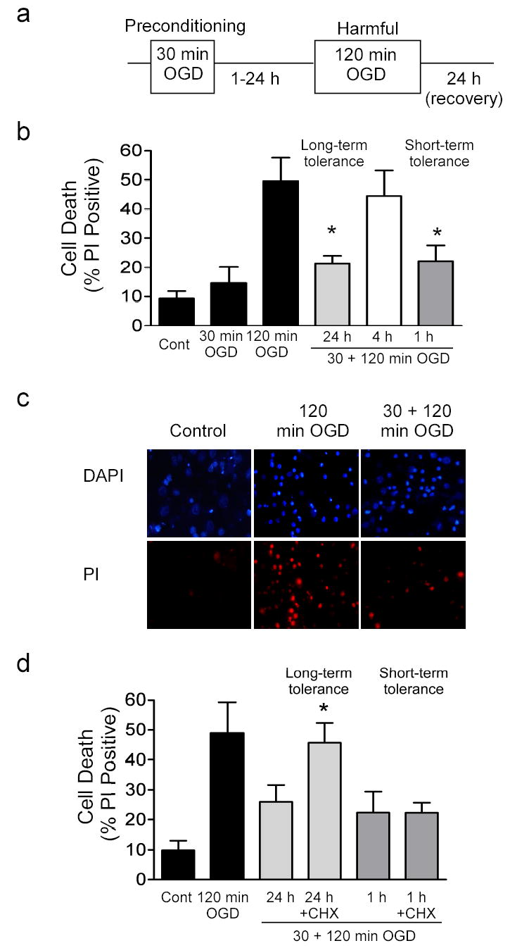
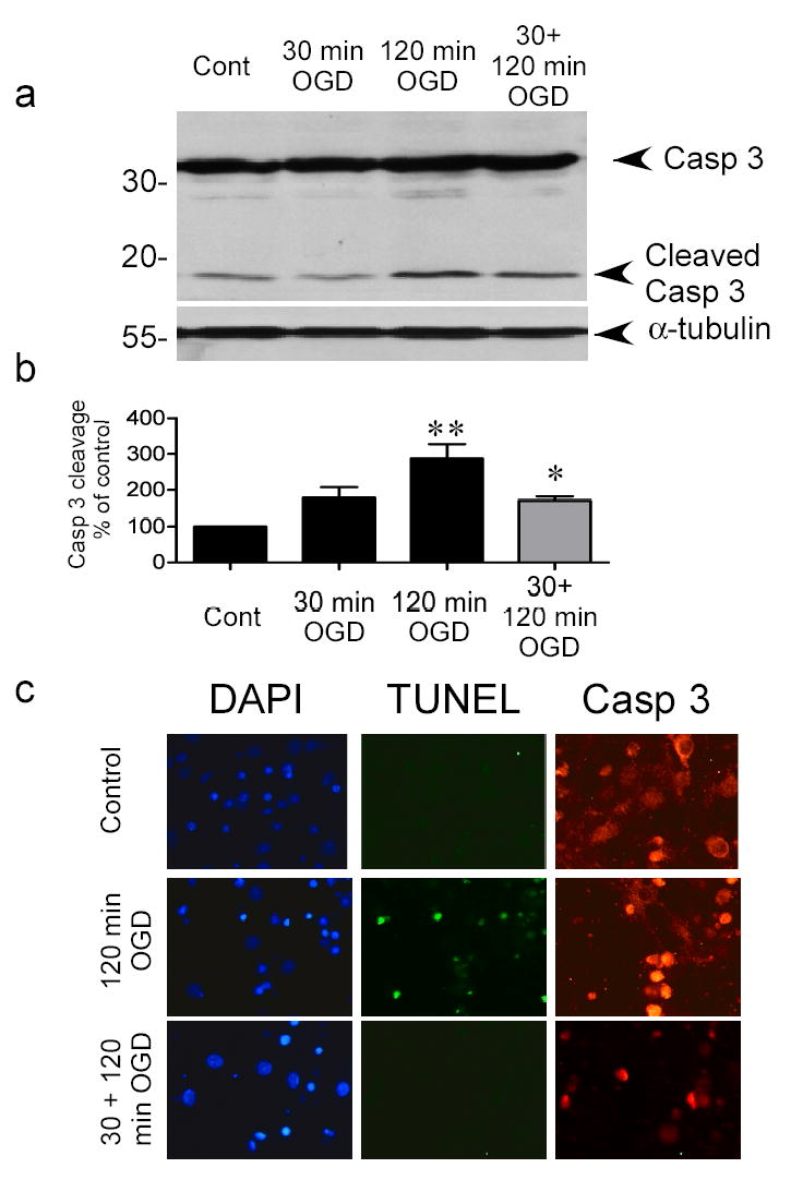
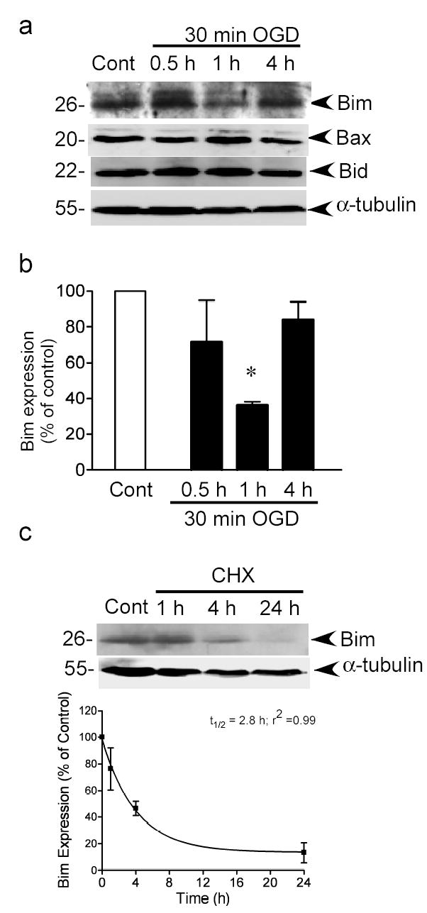
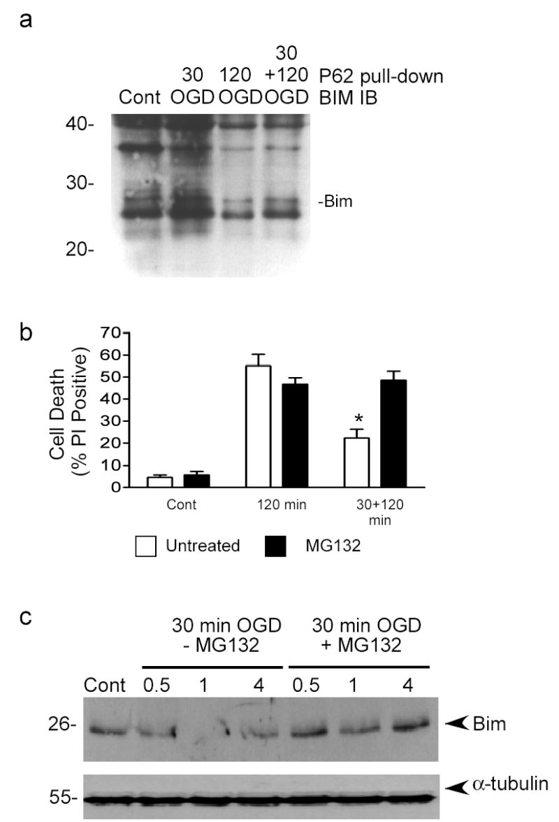
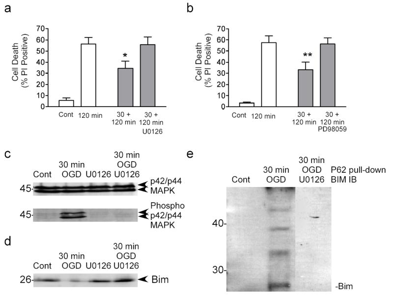
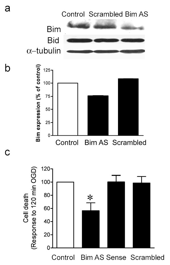
A previous exposure to a non-harmful ischemic insult (preconditioning) protects the brain against subsequent harmful ischemia (ischemic tolerance). In contrast to delayed gene-mediated ischemic tolerance, little is known about the molecular mechanisms that regulate rapid ischemic tolerance, which occurs within 1 h following preconditioning. Here we have investigated the degradation of the pro-apoptotic Bcl-2 family member Bim as a mechanism of rapid ischemic tolerance. Bim protein levels were reduced 1 h following preconditioning and occurred concurrent with an increase in Bim ubiquitination. Ubiquitinated proteins are degraded by the proteasome, and inhibition of the proteasome with MG132 (a proteasome inhibitor) prevented Bim degradation and blocked rapid ischemic tolerance. Inhibition of p42/p44 mitogen-activated protein kinase activation by U0126 reduced Bim ubiquitination and Bim degradation and blocked rapid ischemic tolerance. Finally, inhibition of Bim expression using antisense oligonucleotides also reduced cell death following ischemic challenge. Our results suggest that following preconditioning ischemia, Bim is rapidly degraded by the ubiquitin-proteasome system, resulting in rapid ischemic tolerance. This suggests that the rapid degradation of cell death-promoting proteins by the ubiquitin-proteasome pathway may represent a novel therapeutic strategy to reduce cell damage following neuropathological insults, e.g. stroke.


Figures

Figure 1
Figure 1. Rapid ischemic tolerance in vitro
a) Outline of oxygen and glucose deprivation (OGD) protocol. Cells are subject to 30 min OGD (preconditioning), recovered for variable time periods (1–24 hours) and then subject to 120 min OGD (harmful). For rapid ischemic tolerance an interval of one hour was used between preconditioning ischemia and harmful ischemia. Cell death was assessed by propidium iodide (PI) staining 24 hours following ischemia. Some cells receive 30 min or 120 min OGD only, or no OGD (control). All drug treatments were applied following 30 min OGD and prior to 120 min OGD. b) Effect of various recovery times following 30 min OGD preconditioning on 120 min OGD -induced cell death. Gray blocks denote preconditioned cells. Data shown are mean ± SEM, (n=5). c) Representative images of PI stained cells following no ischemia (control), 120 min OGD and cells preconditioned with 30 min OGD, and then subject to 120 min OGD 1 hour later. Cells were stained and images were acquired 24 hours following last ischemic insult. Note decreased number of PI positive cells in tolerant cultures compared to cells exposed to harmful ischemia without preconditioning. d) Effect of cycloheximide on rapid ischemic tolerance. Cycloheximide was incubated with the cells for 1 hour or 24 hours following preconditioning (Gray denotes long-term tolerance, hatching denotes short-term tolerance). Data shown are mean ± SEM, (n=4) and analyzed by one-way ANOVA with Bonferroni’s post-hoc test (* denotes P<0.05).
Figure 2
Figure 2. Preconditioning ischemia reduces cleavage of caspase 3 following harmful ischemia
a) Cells were preconditioned with 30 min OGD, 1 hour prior to 120 min OGD. Caspase 3 cleavage was determined 24 hours later by immunoblotting with a cleaved caspase 3 P19/17 fragment specific antibody. Blots were reprobed with α-tubulin to control for loading. Image shown is a representative blot of 5 independent experiments. b) Quantification of western blots. Data shown are mean ± SEM (n=5) and analyzed by one-way ANOVA with Bonferroni’s post-hoc test (** denotes P<0.01 vs. control group and * denotes P<0.05 vs. 120 min OGD group). c) Immunocytochemical detection of caspase 3 in control, ischemia-treated and tolerant cortical cells. Cells were subject to various ischemic treatments and caspase 3 levels determined by immunocytochemistry 24 hours later. DNA breaks were also assessed using TUNEL. Note the increased TUNEL staining in 120 min OGD treated cells, and the more nuclear pattern of caspase 3 staining in cells following ischemia. Images are representative of 2 experiments.
Figure 3
Figure 3. Preconditioning ischemia reduces Bim protein levels
a) Bim, Bid and Bax protein levels were determined by immunoblotting. Blots were re-probed with α-tubulin to control for loading. Image shown is a representative image from 3–4 independent experiments. b) Quantification of Bim immunoblots. Data were analyzed by one-way ANOVA with Bonferroni’s post-hoc test, * denotes P>0.05, n=4). c) Cells were incubated with cycloheximide (10 μM) for 1–24 hours and Bim levels were determined by immunoblot. Blots were reprobed for α-tubulin. The optical densities from 3 independent experiments were plotted and data fitted to a mono-exponential decay curve to determine the half-life of Bim (t½ =2.8h, r2= 0.99). Data shown are mean ± sem (n=3).
Figure 4
Figure 4. Bim is ubiquitinated following preconditioning and inhibition of the proteasome blocks rapid ischemic tolerance and Bim degradation
a) Bim ubiquitination was determined by pull-down with a ubiquitin-binding domain conjugated to agarose beads (P62 UBA-agarose). Experiments were run in the presence of MG132 (0.5 μM) to prevent degradation of ubiquitinated Bim. Pulled-down proteins were immunoblotted (IB) for Bim. Image shown is representative of 3 independent experiments. The weight of native Bim is marked. b) Cells were preconditioned with 30 min OGD and then incubated with MG132 (0.5 μM) for 1 hour. Cell death was assessed using propidium iodide (PI) staining. Note that MG132 blocks short term tolerance. Data shown are mean ± SEM (n=6). Data are analyzed by one-way ANOVA, with Bonferroni’s post hoc test (* denotes P<0.01). c) Cells were preconditioned with 30 min OGD and then recovered for 0.5, 1 or 4 hours with or without MG132 (0.5 μM). Bim protein levels were determined by immunoblot. Blots were re-probed with α-tubulin to control for loading. Data shown are representative blot of 3 independent experiments.
Figure 5
Figure 5. p42/ p44 MAPK regulates Bim degradation following preconditioning
a) Cells were preconditioned with 30 min OGD and then incubated for 1 hour with U0126 (10 μM). Cell death was assessed 24 hour following 120 min OGD using propidium iodide staining. Data shown are mean ± SEM (n=8). Data were analyzed by one-way ANOVA, with Bonferroni’s post-hoc test (* denotes P<0.05). b) Cells were preconditioned with 30 min OGD and then incubated for 1 hour with PD98059 (10 μM). Cell death was assessed 24 hour following 120 min OGD using propidium iodide staining. Note that both U0126 and PD98059 block ischemic tolerance. Data shown are mean ± SEM (n=5). Data were analyzed by one-way ANOVA, with Bonferroni’s post-hoc test (** P<0.01). c) Cells were preconditioned with 30 min OGD and the recovered in the presence of U0126 for 1 hour. p42/ p44 MAPK phosphorylation were determined by immunoblot. Blots were reprobed for total p42/ p44 MAPK expression. d) Cells were subject to preconditioning ischemia and then recovered in the presence of U0126. Bim expression was determined by immunoblot. Note that U0126 blocks tolerance-induced Bim degradation. Data shown are representative of 3 independent experiments. e) Cells were preconditioned with 30 min OGD and then recovered in the presence of U0126 for 1 hour. Bim ubiquitination was assessed by p62 UBA precipitation. Bim levels were then detected by immunoblot. The precipitation of ubiquitinated Bim was reduced by U0126 (composite image).
Figure 6
Figure 6. Antisense oligonucleotides reduce Bim expression and ischemia-induced cell death
Cells were incubated for 48 hours with Bim antisense (AS) or scrambled control (SC) oligonucleotides. Bim and Bid protein levels were determined by immunoblot. To control for loading, blots were re-probed for α-tubulin. b) Quantification of immunoblots. c) Cells were treated for 48 hours with Bim antisense, sense or scrambled control oligonucleotides prior to 120 min OGD. Cell death was assessed 24 hours later by propidium iodide staining. Note that Bim antisense but not sense or scrambled control blocks 120 min OGD-induced cell death. Data shown are mean ± SEM (n=7). Data are analyzed by one-way ANOVA, with Bonferroni’s post-hoc test (* denotes P<0.05 vs. Control untreated cells).

References

    1. Dirnagl U, Simon RP, Hallenbeck JM. Trends Neurosci. 2003;26:248–254. - PubMed
    1. Ishida T, Yarimizu K, Gute DC, Korthuis RJ. Shock. 1997;8:86–94. - PubMed
    1. Kitagawa K, Matsumoto M, Tagaya M, Hata R, Ueda H, Niinobe M, Handa N, Fukunaga R, Kimura K, Mikoshiba K, et al. Brain Res. 1990;528:21–24. - PubMed
    1. Dave KR, Saul I, Busto R, Ginsberg MD, Sick TJ, Perez-Pinzon MA. J Cereb Blood Flow Metab. 2001;21:1401–1410. - PubMed
    1. Qi S, Zhan RZ, Wu C, Fujihara H, Yamakura T, Baba H, Taga K, Shimoji K. Neurosci Lett. 2001;315:133–136. - PubMed

Publication types

MeSH terms

LinkOut - more resources